Uterine fibroids - malignant or not
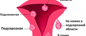
Uterine fibroids are estrogen-dependent benign tumors, but are unpredictable in nature. It develops differentially and can quickly increase, decrease, or even disappear regardless of treatment. In rare cases, it can degenerate into an oncological disease - a fast-growing sarcoma, accompanied by heavy uterine bleeding.
The opinions of experts regarding the degeneration of uterine fibroids into cancer are ambiguous. Some insist that this is still a tumor and favorable factors can give impetus to the transition to oncology. Others defend the benign nature of the formation, and consider fears about whether uterine fibroids can develop into cancer to be unfounded.
We can definitely say that myosarcoma, formed from smooth muscle cells of the myometrium, belongs to oncological diseases. It is believed that the neoplasm develops independently, sometimes simultaneously with fibroids.
Diagnostic algorithm for suspected uterine sarcoma
It doesn’t matter what position the gynecologist takes regarding benign education. If the doctor has the slightest suspicion that a dangerous tumor is hiding under the guise of fibroids, he must conduct a full examination and make an accurate diagnosis. In this case, it is not critical whether the fibroid has degenerated into cancer or whether the malignant neoplasm arose without previous myometrial pathology.
Examination scheme for detecting sarcoma:
Gynecological examination
Mandatory, but not very informative research in this case. The doctor must examine the woman in the chair, but all he will determine is the presence of a formation in the uterus. Sarcoma is supported by its non-displaceability and cyanosis of the mucous membrane of the cervix, however, these signs are not very accurate and cannot be the basis for making a diagnosis.
During a gynecological examination, the doctor can determine the presence of a tumor, but not differentiate it.
If sarcoma is suspected, the gynecologist performs a rectovaginal examination to assess the condition of the tissues of the vagina and rectum. This method allows you to determine the size and location of the node, as well as identify metastases of a malignant tumor.
https://youtu.be/O-9FDRzf6uU
When it turns into cancer
The development of cancer in the body occurs under the influence of a number of factors: excess weight, frequent stress, excessive physical activity, injuries, unhealthy lifestyle, unbalanced diet, heating the area of tumor formation.
Active enlargement of a myomatous node is not a reason to believe that the tumor is developing into a malignant one. Most often this occurs due to swelling or against the background of degenerative processes.
Any changes in a woman’s condition are a signal to go to the doctor. Negative signs include: acyclic bleeding, uncharacteristic vaginal discharge, pain, unreasonable abdominal growth, menstrual irregularities, deterioration in general health, problems with urination and defecation. Many diseases exhibit similar symptoms, so don’t panic and think about the worst.
Therapy
Treatment depends on the stage of cancer growth. With sarcoma, part or all of the uterine body, as well as the appendages and ovaries are removed. The later the disease is diagnosed, the less chance of life. The therapy is prescribed as follows:
- Treatment is combined, using radiation before surgery to reduce the growth and proliferation of the formation.
- Radiation therapy if surgery cannot be used.
- The use of drugs with antitumor effect for third and fourth stage cancer.
- They use the opportunity to improve the quality of life of patients at the last stage of oncology. Chemical treatment and painkillers are used. Then a smear from the pelvic organs is taken for analysis.
- If re-growth of the tumor occurs, the cervical canal and body of the uterus, vagina, part of the intestine, and bladder are removed.
Upon clinical examination, sarcoma and fibroids are similar; it is difficult to determine the disease. For an accurate determination, histological analyzes are used. It is impossible not to treat a small nodule, since as it grows it can negatively affect your health!
Prevention and constant examination in the gynecological office will help prevent serious consequences. You should not neglect diseases of the cervix and uterine body. It is recommended to adhere to a healthy lifestyle and refrain from destructive addictions. A woman should visit a doctor more than once a year. Leiomyoma is not considered a dangerous neoplasm, but oncology occurs in the depths of the layer. Therefore, cancer must be treated at the initial stage of development.
Any change in the functioning of the body is the first signal to contact a specialist. This will allow the disease to be caught at an early stage and will not allow it to progress to the cancerous stage.
Myoma is a benign tumor of the uterus that grows from the muscle layer. The disease is accompanied by the appearance of chronic pelvic pain, intermenstrual discharge and other cycle disorders. Nodules are found mainly in women over 35 years of age, and every woman is primarily concerned about one thing: can fibroids turn into cancer? By and large, the entire tactics of diagnosis, treatment and monitoring of a patient with identified pathology depends on the answer to this question.
The first thing every woman should remember: uterine fibroids are not cancer, but under certain conditions it is possible to develop a malignant tumor in the tissues of the reproductive organ. Knowing how and why oncology occurs, you can notice the first signs of the insidious disease in time, begin treatment and prevent the development of deadly complications.
What types of nodes can progress to tumor
Malignant formations are distinguished by a common feature - rapid cell division and proliferation of pathogenic tissues (proliferation). According to morphological characteristics, myomatous nodes are divided into:
- simple - benign with a low rate of cell division;
- proliferating – fast-growing fibroids with benign morphogenetic criteria and the level of pathological mitoses not exceeding 25%;
- presarcoma - the last step on the path to degeneration into a cancerous tumor, characterized by a large number of foci of development of myogenic cells with obvious signs of atypia.
According to statistics, the full transition of fibroids from benign to malignant occurs in less than 1% of all registered cases.
It is impossible to distinguish oncology by sight with a 100% guarantee. This requires laboratory testing of the tumor. The rate of cell division, the number of myomatous nodes, structural features, and signs of atypia are taken into account. Histological classification identifies the following types of uterine fibroids:
- miotically active – characterized by a complete absence of cell atypia and their rapid growth;
- cellular – develops slowly, there are no atypical signs, smooth muscle tissue predominates in the structure;
- epithelioid - consists of epithelial tissue, forms several subtypes;
- bizarre - grows slowly, does not show atypia, is characterized by degeneration of tumor tissue, occurs during pregnancy and taking hormonal contraceptives;
- vascular – stitched with a large number of large blood vessels, difficult to diagnose;
- apoplectic (hemorrhagic) - appears in pregnant women, women who are addicted to hormonal contraception or after childbirth, accompanied by swelling and hemorrhages;
- leiomyolipoma - distinguished by a large percentage of mature fatty structures, observed before and during menopause;
- polysadoid - extremely rare, characterized by an uncharacteristic arrangement of muscle fibers;
- leiomyoma with infiltration of lymphocytes - often raises suspicions of oncology due to its slight difference with lymphoma, causing inflammatory processes in the female body;
- myxoid - exhibits infiltrative growth; between its smooth muscle tissue there is a lot of amorphous substance resembling mucus. There are no obvious signs of cellular atypia, but at the same time, the chances of a positive outcome of the disease are insignificant.
There are also rare myomatous formations, classified as a separate group, which are characterized by certain growth patterns. Diffuse leiomyomatosis affects young women under 35 years of age. In this case, there is a significant increase in the size of the uterus due to the diffuse proliferation of tumor tissues.
Benign metastatic leiomyoma, consisting of smooth muscle tissue, can extend beyond the uterus. It grows into the lungs and lymph nodes. Parasitic uterine fibroids are capable of separating from the reproductive organ and using blood vessels to infect the omentum and other pelvic organs.
In medical practice, there have been cases where nodes developed in large numbers on the surface of the peritoneum. This pathology is called disseminated peritoneal leiomyomatosis, refers to benign neoplasms, stimulates metastatic carcinoma.
What's happened
Fibromyoma is a pathological process characterized by the formation of benign tumor formations consisting of muscle structures and fibrous tissue.
Its main feature is its slow growth and the absence of transition to adjacent tissues. More often, the pathology is diagnosed in women of reproductive age.
If previously the disease affected patients aged 30-40 years, recently there has been a tendency for the disease to rapidly rejuvenate, since it has increasingly begun to appear in girls over 20 years of age. This is explained by the increased impact of negative factors on a young body.
Mechanical factors and operations that may result in damage to the genital organs should also be taken into account.
Treatment of malignancy
The benign nature of the tumor should not provoke a woman to neglect her own health. At the initial stage, symptoms of the disease rarely appear, but an overgrown node causes many problems. Patients with uterine fibroids should undergo regular gynecological examinations and pelvic ultrasound.
In order not to worry about whether the tumor will turn malignant or not, after diagnosis, you need to immediately begin treatment.
Modern medicine offers two ways to get rid of uterine fibroids: conservative and surgical. In the first case, medications are prescribed to eliminate the main symptoms and stop the growth of the tumor. The second method is more radical and involves instrumental treatment methods: FUS ablation, UAE, myomectomy, hysterectomy, etc.
When a malignant neoplasm is diagnosed, immediate removal of the tumor is necessary. If the disease is advanced, removal of the uterus and sometimes the ovaries will be required. After the operation, the woman is prescribed a course of chemotherapy.
Symptoms
Depending on the location of fibroids, symptoms may differ slightly. But in any case, the early stages of the pathological process are often asymptomatic.
On this topic
- Female reproductive system
Do I need to remove an ovarian cyst if it doesn’t bother me?
- Olga Vladimirovna Khazova
- December 4, 2020
So, with a disease localized in the mammary glands, a woman will complain of symptoms such as:
- mobility of the seal upon palpation;
- formation of a single node;
- no pain .
If the tumor affects the ovaries, the process is accompanied by:
- tachycardia;
- ascites;
- weakness;
- anemia;
- bloating and frequent pain in the lower abdomen;
- shortness of breath.
Since the initial symptoms of uterine fibroids are mild, it is practically impossible to independently determine the presence of the disease. Only a doctor can determine the development of the disease during a gynecological examination.
The symptoms of fibroids will depend on the stage of the process, which is most often accompanied by:
- heavy menstrual bleeding;
- nagging pain
- increase in abdominal circumference;
- bloody discharge during the absence of menstruation;
- pain syndrome during sexual intercourse.
A distinctive feature of uterine fibroids is that during menstruation a woman experiences cramping pain. Prolonged menstruation can cause anemia.
Myths
The most common myth about fibroids is that they are cancer or can develop into it. Among other women's misconceptions regarding neoplasms, the following erroneous opinions are found: with fibroids it is impossible to conceive, the only method of treatment is surgery, the pathology will go away on its own, it is necessary to remove even the smallest tumor, preventive measures will not protect against uterine fibroids.
Sometimes, histologically benign neoplasms “masquerade” as cancerous tumors. Therefore, it is very important that the doctor correctly interprets the results of laboratory tests and prescribes reasonable therapy.
