Keywords
- Gonadotropin-releasing hormone agonists;
- Pregnancy;
- Levonorgestrel intrauterine system;
- Magnetic resonance imaging;
- Menopause;
- Uterine fibroids;
- Non-steroidal anti-inflammatory drugs;
- Tranexamic acid;
- Ultrasonography;
- Ulipristal acetate;
- FUS ablation;
- In vitro fertilization;
- Embolization of the uterine arteries.
https://youtu.be/g3QXWS6mzbk
Bibliography
GnRH agonists - gonadotropin-releasing hormone agonists
ART - assisted reproductive technologies
GnRH - gonadotropin-releasing hormone
SHBG - sex hormone binding globulin
BMI - body mass index
COCs - combined oral contraceptives
CT – computed tomography
LNG-IUD – levonorgestrel-containing intrauterine system
MHT - menopausal hormone therapy
ICD 10 - International Classification of Diseases, Tenth Revision
MMK - intermenstrual uterine bleeding
MRI - magnetic resonance imaging
https://www.youtube.com/watch?v=ytcopyrightru
MSCT - multislice spiral computed tomography
NSAIDs - non-steroidal anti-inflammatory drugs
OMC - heavy menstrual bleeding
SPRM (ulipristal acetate) - selective progesterone receptor modulators
SERM - selective estrogen receptor modulators
Ultrasound - ultrasound examination
FUS – ablation – focused ultrasound ablation
Color Doppler mapping
UAE - uterine artery embolization
ESHRE – European Society for Human Reproduction
MRgFUS—ultrasound ablation under magnetic resonance imaging control (FUS ablation)
- Adamyan L.V. “The state of the reproductive system of patients with benign tumors of the internal genital organs and the principles of its restoration after reconstructive plastic surgery” Dissertation for the academic degree. Doctor of Medicine degree Sciences, Moscow, 1985.
- Operative gynecology (2nd edition, revised) Krasnopolsky V.I., Buyanova S.N., Shchukina N.A., Popov A.A. -Moscow, Publishing house MEDpress -inform – 2013. – P.320
- Andersen J. Factors in fibroid growth. // Baillieres Clin. Obstet. Gynaecol. 1998. –Jun – 12(2).–P.225–243.
- Bulun SE. Uterine fibroids.//N Engl J Med.–2013.–369(14).–P.1344-55.
- Adamyan L.V., Kiselev S.I., Zurabiani Z.R. Endoscopy in the diagnosis and treatment of uterine pathology. M., 1997. – T. 1. – P. 200–204.
- Wamsteker K, de Blok S. Diagnostic hysteroscopy: technique and documentation // Sutton C (ed), Endoscopic surgery for gynecologists. 1993a:263-276.
- AAGL Practice Report: Practice Guidelines for the Diagnosis and Management of Submucous Leiomyomas 2012 AAGL.doi:10.1016/j.jmig.2011.09.005
- H.Marret, X.Fritel, L.Ouldamer et al. Therapeutic management of uterine fibroid tumors: updated French guidelines//European Journal of Obstetrics {amp}amp; Gynecology and Reproductive Biology. –2012.–165.–P.156–164.
- National Guideline ClearinghouseACR Appropriateness Criteria® radiological management of uterine leiomyomas. 2012
- Atlas. Spiral computed tomography in gynecology. Kulakov V.I., Adamyan L.V., Makarenko V.N., Muratov K.D. - M. Medicine - Antidor, 2001. - P. 321.
- Adamyan L.V., Murvatov K.D. Magnetic resonance imaging in the diagnosis of pathology of the uterus and appendages / Materials / International Congress on Endometriosis: Moscow, April 22-26, 1996. - M., 1996. P. 190 – 191.
- Atlas. Magnetic resonance imaging in gynecology.— Kulakov V.I., Adamyan L.V., Murvatov K.D. M.: Antidor, 1999. - P. 193.
- Guidelines for the Management of Uterine Leiomyoma HKCOG GUIDELINES NUMBER 13 (November 2009)
- Critchley H, Broder M, Fraser I; FIGO Working Group on Menstrual Disorders. FIGO classification system (PALM-COEIN) for causes of abnormal uterine bleeding in nongravid women of reproductive age. // International Journal of Gynecology and Obstetrics. –2011 –113(1).- P 3–13.
- Modern aspects of myomectomy using surgical energies / Vardanyan V.G., Adamyan L.V., Kiselev S.I., Khachatryan A.K.. M.: Medicine, 2002.
- Intrauterine pathology. Clinic, hysteroscopic diagnosis and treatment. Educational and methodological manual, 2nd ed. Rudakova E.B., Kurilenko T.Yu., Davydov V.V., Davydov P.V. -M. MEDpress-inform. - 2012.
- Zaraq Khan, Elizabeth A., Stewart. Benign Uterine Diseases. Yen{amp}amp; Jaffe's Reproductive Endocrinology (Seventh Edition).–2014. – P.586-603.
- Sizzi O. et al. Italian multicenter study on complications of laparoscopic myomectomy // J Minim Invasive Gynecol. – 2007. – Vol. 14.– No. 4.– P.453–462.
- Tikhomirov A.L. Myoma, pathogenetic rationale for organ-preserving treatment. – M., 2013–P.319. ill.
- Bezhenar V.F., Tsypurdeeva A.A., Dolinsky A.K., Bochorishvili R.G. Laparoscopic hysterectomy - seven years of experience.//J. obstetrics and women's diseases. – 2011 –. N.4. –C.12 – 19.
- Doridot V., Dubuisson JB, Chapron C. et al. Recurrence of leiomyomata after laparoscopic myomectomy // J Am Assoc Gynec Laparoscop.- 2001.-Vol.8.-No.4. R. - 495-500.
- Guriev T.D., Sidorova I.S., Unanyan A.L. Combination of uterine fibroids and adenomyosis. M: MIA. –2012. -WITH. 256.
- Kongnyuy EJ, Wiysonge CS. Interventions to reduce haemorrhage during myomectomy for fibroids.Cochrane Database Syst Rev. 2009 Jul 8(3).
- Dolinsky A.K. The effectiveness of infertility treatment when performing myomectomy using various surgical approaches. abstract dis. Ph.D., St. Petersburg. – 2013. – P.23.
- Vilos G, Allaire C, Laberge P, Leyland N; Vilos A, Murji A, Chen I. The management of uterine leiomyomas. J Obstet Gynaecol Can. -2015.-Vol.37.No.2.-R.-157-81.
- Kulakov V.I., Savelyeva G.M., Manukhin I.B. Gynecology. National leadership. M.: Geotar-Media, 2009.
- National guidelines for radiation diagnostics and therapy” / Ch. ed. series S.K. Ternova. Chapter: Radiation diagnostics and therapy in obstetrics and gynecology: National guidelines / Ch. ed. volumes L.V. Adamyan, V.N. Demidov, A.I. Gus, I.S. Obelchak. – M.: GEOTAR-Media – 2012. –P. 656.
- Berkane N, Moutafoff-Borie S. Impact of previous uterine artery embolization on fertility // Curr. Opin. Obstet. Gynecol. – 2010. – Vol.22. – No. 3. – P. 242–247
- Sanne M, Willem M. et all. Review of nonsurgical/minimally invasive treatments for uterine fibroids // Curr Opin Obstet Gynecol. -2012.-Vol.24.-No.6.-P.368-75.
- Scheurig-Muenkler C., Lembcke A., Froeling V., Maurer M., Hamm B., Kroencke TJ Uterine artery embolization for symptomatic fibroids: long-term changes in disease-specific symptoms and quality of life.Hum Reprod.-2011 .-Vol.26.-No.8.P.2036–42.
- Kurashvili Yu.B., Salamadina G.E., Chmyr E.N.. Batarshina O.I. The effectiveness of focused ultrasound under magnetic resonance imaging control for uterine fibroids. Obstetrics and Gynecology, 2010, No. 3. P.61-65.
- Boosz A, Reimer P, Matzko M, R?mer T, M?ller A. The conservative and interventional treatment of fibroids. Dtsch Arztebl Int. -2014.-Vol.111.No.51-52.-R.877-83.
- Maksutova D.Zh. Application of focused ultrasound under the control of magnetic resonance imaging (literature review) // Problems of reproduction, 2009, No. 2, pp. 30-36.
- Lyadov K.V., Kurashvili Yu.B., Stepanov AB, Bogomazova S.Yu., Vishninsky AA Prediction of the effectiveness of ablation of uterine fibroid nodes according to magnetic resonance imaging. Obstetrics and gynecol. – 2007. – No. 6. – P.72-75.
- Tempany C, Stewart E, McDannold N. et al. MR imaging-guided focused ultrasound surgery of uterine leiomyomas: a feasibility study. Radiology -2003.Vol.226.-No.3.-R.897-905.
- Duhan N. Current and emerging treatments for uterine myoma – an update // Int J Womens Health. -2011.-No.3.-R.231-41.
- Zairatyants O.V. PAEC (Progesterone receptor modulator Associated Endometrial Changes). Endometrial changes associated with progesterone receptor modulator. A new type of reversible morphological changes in the endometrium during treatment of uterine leiomyomas with Esmya? (ulipristal acetate, pharmaceutical). Guide for pathologists and obstetricians-gynecologists. – M., 2013. –S. thirty.
- Kogan I.Yu., Bezhenar V.F., Dolinsky A.K., Chmaro M.G. The effectiveness of assisted reproduction methods in patients with uterine fibroids. // Journal of Obstetrics and Women’s Diseases – 2012. –N 4.–P.113–118.
- Litsova, A.O. The role of natural killer cells in the pathogenesis of the rapid growth of uterine fibroids, the influence of agonists of gonadotropic releasing hormones on their content and functional state: abstract. dis. Ph.D. honey. Sci. -Ivanovo. – 2013. – P.24.
- Protopopova N.V., Safonova O.M., Portnyagina T.V., Kravchuk N.V. Tactics of management of pregnant women with uterine fibroids. // Bulletin of the East Siberian Scientific Center of the Siberian Branch of the Russian Academy of Medical Sciences. – 2002-No.5. -T. 2.-P.149-150.
- Rational pharmacotherapy in obstetrics and gynecology. Guide for practicing doctors / V.I. Kulakov, V.N. Serov, P.R. Adakarov, A.G. Antonov and others. Under the general direction. ed. IN AND. Kulakova, V.N. Serova. – M.: Litera. – 2005.– P.1152.
- Samoilova T. E. Non-operative methods of treating uterine fibroids // Attending physician. – 2010. – N 3. – P.53–60.
- Yarmolinskaya M.I., Tarasova M.A. Efficacy of phytoestrogens in the treatment of menopausal syndrome in women with uterine fibroids // Journal of Obstetrics and Women's Diseases. – 2009. – T.LVIII.–N 1.– P. 71 – 77.
- Croxtall JD Ulipristal acetate: in uterine fibroids // Drugs. – 2012. – Vol.72N8. – P.1075–1085.
- Donnez J., Vezquez F., Tomaszewski J., Nouri K. et al. Long-term treatment of uterine fibroids with ulipristal acetate. // Fertil Steril. 2014 Jun;101(6):1565-73.
- Eder S, Baker J, Gersten J, Mabey RG, Adomako TL. Efficacy and safety of oral tranexamic acid in women with heavy menstrual bleeding and fibroids. //Women's Health. – 2013.-Jul;9(4).-P. 397-403.
- Giraudet G., Lucot JP et al.Except fertility, place of myomectomy in perimenopause and after menopause //Gynecol Obstet Biol Reprod.–2011.–Vol. 40.–N 8.– P.902–917.
- Gunes M, Ozdegirmenci O, Kayikcioglu F, Haberal A, Kaplan M. The effect of levonorgestrel intrauterine system on uterine myomas: a 1-year follow-up study. // J Minim Invasive Gynecol. -2008- 15.-P. 735–738.
- Klatsky PC, Tran ND, Caughey AB, Fujimoto VY. Fibroids and reproductive outcomes: a systematic literature review from conception to deliver.// Am J Obstet Gynecol. –2008. – 198. – P.357-366.
- LethabyAnne E.,VollenhovenBeverleyJ. An evidence-based approach to hormonal therapies for premenopausal women with fibroids Best Practice {amp}amp; Research Clinical Obstetrics and Gynecology. –2008. – Vol. 22– N. 2– p. 307–331.
- Radosa MP, Winzer H. et al. Laparoscopic myomectomy in peri- and post-menopausal women is safe, effective and associated with long-term patient satisfaction // Eur J Obstet Gynecol Reprod Biol. – 2012. – Vol.162–N 2. – P. 192 – 196.
- Islam, O. Protic, S. R. Giannubilo, P. Toti, A. Luigi Tranquilli, F. Petraglia, M. Castellucci, P. Ciarmela. Uterine leiomyoma: available medical treatments and new possible therapeutic options.// J Clin Endocrinol Metab, 2013–98(3). –P.921–934.
Pharm Group
| Adamyan Leila Vladimirovna | Academician of the Russian Academy of Medical Sciences, Deputy Director of the Federal State Budgetary Institution Scientific Center for Obstetrics, Gynecology and Perinatology named after. V.I. Kulakov" of the Ministry of Health of Russia, Doctor of Medical Sciences, Professor, Honored Scientist of Russia, Head of the Department of Reproductive Medicine and Surgery, Moscow State Medical and Dental University. A. I. Evdokimova" of the Ministry of Health of Russia, Chief freelance specialist of the Ministry of Health of Russia in obstetrics and gynecology |
| Andreeva Elena Nikolaevna | Head of the Department of Endocrine Gynecology of the Federal State Budgetary Institution "Endocrinological Research Center" of the Ministry of Health of Russia, Professor of the Department of Reproductive Medicine and Surgery of the State Budgetary Educational Institution of Higher Professional Education "Moscow State Medical and Dental University named after. A. I. Evdokimova" of the Ministry of Health of Russia, Doctor of Medical Sciences |
| Artymuk Natalya Vladimirovna | Head of the Department of Obstetrics and Gynecology No. 2 of the Kemerovo State Medical Academy, President of the Kemerovo regional public organization “Association of Obstetricians and Gynecologists”, Doctor of Medical Sciences, Professor |
| Belotserkovtseva Larisa Dmitrievna | Head of the Department of Obstetrics, Gynecology and Perinatology, Surgut State University, Chief Physician of the Surgut Clinical Perinatal Center, Doctor of Medical Sciences, Professor |
| Bezhenar Vitaly Fedorovich | Head of the Clinic of Obstetrics, Gynecology and Neonatology of the Pediatric Faculty of the First St. Petersburg State Medical University named after Academician I.P. Pavlov, Deputy Chief Obstetrician-Gynecologist of the Health Care Committee of the Government of St. Petersburg, Doctor of Medical Sciences, Professor |
| Gevorkyan Mariyana Aramovna | Professor of the Department of Obstetrics and Gynecology, Faculty of Medicine, Moscow State Medical and Dental University. A. I. Evdokimova" of the Ministry of Health of Russia, Doctor of Medical Sciences |
| Glukhov Evgeniy Yurievich | Candidate of Medical Sciences, Associate Professor of the Department of Obstetrics and Gynecology, Ural State Medical University, Deputy Chief Physician for Obstetrics and Gynecology, Central City Hospital No. 7, Yekaterinburg |
| Gus Alexander Iosifovich | Head of the Department of Functional Diagnostics of the Federal State Budgetary Institution Scientific Center for Obstetrics, Gynecology and Perinatology named after. V. I. Kulakova" of the Ministry of Health of Russia, Doctor of Medical Sciences, Professor |
| Dobrokhotova Yulia Eduardovna | Head of the Department of Obstetrics and Gynecology No. 2, Faculty of Medicine, State Budgetary Educational Institution of Higher Professional Education "Russian National Research Medical University named after. N.I. Pirogov" of the Ministry of Health of Russia, Doctor of Medical Sciences, Professor |
| Jordania Kirill Iosifovich | Leading researcher at the Russian Oncological Research Center named after N.N. Blokhin, professor at the Department of Reproductive Medicine and Surgery, Moscow State Medical and Dental University. A. I. Evdokimova" of the Ministry of Health of Russia, Chairman of "OSORS - Society of Oncologists Specialists in Tumors of the Reproductive System Organs", Doctor of Medical Sciences, Professor |
| Zairatyants Oleg Vadimovich | Head of the Department of Pathological Anatomy, Moscow State Medical and Dental University. A.I. Evdokimov” of the Ministry of Health of Russia, Chief Pathologist of Roszdravnadzor for the Central Federal District of the Russian Federation, Vice-President of the Russian and Chairman of the Moscow Society of Pathologists, Laureate of the Prize named after. A.I.Strukova, Doctor of Medical Sciences, Professor |
| Kozachenko Andrey Vladimirovich | Leading Researcher of the Gynecological Department of the Department of Operative Gynecology and General Surgery of the Federal State Budgetary Institution “Scientific Center of Obstetrics, Gynecology and Perinatology named after V.I. Kulakov” of the Ministry of Health of Russia, Associate Professor of the Department of Reproductive Medicine and Surgery, Moscow State Medical and Dental University. A. I. Evdokimova" of the Ministry of Health of Russia, Doctor of Medical Sciences |
| Kiselev Stanislav Ivanovich | Professor of the Department of Reproductive Medicine and Surgery, Moscow State Medical and Dental University. A. I. Evdokimov" of the Ministry of Health of Russia, laureate of the Russian Government Prize in the field of science and technology, Doctor of Medical Sciences |
| Kogan Evgenia Altarovna | Head of the 1st Pathological Department of the Federal State Institution FGP named after. V.I. Kulakova of the Ministry of Health of Russia, Professor of the Department of Pathanatomy of the National Research Center GBOU HPE “First Moscow State Medical University named after I.M. Sechenov", Doctor of Medical Sciences, Professor |
| Kuznetsova Irina Vsevolodovna | Chief Researcher, Research Institute for Women's Health, National Research Center, State Budgetary Educational Institution of Higher Professional Education, First Moscow State Medical University named after I.M. Sechenov" of the Ministry of Health of Russia, Doctor of Medical Sciences, Professor |
| Kurashvili Yulia Borisovna | Professor of the Department of Medical Physics of National Research Nuclear University "MEPhI", Doctor of Medical Sciences |
| Levakov Sergey Alexandrovich | Head of the Department of Obstetrics and Gynecology, First Moscow State Medical University. I.M. Sechenova, Doctor of Medical Sciences, Professor |
| Malyshkina Anna Ivanovna | Director of the Federal State Budgetary Institution “Ivanovo Research Institute of Motherhood and Childhood named after V.N. Gorodkov" of the Ministry of Health of Russia, Doctor of Medical Sciences |
| Maltseva Larisa Ivanovna | Doctor of Medical Sciences, Professor, Head of the Department of Obstetrics and Gynecology, HBO DPO “Kazan State Medical Academy”, Chief freelance obstetrician-gynecologist of the Volga Federal District, Honored Scientist of the Russian Federation |
| Marchenko Larisa Andreevna | Leading researcher at the Department of Gynecological Endocrinology of the Federal State Budgetary Institution Scientific Center for Obstetrics, Gynecology and Perinatology named after. V.I. Kulakova" of the Ministry of Health of Russia, Doctor of Medical Sciences, Professor |
| Murvatov Kamoljon Jamolkhonovich | Head of the gynecological department of the Main Military Clinical Hospital of the Military Forces of the Ministry of Internal Affairs of the Russian Federation, Colonel of the Medical Service, Honored Doctor of the Russian Federation, Associate Professor of the Department of Reproductive Medicine, Moscow State Medical and Dental University. A. I. Evdokimova" of the Ministry of Health of the Russian Federation, Candidate of Medical Sciences |
| Pestrikova Tatyana Yurievna | Head of the Department of Obstetrics and Gynecology, Far Eastern State Medical University, Ministry of Health of the Russian Federation (Khabarovsk), Honored Doctor of the Russian Federation, Chief Freelance Obstetrician-Gynecologist of the Ministry of Health of the Russian Federation in the Far Eastern Federal District, Doctor of Medical Sciences, Professor |
| Popov Alexander Anatolievich | Head of the Endoscopy Department of the Moscow Regional Research Institute of Obstetrics and Gynecology, Doctor of Medical Sciences, Professor |
| Protopopova Natalya Vladimirovna | Head of the Department of Perinatal and Reproductive Medicine, Irkutsk State Medical Academy of Postgraduate Education, Honored Doctor of the Russian Federation, Doctor of Medical Sciences, Professor |
| Samoilova Alla Vladimirovna | Deputy Chairman of the Cabinet of Ministers of the Chuvash Republic - Minister of Health and Social Development of the Chuvash Republic, Head of the Department of Obstetrics and Gynecology, Chuvash State Medical University. I.N. Ulyanova. Doctor of Medical Sciences, Professor |
| Sonova Marina Musabievna | Head of the Department of Gynecology, Department of Reproductive Medicine, Moscow State Medical and Dental University. A. I. Evdokimova" of the Ministry of Health of Russia, Doctor of Medical Sciences, Professor |
| Tikhomirov Alexander Leonidovich | Doctor of Medical Sciences, Professor of the Department of Obstetrics and Gynecology, Faculty of Medicine, Moscow State Medical and Dental University. A. I. Evdokimov" of the Ministry of Health of Russia |
| Tkachenko Lyudmila Vladimirovna | Head of the Department of Obstetrics and Gynecology, Volgograd State Medical University, Honored Doctor of the Russian Federation, Doctor of Medical Sciences, Professor, Chief Freelance Obstetrician-Gynecologist of the Volgograd Region |
| Urumova Lyudmila Tatarkanovna | Head of the gynecological department of the Federal State Budgetary Institution "Clinical Hospital No. 123 of the Federal Medical and Biological Agency" of Russia, Honored Doctor of the Russian Federation, Candidate of Medical Sciences |
| Filippov Oleg Semenovich | Deputy Director of the Department of Medical Care for Children and Obstetrics Service of the Ministry of Health of Russia, Professor of the Department of Obstetrics and Gynecology of the National Research Center GBOU HPE “First Moscow State Medical University named after I.M. Sechenov" of the Ministry of Health of Russia, Doctor of Medical Sciences, Professor |
| Khashukoeva Assiyat Zulchifovna | Professor of the Department of Obstetrics and Gynecology, Faculty of Medicine, Russian National Research Medical University. N.I. Pirogov" of the Ministry of Health of Russia, Doctor of Medical Sciences |
| Chernukha Galina Evgenievna | Head of the Department of Gynecological Endocrinology, Federal State Budgetary Institution Scientific Center for Obstetrics, Gynecology and Perinatology named after. V. I. Kulakova" of the Ministry of Health of Russia, Doctor of Medical Sciences, Professor |
| Yarmolinskaya Maria Igorevna | Leading researcher of the department of endocrinology of reproduction of the Federal State Budgetary Institution “Research Institute of Obstetrics and Gynecology named after D.O. Ott”, Professor of the Department of Obstetrics and Gynecology No. 2 of the State Budgetary Educational Institution of Higher Professional Education “North-Western State Medical University named after. I.I. Mechnikov" of the Ministry of Health of Russia, Doctor of Medical Sciences |
| Yarotskaya Ekaterina Lvovna | Head of the Department of International Cooperation of the Federal State Budgetary Institution Scientific Center for Obstetrics, Gynecology and Perinatology named after. V. I. Kulakova" of the Ministry of Health of Russia, Doctor of Medical Sciences |
Conflict of interest: Members of the working group are members of the Russian Society of Obstetricians and Gynecologists and the Russian Association of Gynecologists and Endoscopists.
All members of the working group declare that they have no conflict of interest.
• Hormones of the hypothalamus, pituitary gland, gonadotropins and their antagonists. • Antitumor hormonal agents and hormone antagonists.
Pharmacological action – antiandrogenic, antitumor, antigonadotropic, antiestrogenic. Competitively binds to gonadotropin-releasing hormone receptors in the cells of the anterior pituitary gland. With initial or intermittent administration, buserelin stimulates the release of gonadotropins (LH and FSH) by the pituitary gland, which, in turn, causes a short-term increase in the level of sex hormones in the blood plasma.
Chronic use leads to blockade of the gonadotropic function of the pituitary gland and cessation of the release of LH and FSH (on average after 12–14 days). As a result, there is a suppression of the synthesis of sex hormones in the ovaries and a decrease in the concentration of estradiol in the blood to postmenopausal values. Long-term daily use of buserelin in the treatment of prostate cancer leads to a decrease in testosterone concentration to a level characteristic of the condition after orchiectomy, etc.;
to “medicinal castration”. After discontinuation of buserelin, the levels of testosterone and estrogen in the blood are gradually restored. Preoperative treatment for endometriosis reduces inflammation, the size of pathological lesions and their blood supply. As a result of postoperative therapy, the frequency of relapses and the severity of adhesions decreases.
It is well absorbed from the nasal mucosa and after subcutaneous administration. Cmax with intramuscular administration of 3.75 mg is (0.32±0.12) ng/ml, bioavailability when administered once a month is 53%. Excreted in small quantities into breast milk. In vitro studies using bacterial systems (Ames test with Salmonella typhimurium and Escherichia coli) and mammalian cell systems (micronuclear test in mice) did not reveal a mutagenic effect.
In two-year studies on rats with daily subcutaneous administration in doses of 0.2–1.8 mcg/kg, no signs of carcinogenicity were detected. Suppression of testosterone secretion leads to impaired fertility. It is not known whether fertility is restored after discontinuation of buserelin (it is restored after discontinuation of its analogues).
ICD-10 code
D26 Other benign neoplasms of the uterus
Pharm. gr. Substance Drugs for treatment
| Other immunomodulators | Sodium deoxyribonucleate | Derinat | Deoxynate | Ferrovir | Panagen | Sodium deoxyribonucleate |
| Other miscellaneous means | Sodium aminodihydrophthalazindione | Galavit |
| Antitumor hormonal agents and hormone antagonists | Buserelin | Buserelin | Buserelin-depot | Buserelin-long FS | Buserelin FSintez | Suprefact depot | Suprefact | Buserelin-long | Buserelin acetate |
| Goserelin | Zoladex | Goserelin acetate | |
| Lanreotide | Somatulin Autogel | Somatulin | |
| Leuprorelin | Lucrine depot | Prostap | Eligard | |
| Testosterone [ester mixture] | Omnadren 250 | Sustanon-250 | Testenate for injection 10% | |
| Triptorelin | Diferelin | Decapeptyl | Decapeptyl Depot | Triptorelin-long | Triptorelin acetate | |
| Estrogens, gestagens; their homologues and antagonists | Norethisterone | Norkolut | Primolut-Nor |
| Supplements - proteins, amino acids and their derivatives | Substance not described | Estro Pause |
| Dietary supplements are natural metabolites | Substance not described | Estro Pause |
| Dietary supplements - fats, fat-like substances and their derivatives | Substance not described | Estro Pause |
| Supplements - macro- and microelements | Substance not described | Estro Pause | Epigallate |
| Dietary supplements are products of plant, animal or mineral origin | Substance not described | Mylife | Indinol | Estro Pause | Epigallate |
Appendix B: Patient Information
Uterine fibroids are one of the most common tumors of the female genital organs; they account for 27% of gynecological diseases. In addition, it can occur again in 7-28% of patients, sometimes even degenerating into a malignant tumor. If previously it was believed that uterine fibroids mainly affected 40-year-old women, today the problem is even more pressing, since fibroids are increasingly being found in eighteen- and twenty-year-olds. Who has yet to give birth to children? And uterine fibroids can become a serious obstacle in this.
https://www.youtube.com/watch?v=ytcreatorsru
The development of this benign tumor is largely influenced by inflammatory diseases and abortions. In a woman who has had 10 abortions by the age of 30, the risk of developing uterine fibroids by the age of 40 increases by 2 times. It is absolutely certain that uterine fibroids are much more common in nulliparous women.
In 30-40% of cases, the tendency to form uterine fibroids is still inherited: from mother to daughter. There are so-called “familial forms” of uterine fibroids, when all women in the family - grandmothers, mothers, aunts, sisters - had fibroids.
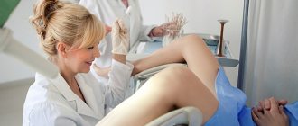
In some cases, at first the fibroid does not manifest itself at all and does not bother the woman. Therefore, fibroids are most often detected during a medical examination by a gynecologist. In this case, the doctor can only state the fact of the presence of uterine fibroids. In other situations, the first signs of fibroids may be pain in the lower abdomen or lumbar region, bleeding, changes in the menstrual cycle, dysfunction of neighboring organs, for example, frequent urination.
Fortunately, in most cases, you can really only get by with an ultrasound. But sometimes, with an atypical location of fibroids, ultrasound results can be unreliable.
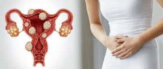
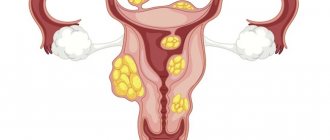
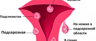
In general, fibroids can be located in the outer, middle and inner layers of the uterus. The most “unpleasant” ones are those located in the inner layer. Such fibroids deform the uterine cavity and, as a rule, cause heavy monthly bleeding with a sharp decrease in hemoglobin. Large fibroids, especially those located in the outer layer, put pressure on neighboring organs, disrupting their functions.
The presence of fibroids in itself is not always an indication for surgery. It all depends on the patient’s condition, the severity of the symptoms of the disease and the size of the tumor.
Indications for surgery to remove uterine fibroids are pain, rapid growth in the size of the node, suspicion of degeneration of uterine fibroids into a malignant tumor, impaired blood supply to the fibroid nodes and inflammatory changes in them, dysfunction of neighboring organs (bladder, intestines), infertility (when all other reasons have already been excluded). If surgery is necessary, then indeed, the sooner it is done, the better.
Small uterine fibroids usually respond well to conservative treatment. Uterine fibroids can be observed dynamically (using ultrasound with a vaginal probe). If symptoms are present, conservative therapy can be carried out - with gestagens, ulipristal, gonadotropin-releasing hormone agonists.
Hormone therapy is carried out to prepare the endometrium (inner layer of the uterus) only in cases where the fibroid node is located in the inner layer of the uterus, and a resectoscopy operation is to be performed (removal of the node from below through the uterus). Hormone therapy is prescribed for 3 months, preferably under ultrasound control, in order to prepare for removal of the myomatous node while preserving the uterus.
lasers (carbon dioxide, argon), special ultrasonic scalpels, etc. After laparoscopy, only small “dots” of 0.5-1 cm are noticeable, since only 5 mm punctures are made to insert instruments into the abdomen. The length of stay in the hospital is reduced to 1-2 days, and immediately after discharge the woman is able to work.
If it is possible to perform the operation through vaginal access or using a hysteroscope, there are no external traces of the surgical intervention left at all. Apart from the woman herself and the gynecologist, no one will ever know what kind of operation she underwent.
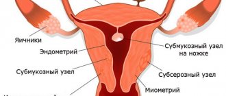
Uterine fibroid (UF) is a limited benign tumor consisting of smooth muscle and fibrous connective tissue elements. The tumor can be single, but more often multiple nodes are detected (sometimes up to 10 or more); hormonal dependent. Statistical data. Found in 4–11% of all women, 20% of women over 30 years of age and 40% of women over 50 years of age.
Uterine fibroids: Diagnosis
Diagnostics
• Palpation of the abdomen. FM are palpated in the form of dense irregularly shaped nodes. With sarcoma, edema and degeneration of FM, the nodes are usually soft and painful. During pregnancy, the uterus has a soft consistency.
• Examination of the pelvic organs (bimanual vaginal examination, speculum examination) is performed every 3–6 months. The most characteristic symptom is an enlarged uterus. It is usually asymmetrical, with irregular outlines and mobile in the absence of adhesions • Submucous FM is characterized by symmetrical enlargement of the uterus • Some subserous myomatous nodes may not be associated with the body of the uterus and move freely in the abdominal cavity. In these cases, it is necessary to carry out differential diagnosis with tumors of the uterine appendages and extrapelvic neoplasms • FM localized in the cervix and submucosal FM on the stalk can protrude far into the lumen of the cervical canal; sometimes submucosal FM may be visible at the level of the cervical pharynx or vaginal opening.
• Palpation of the rectal uterine ligaments is carried out to identify endometriosis, which is often combined with FM.
• In FM, blood loss during menstruation is increased, so patients undergo regular blood tests. To correct IDA, iron supplements are prescribed.
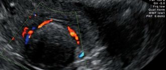
• Pelvic ultrasound is used in cases where an enlarged uterus fills the pelvic cavity, complicating physical examination, and also to detect hydronephrosis resulting from compression of the urethra by uterine fibroids.
• If submucosal FM is suspected, hysterosalpingography or hysteroscopy is usually performed.
• CT/MRI.
• With the rapid growth of myomatous nodes, it is necessary to exclude malignant degeneration.
Differential diagnosis
Uterine pregnancy • Ovarian tumor • Tumor of the cecum or sigmoid colon • Appendiceal abscess • Diverticulitis • Nephroptosis.
Pathogenesis
First, under the influence of provoking factors near the microvessels, an active zone is formed in the muscular layer of the uterus, in the cells of which metabolic processes are accelerated and tissue permeability increases. Subsequently, the smooth muscle cells of this zone begin to accumulate into microscopically and macroscopically defined nodes.
Over time, the sensitivity of proliferating tissue to the action of hormones decreases, and its own autocrinoparacrine growth mechanisms are launched. Submucous fibroids are nourished by vessels that have lost their adventitia. A growing submucosal node increases the area of endometrial rejection and reduces the ability of the myometrium to contract, which provokes more heavy, prolonged and painful menstrual bleeding.
Uterine fibroids, being a progesterone-dependent pathological process, develops, as a rule, during the ovulatory menstrual cycle. Tumor myocytes of leiomyomas differ from unchanged myometrium by higher expression of both estrogen and progesterone receptors. Its growth depends on cyclical hormonal changes in a woman’s body.
Description
Uterine fibroids are a benign, hormone-dependent tumor arising from the smooth muscles of the uterus. Uterine fibroids are found in 15–17% of women over 35 years of age. The term uterine fibroids is the most recognized and widely used, since it characterizes the morphogenesis of the tumor - its development from the muscle tissue of the uterus. Other names (fibroma, fibromyoma) are recommended to be used to clarify its histostructure. With uterine fibroids, elements of the connective tissue stroma predominate; with fibroids, there is an equal ratio of muscle and connective tissue cells. Each uterine fibroid, as a rule, is multiple, so the term multiple uterine fibroids has no logical meaning.
Use during pregnancy and breastfeeding
- It is recommended to carry out conservative treatment of pain associated with degenerative changes in the myomatous node in the second trimester: bed rest, hydration, analgesics [56,57].
Strength of recommendation: C (level of evidence: 4)
Hormone-dependent prostate cancer stages III and IV (if it is necessary to inhibit testosterone production in the testes); breast cancer in women with a preserved menstrual cycle and the presence of estradiol/progesterone receptors; hormone-dependent pathology of the reproductive system. Caused by absolute or relative hyperestrogenism (endometriosis.
Hypersensitivity.
Contraindicated during pregnancy. During treatment you should stop breastfeeding.
https://www.youtube.com/watch?v=upload
IM, s/c, intranasal.
Before starting therapy, it is necessary to exclude pregnancy and stop taking oral contraceptives; during the first 2 months of treatment for endometriosis, endometrial hyperplasia and uterine fibroids, barrier methods of contraception (condoms) must be used. When administered intranasally against the background of rhinitis, it is necessary to clean the nasal passages before administration;
The use of vasoconstrictor intranasal drugs should be avoided before and for 30 minutes after the administration of buserelin. Use with caution in patients with depression. For ovulation induction, it can only be prescribed by a doctor experienced in the treatment of infertility. Use with caution during work for vehicle drivers and people whose activities involve increased concentration.
Classification
Submucosal fibroids differ in the number of nodes, location, size and structure. The following criteria are used to classify the disease: • Number of nodes. There are single and multiple submucous fibroids. • Dimensions. Submucosal tumors up to 20 mm in size (up to 4-5 weeks of pregnancy) are considered small, from 20 to 60 mm (from 4-5 to 10-11 weeks) - medium, more than 60 mm (12 weeks or more) - large.
1.5.1 Clinical and anatomical classification (localization in various parts of the uterus and tumor growth in relation to the muscular layer of the uterus):
- intramural;
- submucosal;
- subserous;
- interligamentous;
- cervical;
- parasitic.
0. Pedicled submucosal nodes without an intramural component;
I. Submucosal nodes on a broad base with an intramural component of less than 50%;
II. Myomatous nodes with an intramural component of 50% or more [2,3].
1.5.4 Classification of uterine fibroids according to the recommendations of the European Society of Human Reproduction (ESHRE):
- Small fibroids: up to 5 cm;
- Large fibroids: more than 5 cm [5,6].
Side effects
From the nervous system and sensory organs. Headache (with intranasal administration), mood lability, sleep disturbance, depression, symptoms of eye irritation (when wearing contact lenses). From the gastrointestinal tract. Changes in appetite, nausea, vomiting. From the genitourinary system. Decreased libido, impotence, vaginal dryness, ovarian cysts, pain in the lower abdomen, menstrual bleeding.
Allergic reactions. Urticaria, skin hyperemia, angioedema. Other. Tides. Irritation of the nasal mucosa and nosebleeds (with intranasal administration). Increased sweating (with intranasal administration). Acne. Dry skin and mucous membranes. Demineralization of bones. Gynecomastia. Thrombosis.
Symptoms
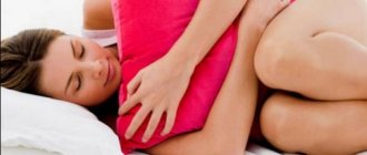
In the initial stages of development, uterine fibroids may not make themselves felt. However, you should pay attention to heavy menstruation, as this may be the first sign of the development of fibroids. In this case, you should definitely contact a gynecologist.
With active development, the following symptoms are observed:
- Pain syndrome in the lumbar region, lower abdomen.
- Development of anemia.
- Cutting pain during intimacy.
- Frequent urge to urinate.
- Feeling sluggish.