Physiological side of the process
Myoma always leads to a lack of iron (Fe) in the body in a hidden (latent) form. There are practically no symptoms. A woman experiences frequent fatigue even in the absence of heavy physical activity. Another symptom that indicates a drop in Fe levels in the body is frequent headaches, general sweetness, and psycho-emotional overexcitement. An increase in iron deficiency in the blood leads to a proportional decrease in the volume of oxygen that reaches organs and tissues. O² molecules are carried by Fe, so deficiency leads to more severe health problems.
The standard value of hemoglobin level in women ranges from 120 to 140 g per 1 liter. The threshold value is determined taking into account the woman’s age, the presence of chronic diseases or inflammatory processes. Taking potent drugs can affect the indicator, so before conducting a general blood test, you need to tell your doctor about everything. Its task is to distinguish the initial form of the disease from a one-time and short-term increase in Fe levels under the influence of external factors:
- prolonged physical activity;
- addiction to smoking;
- stay in high mountain areas;
- working in an environmentally unsafe enterprise.
Iron can be elevated in the female body due to natural reasons that do not require medical intervention.
On a note!
The body of professional athletes consumes oxygen more actively, so there are more red blood cells in the bloodstream.
Iron deficiency conditions in gynecological diseases and methods for their correction
Iron deficiency anemia (IDA) is a hematological syndrome characterized by impaired hemoglobin synthesis due to iron deficiency. Anemia is based on tissue hypoxia, which develops as a result of a decrease in the amount of hemoglobin due to blood loss, impaired formation of red blood cells, their destruction, or a combination of these reasons [1, 2].
Iron deficiency occurs in almost 1/3 of the world's inhabitants, and iron deficiency anemia accounts for 80–90% of all anemias. The prevalence of anemia varies depending on gender, age, climatic-geographical and environmental-industrial causes [1, 3, 4].
A corresponding deficiency develops when iron losses exceed iron intake of 2 mg/day, and is observed in various physiological conditions and diseases [5, 6].
Iron deficiency (hypochromic, microcytic) anemia usually occurs due to a decrease in iron resources in the body due to chronic blood loss or insufficient external iron supply.
The most vulnerable to the development of iron deficiency are women of reproductive age due to monthly physiological blood loss during menstruation, pregnant women due to their high need for microelements, children and adolescents due to the high needs of a growing body, elderly people suffering from chronic diseases and having poor nutrition.
Iron is an essential microelement that plays an important role in the functioning of cells in many body systems, the main of which is the participation of iron in the processes of tissue respiration. The total amount of iron in a woman’s body reaches 2–3 g, and its concentration is 40–50 mg per kg of body weight.
There are two types of iron: heme and non-heme. Heme iron is part of hemoglobin, is found only in meat products, is easily absorbed, and its absorption is practically not affected by the composition of food.
Non-heme iron is found in free ionic form - ferrous or ferric iron. The absorption of non-heme iron (found primarily in vegetables), which accounts for up to 90% of the iron in the total diet, depends on a number of factors. The main part of iron is included in hemoglobin and myoglobin - 70%; iron depot - 18% (intracellular accumulation in the form of ferritin and hemosiderin); functioning iron - 12% (myoglobin and iron-containing enzymes); transported iron - 0.1% (iron bound to transferrin) [7–9].
The most important iron-containing compounds include: hemoproteins, the structural component of which is heme (hemoglobin, myoglobin, cytochromes, catalase, peroxidase), non-heme enzymes (succinate dehydrogenase, acetyl-CoA dehydrogenase, xanthine oxidase), ferritin, hemosiderin, transferrin.
In the mucous membrane of the small intestine, only divalent non-heme iron is absorbed from food. In order for iron to contact proteins and enter the cell, ferric iron is reduced to divalent iron in the presence of an acidic environment; in most cases, ascorbic acid plays the main role in this process [10–13]. Then, to enter the blood plasma and bind to transferrin, iron is again oxidized to trivalent. In the transferrin-bound state, iron is delivered to tissues through endocytosis, where it is either utilized by the cell or deposited in ferritin. Ferritin stores iron in an easily accessible and non-toxic form. Ferritin level is the “gold” standard indicator of the amount of stored iron in the body. Each microgram of ferritin corresponds to 8 mg of deposited iron and for women is normally 15–150 mg/ml [2, 14–16]. The concentration of serum iron is subject to significant daily fluctuations and averages 6.6–26 µmol/l in women. Therefore, its definition is not of paramount importance [2].
Hemoglobin is an oxygen transport protein containing heme iron. The hemes are combined into a protoporphyrin framework, in the center of which there is one divalent iron atom. One hemoglobin molecule can carry a maximum of 4 oxygen molecules. Thus, the number of red blood cells and hemoglobin plays an important role in oxygen transport [2].
During iron deficiency, successive stages are distinguished: latent iron deficiency, in which iron losses exceed iron intake without changes in hemoglobin concentration, and iron deficiency anemia itself, which, depending on the severity, is divided into compensated, subcompensated and decompensated forms [3].
Hemoglobin concentration is expressed in grams per liter. The normal hemoglobin level in women is considered to be 115-145 g/l (MCHC, Mean Corpuscular Hemoglobin Concentration - the average concentration of hemoglobin in a cell is 320-360 g/l); anemia is indicated when hemoglobin decreases below 110 g/l. The severity of anemia is judged by the level of hemoglobin. According to the severity of anemia, there is a mild degree - 90-109 g/l, a moderate degree - 70-89 g/l and a severe degree - less than 70 g/l [3, 14, 15].
In addition, iron deficiency anemia is characterized by a decrease in hematocrit, hypochromia (MCH, Mean Corpuscular Hemoglobin - the average absolute content of hemoglobin in one erythrocyte), microcytosis (MCV, Mean Corpuscular Volume - the average volume of an erythrocyte), hyposiderosis (a decrease in the amount of serum iron, an increase in total iron-binding ability of blood serum, decrease in ferritin level in blood serum and in hemolysate) [3, 5]. The number of red blood cells, hematocrit and hemoglobin constitute the measured values, and MCV, MCH, MCHC are derivatives of these values [2].
The clinical picture of anemia is caused by oxygen starvation of tissues, progressive hemic hypoxia with the subsequent development of secondary metabolic disorders. Clinical symptoms appear as the severity of the disease increases: general weakness, dizziness, headache, palpitations, shortness of breath, fainting, decreased performance, insomnia. Symptoms characteristic of IDA include changes in the skin, nails, hair, muscle weakness, and distortion of the sense of taste. The skin becomes dry and cracks appear on it. Due to impaired carotene metabolism, iron deficiency causes yellowness of the skin. The shape of the nails changes, they flatten, become concave and brittle. Hair becomes thinner, breaks and falls out profusely, and gray hair appears. With IDA, symptoms of damage to the cardiovascular system are also possible: palpitations, shortness of breath, chest pain and sometimes swelling in the legs [6, 14].
The main compensatory mechanisms are circulatory and ventilatory adaptation, including an increase in cardiac output, vasodilation, a decrease in vascular resistance, an increase in tissue perfusion, blood redistribution, an increase in minute volume of respiration, and an increase in erythropoietin activity. Acute, severe and uncompensated anemia can lead to circulatory collapse and shock [2].
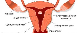
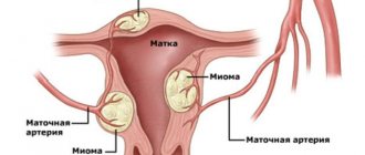
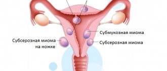
The main cause of the development of iron deficiency anemia in women is blood loss of various etiologies. Iron deficiency is 6 times more common in women than in men. A significant amount of blood is lost during menstruation, pregnancy, and childbirth. With heavy menstruation, iron loss of 50–250 mg of iron is possible [2]. Most often, hyperpolymenorrhea is associated with the presence of uterine fibroids, adenomyosis, hyperplastic processes of the endometrium, and dysfunctional uterine bleeding. Metrorrhagia with uterine fibroids is most often associated with submucosal localization of nodes, in which the menstrual surface increases, the permeability of blood vessels supplying the fibroids increases, and the contractility of the uterus decreases. With fibroids, the regeneration of the bleeding surface of the uterus slows down after desquamation of the endometrium. With adenomyosis, the uterine myometrium is affected, which leads to prolonged heavy menstruation and secondary iron deficiency anemia.
Menstrual irregularities occur in women at different ages. Various reasons can lead to the development of hypermenstrual syndrome - severe emotional shocks, malnutrition, vitamin deficiencies, obesity, occupational hazards, infectious and septic diseases, immaturity of hypothalamic structures in puberty and involutive changes in premenopause. In a significant proportion of cases of menometrorrhagia in women, they are accompanied by anemia of varying severity, which contributes to the development of trophic disorders in various organs and tissues. Prevention and treatment of anemia in women with menstrual irregularities and organic gynecological diseases are the most important factors in restoring their health [1, 4, 17].
Regardless of the cause of menometrorrhagia (fibroids, endometriosis, ovarian dysfunction) and the need to influence the corresponding factor, long-term therapy with iron preparations for oral administration is required. The dose, dosage regimen and specific drug are selected individually, taking into account the iron content in the drug, its tolerability, etc.
When choosing a specific drug and the optimal dosage regimen, it is necessary to keep in mind that an adequate increase in hemoglobin levels in the presence of IDA can be ensured by the intake of 30 to 100 mg of ferrous iron into the body [18]. Dietary measures alone cannot compensate for iron deficiency and achieve a therapeutic effect.
Antianemic therapy should be carried out with oral medications and should not be stopped after hemoglobin normalization. Indications for parenteral administration are quite limited: intestinal pathology with malabsorption, intolerance to oral drugs, social reasons (use in patients with personality changes, the mentally ill). According to many studies, parenteral forms do not have advantages over oral ones, and a large number of serious side effects are noted [4, 10, 19]. Blood transfusions for IDA should be carried out only for health reasons.
Modern oral ferrous preparations are salts of ferrous iron (ferric ions are not absorbed in the digestive tract) or compounds consisting of a hydroxide-polymaltose complex of ferric iron (the absorption mechanism differs from that of ionic preparations) [20].
Ferric sulfate salt has the highest bioavailability, therefore ferrous sulfate is mainly contained in ferrous preparations [19, 21]. Ferrous sulfate is the most studied, proven form, which has proven itself with long-term use, therefore it is most often included in preparations for oral administration. It has the highest degree of absorption of all iron preparations.
In the process of absorption of divalent iron in the intestine, ascorbic acid is of great importance, which helps maintain iron in divalent form, so its presence in the preparation is very important [22]. In hematopoiesis, folic acid plays a significant role, enhancing nucleic acid metabolism. For normal folic acid metabolism, cyanocobalamin is necessary, which promotes the formation of its active form. Deficiency of these substances, which often occurs in anemia associated with blood loss, leads to disruption of DNA synthesis in hematopoietic cells, while the inclusion of these components in the drug increases the active absorption of iron in the intestine and its further utilization. The presence of ascorbic and folic acids, as well as cyanocobalamin in the drug significantly increases the rate of hemoglobin synthesis and increases the effectiveness of therapy for iron deficiency conditions and iron deficiency anemia [14, 23, 24].
The listed components that increase the bioavailability of iron are part of the complex antianemic drug Ferro-Folgamma® [24], which was developed and produced in accordance with the recommendations of the World Health Organization (1998).
1 capsule contains 112.6 mg of ferrous sulfate (elemental iron 37 mg), which is the optimal dose for therapy and reduces the incidence of side effects.
Absorption of iron from salt preparations (Fe2+) occurs in the form of passive diffusion according to a concentration gradient, does not depend on pH and motor activity of the gastrointestinal tract (GIT), which ensures rapid saturation.
Ascorbic acid improves the absorption of iron in the intestine, prevents its transition from divalent to trivalent form, accelerates the transport of iron and its inclusion in heme, and also participates in the process of releasing iron from the depot.
Ascorbic acid is necessary for the formation and preservation of the reduced form of folic acid - tetrahydrofolic acid.
In turn, folic acid is an essential factor for the synthesis of DNA and RNA, protein metabolism and the formation of red blood cells, and also acts as an additional stimulator of erythropoiesis and hematopoiesis [21, 23, 25].
Cyanocobolamine is necessary for the formation of hemoglobin, red blood cells, metabolism of proteins, fats, carbohydrates, and energy production.
Ferro-Folgamma® is highly effective, providing an average increase in hemoglobin of 2.5 g/l/day (the highest rate of increase in hemoglobin among ferrous sulfate preparations). When using the drug Ferro-Folgamma®, a weakening of the clinical symptoms of posthemorrhagic anemia is observed during the first 10 days of use [6].
Ferro-Folgamma® is prescribed 1 capsule 3 times a day after meals for 3–4 weeks for mild forms of anemia, for moderate forms — 1 capsule 3 times a day for 8–12 weeks, and for severe forms of anemia — 2 capsules 3 times a day for 16 weeks or more. During pregnancy, it is prescribed to prevent folic acid and iron deficiency, 1 capsule 3 times a day in the II and III trimesters, in the postpartum period during breastfeeding.
After cessation of treatment with Ferro-Folgamma®, the positive effect and stabilization of blood serum parameters (hemoglobin level, red blood cells, serum iron, total iron-binding capacity of blood serum) are guaranteed to persist for at least one month [4].
The active components of Ferro-Folgamma® are in a special neutral shell, which ensures their absorption in the upper part of the small intestine, which eliminates irritating effects on the stomach. The components are dissolved in rapeseed oil, which improves the absorption of iron and at the same time reduces the irritating effect of iron on the gastric mucosa, promoting good tolerability of the drug in the digestive tract [1, 13]. Due to the absence of an aggressive effect on the gastrointestinal mucosa, Ferro-Folgamma® can be successfully used to correct anemic syndrome, with a deficiency of vitamin B12 and folic acid against the background of impaired absorption in the gastrointestinal tract (atrophy of the gastric and duodenal mucosa).
In addition, Ferro-Folgamma® is effectively used for combined iron-folate-B12-deficiency anemia caused by chronic blood loss, chronic alcoholism, infections, taking anticonvulsants and oral contraceptives, anemia during pregnancy and breastfeeding.
The effectiveness of Ferro-Folgamma® for the treatment and prevention of iron deficiency anemia with good tolerability and favorable pharmacoeconomic characteristics (cost/iron dose/efficacy) was proven in several Russian studies that included a wide range of patients, including pregnant women and women with gynecological diseases [4, 18, 19]. Indications for the use of the drug Ferro-Folgamma® are anemia caused by a combined deficiency of iron, folic acid and vitamin B12, occurring against the background of chronic blood loss (menorrhagia and metrorrhagia, etc.), as well as with chronic alcoholism, infectious diseases, taking anticonvulsants and oral contraceptives. The drug is approved for the prevention and treatment of iron and folic acid deficiency in the second and third trimesters of pregnancy, in the postpartum period and during lactation.
Thus, anti-anemic therapy using the drug Ferro-Folgamma® with optimal iron content is highly effective with a rapid increase in hemoglobin (complete clinical and hematological remission in 93% of patients after 3 weeks of therapy). There is good tolerability - the absence of side effects in 95% of patients, allergic reactions and negative effects on the body of women, as well as the stability of the results achieved and the maintenance of a positive effect for a month after treatment, which makes it possible to recommend the drug Ferro-Folgamma® to a wide range of patients.
Literature
- Transcript of the scientific symposium “Iron deficiency conditions in obstetrics and gynecology.” III Russian Forum “Mother and Child”. M., 2001, 29.
- Hook R., Breiman K. Anemia during pregnancy and the postpartum period. M., 2007, 74.
- Gorodetsky V.V., Godulyan O.V. Iron deficiency conditions and iron deficiency anemia: diagnosis and treatment. Guidelines. M.: Medpraktika-M, 2005; 28.
- Konovodova E. N., Dokueva R. S.-E., Yakunina N. A. Iron deficiency conditions in obstetric and gynecological practice // Breast Cancer. 2011; 20: 1228–1231.
- Dolgov V.V., Lugovskaya S.A., Morozova V.T., Pochtar M.E. Laboratory diagnosis of anemia. M., 2001. P. 84.
- Kozlovskaya L.V. Hypochromic anemia: differential diagnosis and treatment // New Med. magazine 1996; 56:8–12.
- Shekhtman M. M. Guide to extragenital pathology in pregnant women. M., 2005, 816, 373–399.
- Johnson-Wimbley TD, Graham DY Diagnosis and management of iron deficiency anemia in the 21st century // Therap. Adv. Gastroenterol. 2011; 4 (3): 177–184.
- UNICEF/UNU/WHO. Iron Deficiency Anemia: Assessment, Prevention, and Control. A Guide for Program Managers. Geneva: WHO/NHD, 2001.
- Arkadyeva G.V. Diagnosis and treatment of iron deficiency conditions. Educational and methodological manual. M.: 1999: 22–25.
- Burlev V. A., Gasparov A. S. et al. Epocrine in the treatment of iron deficiency anemia in patients with uterine fibroids after hysterectomy // Problems of reproduction. 2003; 6:59–64.
- Kasabulatov N.M. Iron deficiency anemia in pregnant women // Breast cancer. 2003; 11, 1: 18–20.
- Lebedev V. A., Pashkov V. M. Principles of treatment of iron deficiency anemia in gynecological patients // Difficult patient. 2013, 11, 11: 3–7.
- Dvoretsky L.I., Zaspa E.A. Iron deficiency anemia in the practice of an obstetrician-gynecologist // Breastfeeding. 2008; 29.
- Kazyukova T.V., Samsygina G.A., Kalashnikova G.V. et al. New possibilities of ferrotherapy for iron deficiency anemia // Klin. pharmacology and therapy. 2000; No. 9 (2): 88–91.
- Pasricha SR, Flecknoe-Brown SC, Allen KJ et al. Diagnosis and management of iron deficiency anemia: a clinical update // Med. J. Aust. 2010; 193(9):525–532.
- Fernandez-Gaxiola AC, De-Regil LM Intermittent iron supplementation for reducing anemia and its associated impairments in menstruating women // Cochrane Database Syst. Rev. 2011. 12. CD009218.
- Burlev V.A., Konovodova E.N., Ordzhonikidze N.V., Serov V.N., Elohina T.B., Ilyasova N.A. Treatment of latent iron deficiency and iron deficiency anemia in pregnant women // Russian Bulletin of Obstetrics- gynecologist. 2006. No. 1. P. 64–68.
- Vertkin A.L., Godulyan O.V., Gorodetsky V.V., Skotnikov A.S. Iron deficiency anemia and the choice of drug for its correction // Russian Medical Journal. 2010. No. 5.
- Gratsianskaya A.N. Iron deficiency anemia: Ferro-Folgamma // Breast cancer. 2013; No. 29.
- Arvas A., Gur E. Are ferric compounds useful in the treatment of iron deficiency anemia? // Turk J Pediatr. 2000. Vol. 42(4). R. 352–354.
- Teucher B., Olivares M., Cori H. Enhancers of iron absorption: ascorbic acid and other organic acids // Int J Vitam Nutr Res. 2004. Vol. 74(6). R. 403–419.
- Konovodova E. N., Burlev V. A. Ferro-Folgamma + Erythropoietin - new possibilities for the treatment of anemia in patients with uterine fibroids // Farmateka. 2004. No. 15 (92). pp. 70–73.
- Ferro-Folgamma. Therapy with iron, folic acid, vitamin B12 and ascorbic acid. Scientific review. Werwag Pharma. M., 2001.
- Ghinea MM Treatment of iron deficiency anemia with Ferro-Folgamma // Rom J Intern Med. 2004. Vol. 42(1). R. 225–230.
A. Z. Khashukoeva1, Doctor of Medical Sciences, Professor S. A. Khlynova, Candidate of Medical Sciences M. V. Burdenko, Candidate of Medical Sciences M. R. Narimanova O. V. Kozlova, Candidate of Medical Sciences, Associate Professor
GBOU VPO RNIMU im. N. I. Pirogova Ministry of Health of the Russian Federation, Moscow
1 Contact information
Abstract. The role of Ferrum in the human organism has been analyzed, mechanisms of its digestion from food, clinical presentations of hypoferric conditions and women's hypoferric anemia, as well as approaches to therapy and prophylaxis of hypoferric anemia with modern oral preparations.
Buy an issue with this article in pdf
Negative effects on the body
Myoma is a disease that is dangerous in itself. At the same time, Fe deficiency is formed. The severity of a woman’s condition cannot be predicted. Regular preventive visits to the doctor's office will help prevent the development of irreversible complications. The recommendation is relevant for women at risk and for those who do not have health problems. Taking a complete blood test (CBC) will allow you to determine at an early stage the negative impact of iron deficiency on vital organs:
- disruption of hematopoietic function in the body;
- pathological changes in the liver;
- increased risk of heart attack;
- increased load on blood vessels and heart;
- pathological change in tissue;
- a sharp decrease in the body's defenses;
- fainting.
Low hemoglobin initially negatively affects the functioning of the gastrointestinal tract, the structure of the skin, and the mucous membranes.
Low hemoglobin in the blood with fibroids: how anemia with fibroids affects life
The effects of low hemoglobin caused by the development of fibroids can also manifest themselves in different ways. These can be very serious conditions, which are accompanied by decreased immunity and even such threatening conditions as liver enlargement. Low hemoglobin, caused by the effect of fibroids on hematopoietic processes, has the following effects on the functioning of the body:
- The development of anemia caused by prolonged iron deficiency.
- A persistent decrease in immune defense, which threatens infections.
- Organs and tissues undergo changes.
- Excessive load on the heart leads to the development of cardiac pathologies.
- Liver dysfunction and changes in its structure.
Particularly affected by low hemoglobin are tissues consisting of epithelium in the form of mucous membranes, as well as skin and gastrointestinal functions.
Primary and secondary symptoms of anemia
Iron deficiency in the body is confused with temporary fatigue. The woman does not attach importance to what is happening. Within 4-6 days, rare dizziness and weakness become more frequent. Other symptoms are added to them:
- hair loss;
- nails become more brittle;
- the corners of the lips are covered with cracks;
- constant dry skin;
- increase in body temperature.
The above symptoms do not always allow us to make an unambiguous conclusion about iron deficiency due to a tumor in the uterus. Only a comprehensive examination can bring clarity. Its results will allow the doctor to make a conclusion about how related the existing symptoms are to each other. In any case, anemia is not an independent disease. Iron deficiency is one of the symptoms of a chronic disease that has led to a decrease in the concentration of red blood cells.
Causes of anemia during pregnancy
The second and third trimester are the most important, so you need to include increased amounts of iron in your diet. The average daily dosage should be 10 mg.
The following factors can be identified that can influence the occurrence of anemia during pregnancy.
- Toxicosis in the early stages, which is accompanied by regular vomiting and nausea.
- Wrong diet. Anemia of the uterine cavity develops as a result of following a vegetarian diet, because the main source of iron is fermented milk and meat products.
- Early problems with iron deficiency are diagnosed if patients have previously been diagnosed with anemia as a result of gynecological pathologies, prolonged bleeding and heavy discharge during menstruation. Most often they occur with the progression of endometriosis and uterine fibroids.
- Miscarriages and frequent childbirth.
- Infectious diseases and pathologies of internal organs that have developed into a chronic form. Liver dysfunction is considered the most dangerous - it can be hepatosis and severe gestosis. In a woman’s body, the processes of absorption and accumulation of iron are disrupted.
During pregnancy, chronic diseases can worsen, so the reserve of nutrients is depleted.
Some women experience relative or false anemia. This is due to the fact that the number of blood components increases unevenly.
Restoring normal iron levels
Treatment involves treating the pathology that caused Fe deficiency. The patient is referred for additional examination to an oncologist. If the size of the tumor does not exceed 12 weeks, and there are no signs of the tumor becoming malignant, the doctor will prescribe a conservative therapeutic course. Hormonal medications are used. The doctor selects the dosage to solve three problems:
- slow down the rate of generation of sex hormones;
- improve the structure of the uterine epithelium;
- prevent the development of a pronounced oncological process.
If the results of the tests indicate an active increase in the size of the fibroids, the doctor recommends immediate surgical intervention. The surgeon assesses the risk associated with the procedure.
On a note! Depending on the severity of the disease, partial or 100% removal of the uterus is performed.
Adviсe
Long, heavy menstruation, spotting, regardless of the menstrual cycle, accompanied by an increasing feeling of weakness and weakness - the first sign that you need to see a gynecologist. Progressive anemia significantly impairs quality of life and can become hazardous to health. The faster the tumor is treated, the easier it will be to cope with the symptoms of anemia.
Increasing hemoglobin by consuming foods containing iron is possible only in the initial stages of anemia.
Hemoglobin levels below 100 g/l require the prescription of antianemic drugs, which must be selected by a doctor.
Rehabilitation course
Having eliminated the tumor in the uterus, the therapist proceeds to stabilize the level of iron in the bloodstream. The diet requires changes. The patient eats more fresh vegetables and fruits. You should not exclude beef, liver and lamb from your diet. They are rich in iron. It is worth eating less green apples and buckwheat. The beneficial substances they contain are processed more slowly by the body. Other recommendations:
- taking ascorbic acid – vitamin C accelerates the processing of Fe;
- citrus;
- black currant;
- sauerkraut;
- bell pepper;
- Blood transfusion is performed for severe anemia.
Vitamin-mineral complexes are allowed to be taken only with a doctor’s recommendation. Errors caused by independent choice will lower hemoglobin in the blood with already cured uterine fibroids.
What are the signs of uterine fibroids?
Most experts claim that fibroids are hormonal in nature. It is believed that an incorrect ratio of sex hormones in a woman’s body affects the pathological condition of the endometrium and metabolic disorders. The causes of the disease currently also include the effect on the female body of psychosomatic factors - depressive states, prolonged stress, feelings of loneliness, hopelessness, depression, family conflicts.
The main risk group for uterine fibroids includes women of childbearing age. After the decline of reproductive function, the incidence rate decreases significantly.
The reasons for the development of uterine fibroids are also operations for artificial termination of pregnancy (abortion), diabetes mellitus, high blood pressure, obesity, lack of a full intimate life, and increased levels of estrogen (female sex hormones). Indirect causes of the disease may be dysfunction of the cardiovascular system.
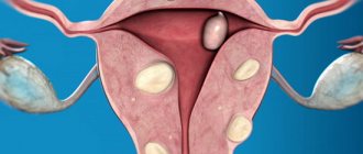
Signs of uterine fibroids
The most characteristic symptoms of uterine fibroids are unusually heavy menstrual bleeding, as well as unexpected intermenstrual bleeding. There are also pain sensations that vary in location and intensity. There is pain in the pelvic area, manifested by a feeling of constant distension and heaviness in the lower abdomen. Tumors located inside the uterus can cause cramping pain.
Acute and paroxysmal pain is a sign of impaired blood supply to uterine fibroids.
Painless, but rather large myomatous nodes can be detected upon independent palpation of the abdominal cavity. The size of uterine fibroids can vary. Neoplasms can be the size of a pea; there are giant tumors weighing more than three kilograms. This may be one large tumor or a cluster of small tumors.
With uterine fibroids, a woman’s body develops overt or hidden anemia, and the level of hemoglobin in the blood decreases. As a result, chronic fatigue syndrome appears, fatigue is observed, and performance and mood decrease. Without treatment, increased blood loss becomes permanent, and this negatively affects the functions of hematopoiesis and leads to disruptions in the functioning of all organs and systems in the woman’s body.
How to detect anemia
The examination traditionally begins with interviewing the patient and collecting anamnesis. The doctor must remember that anemia does not always develop as a result of an underlying disease. There may be hereditary or any other causes that are not related to the tumor process. It is also necessary to establish the type of previous antitumor treatment, the duration and number of courses, and the name of the drugs that were used. At the next stage, the doctor conducts a general examination. Anemia can be manifested by pale skin, weakness, loss of appetite, dizziness and other symptoms. Very often, these manifestations are observed in various types of cancer, which occurs without anemia, so it is impossible to make a diagnosis based only on the patient’s complaints.
Objective information can only be obtained through laboratory research. Anemia is diagnosed using the following tests:
- Reticulocyte count.
- Serum iron level.
- Transferrin and ferritin protein levels.
- Vitamin B12 and folate levels.
Special attention is paid to a general blood test. It determines the level of hemoglobin, the number of red blood cells and hematocrit (the ratio of formed elements to the liquid component of blood). The average volume of the erythrocyte, the average hemoglobin content in the erythrocyte and the average hemoglobin content in the entire erythrocyte mass are also determined.
If necessary, other laboratory tests or special methods may be included in the comprehensive examination program for anemia.
Hemoglobin in women
The diagnosis of iron deficiency anemia must be confirmed by laboratory testing. To do this, you need to take a general blood test, as well as tests to determine the level of transferrin, ferritin and latent iron-binding capacity (LIF).
Standard
Normal blood counts in women are:
- Hemoglobin – 117-155 g/l.
- Iron in blood serum – 8.9 – 30.4 µmol/l.
- Ferritin – 10-120 mcg/l.
- Transferrin – 2.0-4.0, saturation percentage 15-50.
- LVSS – 20-62 mmol/l.
Description of the disease
Fibroids and hemoglobin are closely related.
With a benign neoplasm, a disturbance in the blood supply to the tumor often develops, causing sharp pain of a paroxysmal nature. Myomatous nodes at the beginning of the development of the disease are small and the symptoms they exhibit are almost invisible. As the disease progresses, the size of the tumor increases. If left untreated, the mass of the node sometimes reaches several kilograms. The neoplasm can be single or multiple. Although at the beginning of its development the disease is asymptomatic and does not manifest itself in any way externally, the tumor changes the course of certain processes in the body. As the tumor grows, heavy bleeding begins. Their result is a decrease in hemoglobin levels and the development of anemia. The disease develops clearly or has no visible symptoms.
When anemia occurs, a woman experiences the following symptoms:
- weakness;
- constant fatigue;
- depressed emotional state;
- decreased performance.
Lack of treatment for fibroids leads to further growth of the tumor. Then the symptoms of anemia appear more clearly. Bleeding becomes profuse and becomes constant. Because of this, the body's ability to produce blood decreases. A deficiency of iron-containing protein worsens the condition of all organs and systems.