Myoma on ultrasound is characterized by certain signs, according to which a gynecologist will be able to establish an accurate diagnosis and select the most effective treatment regimen.
In addition to determining the diagnosis, ultrasound diagnostics will help monitor the development of the disease, because the fibroid node may increase in size or become smaller over time. Based on the results of the ultrasound examination, the doctor adjusts the therapeutic regimen or decides to perform surgical removal of the benign tumor.
Prevalence of fibroids
This disease, as a rule, is detected during the next preventive examination in women who have reached 35 years of age (20-40% of the total). It is characteristic that urban women are more susceptible to this pathology than women living in rural areas. At a young age, fibroids are very rare, and they never occur in adolescents until the onset of puberty, that is, puberty. With a decrease in the level of sex hormones in the blood, which entails the onset of menopause, fibroids, as a rule, begin to decrease in size and gradually resolve.
Myoma is a disease of a multifactorial nature. Until now, the true reason for its development is not known to science. Risk factors contributing to the occurrence of myomatous nodes include:
- Late formation of menstrual function, that is, late arrival of the first menstruation;
- Heavy menstrual bleeding;
- History of frequent medical abortions;
- Presence of gynecological diseases;
- Addiction to meat food;
- Change of climatic zones;
- Decreased activity of cellular immunity;
- Addiction to meat products;
- Ethnic factor (African American women have twice the risk of developing fibroids than Caucasian women).
At the same time, clinicians include risk factors for the development of fibroids such extragenital pathologies as overweight and obesity, disorders of the cardiovascular system and digestive tract, neuroses and pseudoneurotic conditions, hormonal disorders (diabetes mellitus), hypertension and frequent stress. Some experts argue that genetic predisposition is also a risk factor for the development of this disease. The cause of fibroids at a young age (in girls under 25 years of age), according to doctors, is damage to the uterine cells that occurred during the antenatal period of fetal development.
Oral contraceptives and pregnancy can provoke increased tumor growth.
When and how do they do it?
When is it better to do an ultrasound for uterine fibroids? The patient is usually interested in which day of the cycle the procedure is performed and what kind of research is used. Women are worried whether ultrasound will show fibroids at all?
If an examination through the abdominal wall is prescribed, it can be performed on any day of the cycle and even during menstruation. But it happens that a small myomatous node can be hidden in the thickness of the myometrium during ovulation, therefore, for the reliability of the examination, it is advisable to carry it out at the beginning of the cycle or on the day prescribed by the doctor.
Video 1. Submucous uterine fibroids.
An exceptional case is submycotic fibroids, the features of which make it advisable to undergo an ultrasound scan on days 19–24 of the cycle. The fact is that at this time the inner mucous surface of the uterus has hyperechoic characteristics and optimal thickness.
But it is best to do an ultrasound on the fifth to tenth days of the female cycle , when the endometrial layer is thinnest, which creates good conditions for searching for possible pathologies. This is a chance to detect tumors at the very beginning of their development, as well as an opportunity to minimize diagnostic errors.
Types and stages of uterine fibroids
In the course of numerous studies, it was found that the rudiments of myomatous nodes appear during the embryonic period of development of the female body. In the antenatal period, the fetal uterus develops from two Müllerian ducts. In this case, smooth muscle cells are formed up to 30 weeks from the mesoderm. However, undifferentiated cells can also proliferate into uterine smooth muscle cells. It is they who, during intrauterine development, under the influence of various endogenous and exogenous factors, become the precursor of myomatous neoplasms. These precursor cells, remaining in the muscular layer of the uterus, begin to grow after menarche (the arrival of the first menstruation). This growth is a consequence of the pronounced secretory activity of hormones produced by the ovaries (estrogens and progesterone).
At the same time, the rudiment of the myomatous node can be formed from the muscular lining of the vessels feeding the uterus, that is, from pericytes.
According to experts, under the influence of local growth factors, sex hormones and somatic mutations, the myometrium can transform into fibroids.
The myomatous node begins its development from a growth zone located around a thin-walled blood vessel.
There are three stages of fibroid morphogenesis:
- Formation of the rudiment (active growth zone) in the myometrium.
- Growth of an undifferentiated tumor.
- Growth and maturation of a tumor with differentiated elements.
Some authors argue that the development of a pathological process can be triggered by a disturbance in the myomatous nodes of neuromuscular regulation, and therefore they are generally considered to be formations devoid of nerve connections with the central nervous system.
Today, uterine fibroids are usually divided into simple and proliferating.
In simple fibroids, the growth zones do not reach large sizes and are located in the perivascular space. Proliferating fibroids are also characterized by a perivascular location, but at the same time, due to their increased “cellularity,” they reach significant sizes. However, the fundamental difference between simple myomatous nodes and proliferating ones is the accumulation of extracellular matrix in the former and its almost complete absence in the latter. It should be noted that when the balance of growth factors changes, a simple fibroid can transform into a proliferating one, and vice versa.
In patients with fast-growing tumors, proliferating fibroids are most often detected, and simple fibroids are most often detected in patients with a slow or moderate tumor growth rate.
Like any neoplasm, fibroids can grow and enlarge, sometimes reaching very significant sizes. The mass of the tumor can often reach 3-5 kg, less often – 15-20 kg.
Due to the fact that myoma is a tumor not only of mesenchymal origin, since it can be formed from coelomic epithelium), a wide morphological variety of types of these neoplasms is encountered in clinical practice.
Decoding the results
When interpreting ultrasound results if fibroids are suspected, the size of the uterus is first measured. The distance between the bottom of the uterine cavity and the internal cervical os is normally from 5 to 8 centimeters. With various formations and nodes in the uterus, the organ on the ultrasound screen appears visually enlarged.
A slight increase in the size of the organ is acceptable if a woman has given birth several times. Possible deviation is 5 millimeters.
The thickness of the walls of the endometrium, the outer layer of the uterus, is normally 3–4 centimeters. The normal width is 4.5–6 centimeters. If the final description indicates heterogeneous myometrium or the presence of myometrial cysts, muscle tissue within the endometrium, the woman is diagnosed with fibroids.
The size of the uterus with fibroids is measured by weeks of pregnancy to determine the rate of tumor growth. The course of the disease is influenced by the location of the uterus. With normal indicators in the description of the ultrasound result, it is located in the center of the small pelvis closer to the front of the abdominal wall.
What does a fibroid look like on an ultrasound image?
Signs of uterine fibroids on ultrasound are considered to be round or oval-shaped neoplasms with a layered structure. Uterine fibroids look different on ultrasound images. This is influenced by the location and type of tumor:
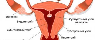
- Intramural nodes are located deep inside the walls of the uterus, in the myometrial layer, practically without changing the shape and size of the organ. In ultrasound images of this type, fibroid nodes appear as homogeneous tissue with reduced density (shown in dark gray on the monitor).
- The subserous node on an ultrasound image appears as a formation with a clear external contour with small tubercles. With large sizes, it changes the shape of the uterus.
- The submucosal node grows inside the uterus. On ultrasound, this type of fibroid is distinguished by the homogeneity and ovality of tissues of low density (gray color on the screen).
- Leiomyoma is better recognized on MRI than on ultrasound. In the latter case, the neoplasm appears as an area with uneven density on the ultrasound image. Tissue density is reduced (dark gray color), a clear limiting contour of the tumor can be seen.
If there is any doubt about the diagnosis, the sonologist prescribes additional examinations.
In addition, watch the video of a large fibroid on an ultrasound:
https://youtu.be/QEGdd1UrUWU
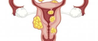
Classification of myomatous neoplasms
Most often (in 95% of cases) fibroids are localized in the body of the uterus, and only in 5% - in the cervix. They can be single or multiple (multiple fibroids are much more common in clinical practice).
Classification of fibroids according to the location of the direction of growth in the uterus
1. Myomatous neoplasms with typical localization:
Subserous (subperitoneal) fibroid is a tumor on a stalk or broad base, located in the peritoneum on the surface of the uterine body and growing towards the abdominal cavity. In this case, myomatous nodes located on the stalk are able to attach to nearby structures (intestine, omentum or mesentery), developing a secondary blood supply. If the tumor loses its primary blood supply, a parasitic leiomyoma is diagnosed, and in the case where the myomatous nodes are located between the broad ligaments, we are talking about intraligamentary myoma.
Submucous (submucosal) fibroids are a neoplasm localized under the endometrial mucosa, the growth of nodes of which is directed towards the uterine cavity. It may also have a stem or a wide base. Pedicled fibroids can sometimes “fall out” of the cervical canal, becoming twisted and infected.
Interstitial (intermuscular) fibroids are a tumor whose nodes are located in the thickness of the uterine wall.
2. Myomatous neoplasms with atypical localization:
Interligamentous (subserous myoma located behind the peritoneum).
Classification of fibroids by localization in relation to the uterine axis
- Corporal fibroids are the most common tumor (90.2%), which is localized in the body of the uterus.
- Isthmic or isthmic fibroids are neoplasms that cause pain in the bladder area.
- Cervical (cervical) fibroids are a tumor that grows in the vagina and provokes the development of various infectious complications.
WHO classification
1. Leiomyoma
A). Ordinary leiomyoma is a mature hormone-dependent tumor, which is a node of dense consistency, clearly demarcated from healthy tissue. This neoplasm, consisting only of smooth muscle cells of the myometrium, is surrounded by a hyalinized connective tissue layer resembling a capsule. Tumor growth occurs towards the soft tissues, without compromising the integrity of the endothelium.
b). Cellular leiomyoma is a tumor of soft consistency with clear boundaries, usually located inside the uterine wall. This tumor can actively grow during pregnancy. Histological sections examined under a microscope reveal cells with enlarged, slightly elongated nuclei and low mitotic activity.
V). Bizarre leiomyoma is a neoplasm consisting of not only round muscle cells. It also includes multinucleated giant cells of a polygonal shape, which is why this tumor is sometimes confused with leiomyosarcoma. However, confirmation of its benign nature is the small number (or complete absence of mitoses), as well as the absence of infiltrative growth.
G). Epithelial leiomyoma, or leiomyoblastoma, is a tumor that is quite rare in clinical practice. It consists of smooth muscle tissue and elements of the vascular walls.
d). Metastatic leiomyoma is also a very rare type of tumor. Histological examination confirms the benign nature of this neoplasm, but at the same time, it is capable of metastasizing into vascular clefts and growing into the lumen of the vessel. When tumor cells break off, they can travel through the bloodstream into the stomach or lungs, where a new myomatous node begins to develop.
e). Proliferating or growing leiomyoma is a neoplasm characterized by slow growth and the presence of proliferation zones located in the thickness or along the periphery of the tumor. At first, the cellular elements contained in them are presented in the form of couplings, and then they gradually transform into smooth muscle cords that grow and merge with nearby tissues.
and). Myoma with symptoms of presarcoma (malignizing leiomyoma) is a neoplasm in which atypical cells and cell nuclei are detected.
2. Fibromyoma. Depending on the age of the fibroid, its microstructure changes. Over time, the neoplasm turns into a clearly defined node, acquiring the character of a fibromyoma (a tumor consisting of connective tissue and muscle elements).
Note: as fibroids age, the connective tissue becomes coarser and hyalinized, and its quantity increases.
3. Rhabdomyoma is a benign neoplasm consisting of striated muscle tissue.
4. Angiomyoma is a formation presented in the form of a myomatous node with a developed network of blood vessels.
Ultrasound view
Ultrasound for this disease is based on standard diagnostic principles. It is carried out through the vagina and the anterior wall of the peritoneum.
If we talk about characteristics, they have the following features:
- roundness of the tumor;
- heterogeneity of structural content;
- clarity of external contours and boundaries;
- the signal weakens closer to the edge of the anomaly and intensifies in the area of its central part.
Video from an ultrasound machine:
Basic parameters for diagnosing the disease:
- if pathologies are localized in the walls, their surface remains unchanged;
- formation concentrated under the mucosa provokes the growth of the uterus;
- when the pathology grows inside the peritoneum, the organ moves slightly, and its surface zone is more convex and lumpy;
- the size of the uterus is larger than normal;
- the contours of the body are slightly deformed in comparison with the original parameters;
- the density is not uniform;
- if you use Doppler during the study for a more accurate clinical picture, you can identify uncharacteristic blood flows;
- fibroids are characterized by a pronounced vascular network, both on the surface of the seal and deep inside the anomaly;
- The pathology is most often heterogeneous, which manifests itself in the form of darker, or, conversely, lighter areas.
In addition, the diseased organ must be measured. This is done in the following way - for a single formation - the standard method, for multiple manifestations - the description is carried out taking into account the largest formation.
What consequences await a woman after removal of the uterus with fibroids? Complications are described here.
Pregnancy with fibroids on ultrasound
During the pregnancy of a woman suffering from fibroids, the organ becomes somewhat deformed under the influence of pathology.
Already at the first ultrasound, which is performed in the initial trimester of gestation, the monitor can clearly distinguish the localization of the pathology and the location of the ovum.
The doctor identifies the number of nodal connections, their growth pattern and location relative to the placental layer. Determine how much the tumor can affect the condition and development of the fetus.
Symptoms of uterine fibroids
The most characteristic symptom of this disease is the development of heavy menstrual bleeding. The abdomen increases in size, almost all patients complain of discomfort and the occurrence of pain of varying intensity. There is also pain in the lower back, constipation develops, and urination becomes frequent. In the case when torsion of the fibroid stalk occurs, symptoms of an acute abdomen are observed.
Clinical manifestations of the disease depend on the location, direction of growth of myomatous nodes, tumor size, age of the patient, as well as the presence of concomitant gynecological pathologies.
In 25-30% of cases, the development of fibroids occurs very slowly, and at first is not accompanied by severe symptoms. Most often, a tumor is first detected during pregnancy (since dense myomatous nodes are quite easily palpable in the softened myometrium). However, fibroids can be detected during a routine examination, installation of an intrauterine device, ultrasound of the pelvic organs and other diagnostic procedures.
In the case when a patient develops a corporal myoma of subperitoneal localization, which has a pedicle or a wide base through which it is nourished, the functional activity of the uterus remains unchanged, and therefore all of the above symptoms are usually absent.
With the development of subperitoneal fibroids localized in the cervical region (or in the isthmus region), patients complain of problems with the bladder and pain of varying intensity developing in this area. In this case, urination becomes difficult, discomfort in the vagina is observed, cystitis develops, and the bladder is not completely emptied. This form of pathology causes cervical deformation, infertility or spontaneous abortion. If pregnancy does occur, symptoms of compression of nearby organs often develop. At the same time, the tumor, due to impaired blood supply in the pelvic area, can provoke venous stagnation and vein thrombosis.
Clinical signs of an acute abdomen are observed when the subperitoneal fibroid has a torsion of the leg.
Note: if sharp pain and signs of irritation of the peritoneum, increased body temperature, nausea and vomiting occur, it is imperative to carry out a differential diagnosis with acute appendicitis, ectopic pregnancy, acute inflammation of the appendages, and torsion of an ovarian cyst.
Aching pain in the sacrum and lower back is one of the most characteristic signs of the occurrence of a myomatous node on the back wall of the uterus. Retrocervical fibroids displace the uterus anteriorly, filling the pelvis and compressing the rectum. As a result, patients develop hemorrhoids or chronic colitis, there is a frequent urge to defecate, and there is also a constant feeling of incomplete emptying of the rectum.
In the case when the tumor grows upward, it begins to compress the ureters, which disrupts the outflow of urine; pyelonephritis can also join the pathological process, and in severe cases, hydronephrosis.
Secondary changes in the myomatous node, resulting from torsion of the tumor stalk or due to the large size of the tumor, cause swelling and subsequent necrosis (in the presence of a clinical picture of an acute abdomen).
Note: the patient requires urgent surgical intervention, as otherwise purulent peritonitis may develop.
Rupture of one of the vessels supplying the myomatous node is one of the rarest complications of fibroids. It can occur after physical activity, or for no apparent reason, as well as during pregnancy.
Technique of the procedure
Diagnosis of the disease is carried out using an ultrasound scanner - a device that emits high-frequency sound waves. Ultrasound is not perceived by the human ear, but it spreads well in the soft tissues of the body. This is a relatively harmless, completely painless and accessible method that allows you to identify uterine fibroids and identify other concomitant pathologies.
There are several options for ultrasound diagnostics:
Transvaginal ultrasound
The examination is carried out through the vagina using a special sensor. No special preparation required. Immediately before the ultrasound, you need to completely empty your bladder. Gynecologists also advise refraining from eating gas-forming foods the day before the procedure.
To perform a transvaginal ultrasound, it is necessary to expose the lower part of the body (remove all clothing, including underwear). During the examination, the patient lies on the couch with her legs spread wide. The doctor inserts a sensor lubricated with a special gel into the vagina and rotates it inside for better visualization of the tumor. It doesn't hurt, but it's a little unpleasant. The whole procedure takes 5-15 minutes.
During a transvaginal ultrasound, the uterine cavity is examined using a special sensor that is inserted through the vagina.
On a note
The speculation that after the ultrasound the fibroid began to bleed has no basis. During the examination, the doctor does not touch the tumor, unless it is a nascent submucous node protruding into the vagina. The appearance of bloody discharge after a transvaginal ultrasound examination may indicate the presence of erosion or polyp of the cervix.
Transabdominal option
The examination is carried out through the abdominal wall, and the woman needs to prepare for the procedure:
- 3 days before the ultrasound, exclude from the diet foods that increase the formation of gases in the intestines,
- On the day of the test, you should eat only light food.
- If constipation is present, a cleansing enema is indicated on the eve of the procedure.
- Two hours before the test, you must drink at least 1.5 liters of plain water without gas. You cannot empty your bladder.
When preparing for a transabdominal ultrasound, it is recommended to drink up to 1.5 liters of water. The bladder should be full.
Transabdominal ultrasound requires careful preparation, so specialists increasingly prefer other research methods. An insufficiently full bladder, accumulation of gases or feces in the intestines - all this makes it difficult to visualize the pelvic organs and can lead to diagnostic errors.
During the examination, the patient lies on the couch. Only the lower abdomen is exposed. The doctor lubricates the ultrasound sensor with gel and moves it over the skin in the projection of the uterus. The duration of the procedure is no more than 15 minutes. You can empty your bladder after the test is completed.
The picture shows a small interstitial uterine fibroid. The tumor is located deep in the muscle layer. No other pathologies were detected during ultrasound:
Diagnosis of uterine fibroids
The presence of uterine fibroids can be demonstrated by palpation examination of the pelvic organs. However, in order to confirm this diagnosis, an ultrasound will be needed. As a rule, a diagnostic examination begins with a history and a thorough gynecological examination of the patient. The specialist must necessarily pay attention to the presence of factors characteristic of this pathology: absence of childbirth in late reproductive age (after 30-35 years), heavy blood loss during menstruation, late onset of menstruation and early menopause, abortion, long-term intrauterine contraception, irregular sexual intercourse life, inflammatory gynecological diseases, etc. At the same time, you should pay attention to the fact that fibroids can be hereditary. As a rule, hereditary fibroids are diagnosed in young women (under 25 years of age), exposed to frequent stress, as well as in those who were often ill in adolescence.
The uterus in patients suffering from fibroids is enlarged and has a dense, tuberous surface. In the presence of a large myomatous node, it is usually inactive and deformed, and sometimes can reach very large sizes.
Ultrasound (echography) of the uterus is one of the most accessible, safe and informative methods of diagnostic research. As a rule, at the present stage, ultrasound diagnostics of the pelvic organs most often involves simultaneous transvaginal and transabdominal examination. Thanks to this technique, the location, size and structure of myomatous nodes are determined. The sensitivity of this method is 80-100%.
With the development of proliferating fibroids, the uterus is the same size as at 14-17 weeks of pregnancy. In the event that a specialist suspects a malignant transformation (malignization) of the endometrium or myomatous node, color Doppler mapping is performed. With its help, blood flow is assessed and the presence of newly formed pathological vessels is detected.
Diagnostic hysteroscopy is a technique that involves the use of a hysteroscope. With its help, the walls of the uterus are examined if the development of submucosal fibroids is suspected (it looks like pale pink oval or round formations).
X-ray television hysterosalpingography is a diagnostic method that helps to visualize deformation of the inner surface of the uterus, submucosal myomatous nodes, intramuscular adhesions, etc.
Note: this technique, which allows obtaining selective images, is not prescribed to all patients, but only according to indications.
If there is a suspicion of the development of a malignant process in the endometrium or in the cervical canal, a cytological examination of the material is performed.
Diagnostic curettage, carried out under the control of hysteroscopy with subsequent histological examination of the scraping, has proven itself very well in gynecological practice. Thus, the condition of the endometrium is diagnosed. As a rule, diagnostic curettage is prescribed for rapid growth of fibroids, before organ-preserving or radical surgery, as well as before prescribing conservative treatment.
It should be noted that before prescribing a surgical operation, the specialist is obliged to conduct a thorough examination of the patient in order to identify concomitant pathologies. To assess the condition of the urinary tract, an ultrasound of the kidneys, as well as other additional research methods, must be performed.
Note: when making a diagnosis, fibroids should be differentiated from leiomyosarcoma and the nodular form of adenomyosis.
Hysteroscopy view
It is prescribed when standard methods of diagnosing and determining the clinical picture of the development of pathology are clearly not enough.
Hysteroscopy is prescribed when, according to the doctor’s preliminary predictions, the size of the nodular formation is no more than 5 cm in diameter.
During the procedure, the doctor receives detailed information about the condition of the fallopian tubes, the density of the pathology, its size, as well as how uniform the soft mucous tissues are. In addition, the doctor clearly sees the nature of the tumor and accurately diagnoses it as fibroids or cancerous oncology.
Treatment of uterine fibroids
It is most advisable to begin treatment for fibroids immediately after diagnosis, since subsequent passive observation can lead to progression of the disease and further growth of fibroids. As a result, pathological uterine bleeding increases, chronic iron deficiency anemia develops, hyperplastic disorders in the endometrium and systemic disorders throughout the body as a whole (decreased immunological activity, hypovolemia (decreased circulating blood volume), hyperplastic processes and vascular disorders).
Currently, according to medical indications, conservative and surgical treatment of fibroids is carried out. As a rule, treatment tactics are chosen depending on the size, location and clinical and morphological variant of the tumor, the hormonal status of the patient, the state of her reproductive systems, etc. Some experts believe that there is no need to rush into surgery, but it is wiser to monitor the woman’s health before the onset of menopause. This is explained by the fact that during menopause the tumor stops growing. Therefore, competent professionals urge, before removing the reproductive organ, to clearly define the indications for the operation.
1. Many patients suffering from infertility or recurrent miscarriage are recommended to undergo conservative myectomy (of course, unless there is a history of more likely causes of infertility or recurrent miscarriage). If the operation is successful, the woman’s uterus and reproductive function are preserved.
When performing a myectomy, only myomatous nodes are removed, and the body of the uterus is preserved. This operation is performed in 4 stages using a laparoscope. First, the myomatous nodes are cut off and enucleated, then the myometrial defects are restored, the cut off nodes are removed, and finally, sanitation of the abdominal cavity is performed (removal of blood clots and endoscopic hemostasis of all existing bleeding areas). This helps prevent the development of adhesions.
Note: according to statistics, a quarter of women who have undergone a myectomy experience re-growth of the tumor, and also develop scarring in the uterus and other pelvic organs, which can cause infertility.
2. If it is impossible to remove the myomatous nodes and a fairly common pathological process is detected, the patient is indicated for a hysterectomy (a radical operation involving amputation of the uterus). However, indications for complete removal of the uterus are heavy, prolonged menstruation with the release of blood clots, anemia resulting from blood loss, severe pain, constant pressure in the lower abdomen or lumbar region, and frequent urge to urinate.
In this case, gynecologists do not recommend wasting time and money on conservative treatment, since it can only provide temporary improvement (or will not help at all).
3. In the case when the patient, according to medical indications, does not need surgical intervention, she is prescribed conservative therapy, which will help slow down the growth of the tumor. Such treatment takes place for asymptomatic or low-symptomatic fibroids that do not cause heavy bleeding, provoking the development of iron deficiency anemia. It is prescribed to women suffering from severe extragenital pathologies and chronic inflammatory processes of the internal genital organs and pelvic tissue, as well as in cases where fibroids are combined with adenomyosis or endometriosis of the cervix.
4. The most basic pathogenetic conservative treatment method is hormonal therapy, which involves the use of androgens and COCs (combined estrogen-progestogen drugs). Thanks to this treatment, menstrual blood loss is reduced, the patient’s general condition improves, the tumor stops growing and its size may even decrease, and water-salt metabolism is normalized.
Conservative treatment includes the administration of iron supplements, minor antipsychotics and sedatives. Adaptogens, immunocorrectors, prostaglandin synthetase inhibitors, combined multivitamins with microelements and iodine preparations have proven themselves well. During the period of exacerbation of the inflammatory process, antibacterial therapy is carried out according to medical indications. At the same time, patients are prescribed a special diet, hardware physiotherapy, exercise therapy, treatment with mineral waters, etc.
It should be noted that in some cases, conservative treatment can avoid uterine amputation. It can be quite effective at any age, up to the onset of menopause. However, at all stages of fibroid treatment, maximum oncological vigilance should be observed.
Survey methods
Ultrasound of the pelvic organs if a benign formation in the uterine cavity is suspected is carried out in three ways:
- Transvaginally. An elongated sensor is inserted into the vagina, onto which a special condom is first placed. The method is considered the most informative, since the smallest changes in the uterine cavity are noticeable.
- Transabdominal. During the research process, a linear type sensor is used. Diagnosis is carried out in the lower abdomen. The bladder should be full during an ultrasound.
- Rectally. In practice, this method for identifying fibroids is used in rare, complex cases. An ultrasound probe is inserted into the rectum. The uterine cavity is clearly visible during this study, since the organ is located in front of the rectum.
You can do an ultrasound during menstruation. But an ultrasound examination is not very informative if there are submucosal nodes in the uterus. To identify them, it is necessary that the endometrial layer be thick. During menstruation, the inner lining of the uterus is too thin, and the nodes are practically invisible on ultrasound.
In the following video, a gynecologist describes in detail various methods of ultrasound examination of the uterus:
How often is an ultrasound performed for fibroids?
When is it better to do an ultrasound for uterine fibroids? Ultrasound examination is used not only for diagnosis, but also to monitor the growth and development of tumors. Modern recommendations recommend performing an ultrasound of the uterus in such patients at least once a year. This will allow timely detection of women who require elective surgery:
- large tumor sizes that exceed the size of the fetus at 14 weeks of pregnancy;
- rapid growth of the tumor (more than 2 cm in transverse size);
- tumor spread to the cervix;
- overlapping processes of more than half of the organ lumen;
- development of reactive inflammation in surrounding tissues.
Long-term stabilization or even regression of tumor size is often observed. This most often occurs in patients over 75 years of age.
In the postoperative period, monitoring the patient’s condition is also necessary to avoid early tumor recurrence. Therefore, everyone undergoes ultrasound control 3, 6 and 12 months after examination by the gynecologist who performed the intervention.