Subserous myomatous node: causes of occurrence
Subserous uterine fibroids are also called subperitoneal fibroids because of their location. As a rule, the neoplasm does not leave the muscular layer of the reproductive organ (interstitial or intramural myomatous node). The beginning of the process of its formation can be facilitated by:
- hormonal disbalance;
- frequent artificial termination of pregnancy;
- menopause;
- ovarian pathologies;
- regular and severe stress;
- chronic diseases of the genital organs of an infectious nature;
- problems with the thyroid gland;
- overweight.
Also, a subserous node of the uterus can occur if a woman refuses to breastfeed. Artificial birth or lack thereof is another predisposing factor to the development of subserous interstitial fibroids.
Doctors tend to believe that such myomatous nodes appear more often in women over 40, however, as practice shows, even patients of reproductive age are not immune from this disease.
Therefore, it is extremely important for young girls to think about natural childbirth and breastfeeding their baby. Terminating a pregnancy or transferring a baby to artificial nutrition are already extremes that must be taken in the absence of an alternative.
Methods for diagnosing fibroids
The presence of a subserous node can be suspected by palpation if it is of sufficient size. However, the main way to diagnose pathology is pelvic ultrasound. This method is used for initial diagnosis and as a method of dynamic observation. It is considered quite subjective, since the results of the examination depend on the skills of the diagnostician and his ability to decipher the echo signs of a particular type of tumor. But for routine diagnostics, as well as regular monitoring, the ultrasound method is ideal because it does not create radiation exposure, is non-invasive, allows for a quick examination and does not require special training for the woman. You will learn about the best time to perform an ultrasound examination for fibroids in our article.
Ultrasound examination is one of the most accurate methods for diagnosing uterine fibroids during the initial diagnosis.
Currently, visualization methods have emerged that make it possible to create three-dimensional 3D images. They can be printed as photos and stored in the medical record. Also, new technologies for obtaining three-dimensional images make it possible to determine the nature of blood flow and find the vessel feeding the node. This helps in planning the subsequent uterine artery embolization procedure.
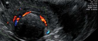
Color Doppler mapping allows you to determine the nature and volume of blood flow in the fibroid. In large nodes, atypical blood flow develops, which can cause severe pain and a feeling of fever.
X-ray methods are of great importance for diagnosing fibroids. However, survey images and intrauterine venography are not widely used due to high radiation exposure. MRI and CT are more informative and safe. These research methods allow us to consider the location of the nodes, their relative placement, the nature of the blood flow, and the effect on neighboring organs. A positive point is the high sensitivity of these methods, which makes it possible to detect very small formations.
Due to the peculiarities of signal recording, MRI reveals several types of tumor:
- Similar to skeletal muscles;
- With a homogeneous structure, but with areas of edema and hyalinosis;
- With a signal similar to the myometrium due to the small amount of collagen;
- With cystic degeneration;
- With degenerative changes and areas of hemorrhage.
Magnetic resonance imaging (MRI) of the uterus allows you to determine the most accurate picture of pathological changes. This diagnostic method is especially valuable if the tumors are small.
Hysteroscopy for subserous localization of the tumor is only important if multiple nodes are suspected, which may have a different location.
Depending on the data obtained during the diagnosis, an individual treatment plan is drawn up.
What does an interstitial subserous myomatous node look like?
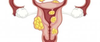
Subserous myomatous node can be single, but it can also be multiple. If the neoplasm is located in the myometrial region, it is called interstitial (intramural). When a tumor-like element is localized in the subperitoneal space, it is called a subserous node.
The subserous-interstitial node of the uterus grows from the myometrium towards the peritoneal space. It can have either a wide base or a rather thin leg. The sizes of such neoplasms vary - from several mm to 10 cm.
The localization of subserous intramural fibroids is different, however, the most favorable and easily treatable tumor is considered to be a tumor located on the anterior or posterior wall of the genital organ. If it is located in the cervix or fallopian tubes, it can lead to infertility. The fact is that such a node significantly narrows the lumen for sperm to penetrate into the cavity of the reproductive organ.
A single subserous intramural node with fibroids looks like a growth enclosed in a muscle capsule. It is attached to one wall of the uterus - posterior or anterior.
Multiple growths can be localized on both walls and have different sizes. In the absence of timely treatment, torsion of the legs of one or several fibroids may occur, which can lead to serious complications.
Why does the tumor grow?
The reasons for the appearance of a tumor in a woman’s body have not been fully established. However, recent studies have found that a major role in maintaining fibroid cell division belongs to the disturbed balance of estrogen and progesterone, with the relative predominance of the former. Estrogens have proliferative activity; they stimulate cell growth and division. But progesterone also plays an important role in this process. It has been proven that after the tumor begins to grow, the number of receptors for this hormone in it increases, which leads to additional stimulation of the cells of which it consists and the maintenance of their division. Progesterone also reduces local immune defense and disrupts the mechanism of apoptosis - programmed cell death. Therefore, cells in fibroids do not die, as should happen normally.
With an imbalance of estrogen and progesterone - hormones that are responsible for a woman’s reproductive health - there is a high risk of tumors.
Numerous studies have found that there are certain factors that can increase or decrease the risk of developing fibroids. Most often, factors predisposing to the appearance of pathology act as a complex.
The following conditions increase the risk of fibroids:
- Early onset of menstruation;
- Absence of childbirth in patients over 30 years of age;
- Late reproductive age (after 35 years);
- Obesity and endocrine pathology;
- Treatment with Tamoxifen and some other drugs that change hormonal levels;
- African American race.
Reduce the risk of developing fibroids:
- A large number of births;
- No miscarriages and no induced abortions;
- Menopause;
- Long-term breastfeeding (more than 6 months).
On a note
There is no exact information about the influence of combined oral contraceptives, hormonal therapy, dietary habits, or region of residence on the development of leiomyoma.
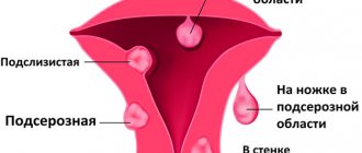
Myoma growth can occur in different directions. Depending on its location relative to the uterine wall, it is divided into several types:
- Submucous - protrudes into the uterine cavity, can be located on a stalk;
- Intramural – grows in the thickness of the muscle layer;
- Subserous - located mainly under the peritoneum or extends beyond the uterus, connecting to it with a thin stalk;
- Interligamentous – located between the layers of the uterine ligament;
- Cervical - grows in the area of the cervix.
Myomatous nodes, depending on their location, are classified into several types, which may differ in symptoms, clinical form, and treatment methods.
Extracorporeal cervical nodes account for only 5% of the total number of myomatous formations. The main location of the tumor is the body of the uterus.
As mentioned above, each form of leiomyoma has its own characteristics of the course of the disease and characteristic symptoms, as well as approaches to treatment. More complete information about the different types of fibroids can be found in a separate article. The most blurred picture of manifestations is in a tumor with a subserous location. In ICD-10 it is assigned code D25.2.
Interstitial subserous fibroids: symptoms and signs
A subserous node with uterine fibroids can cause various ailments, the severity of which depends on its location and size. Thus, an intramural tumor-like growth, which reaches a size of 2-4 mm in diameter, does not cause any abnormal changes in the patient’s well-being.
However, the node can not only enlarge, but also periodically become inflamed, which often causes severe pain in the lower abdomen, pulling sensations in the lower back, and copious vaginal discharge of white, reddish or brownish color. Menstruation becomes more painful and prolonged, sometimes large blood clots are released (if the interstitial node in the uterine cavity has reached a large size).
Myoma, located on the back wall of the reproductive organ, and growing rapidly, begins to put pressure on adjacent internal organs. First of all, the pathological process affects the rectum, as a result of which the patient experiences frequent and prolonged constipation, discomfort or pain during bowel movements.
If the intramural subserous node is located on the anterior wall of the reproductive organ, then its enlargement puts pressure on the bladder. This causes frequent (often false) urges to urinate, and emptying the bladder becomes a rather painful process.
What signs should you be wary of?
Fibroids with subserous growth can remain asymptomatic for a long time. This tumor does not affect the size of the uterine cavity, so it is not characterized by changes in the duration or intensity of menstruation. Endometrial hyperplasia may be present as a concomitant pathology, but in most cases with subserous fibroma the endometrium is not affected. Therefore, intermenstrual bleeding is not typical for this form of fibroids.
Intermenstrual bleeding is not a characteristic symptom of subserous fibroma, since the mucous membrane of the uterine body is not affected by this type of pathology.
To a small extent, symptoms are also expressed in interstitial-subserous fibroids, a tumor that has a wide base. This is due to the lack of its influence on the functional activity of the uterus.
Thus, at first the patient with this pathology does not feel its presence in the body. Symptoms of fibroids begin to appear as the size of the tumor increases and its effect on adjacent organs. You can find out how quickly fibroids grow in our other article. Signs of the presence of pathology will correspond to the location of the node:
- Antecervical localization (isthmus) - the location of the tumor in the isthmus region leads to problems with urination due to compression of the bladder or its nerve plexus;
- Paracervical location - along the lateral surfaces of the cervix. In this case, the patency of the ureters may be impaired, which will be compressed by the tumor on one side. During the operation, problems will arise with catheterization of the ureter;
- Subperitoneal growth of the node leads to detachment of the peritoneum from the posterior surface of the anterior abdominal wall. In this case, the tumor practically does not give any symptoms, so it is rarely diagnosed. Sometimes it can be discovered during surgery when difficulties arise in entering the abdominal cavity. Also, the tumor can put pressure on the bladder and lead to problems with urination, which take a very long time to resolve;
- Retrocervical localization - the node grows behind the cervix towards the rectum. The leading symptoms will be constipation, sometimes with insufficiently formed stool it can take on a ribbon-like shape;
Does a subserous intramural node affect pregnancy?
Uterine fibroids are a real challenge for expectant mothers. Since there are known cases of spontaneous termination of pregnancy due to the rapid growth of a tumor, women’s fears are not unfounded. However, if the subserous node with fibroids is quite small and there are no prerequisites for its enlargement, there is no need to worry. Many women successfully carry and give birth to completely healthy babies, after which they move on to the issue of treating the pathology.
About pregnancy with fibroids >>
If there are multiple subserous nodes located on both walls of the reproductive organ, this means that the expectant mother is at risk of premature birth or miscarriage. In the second and third trimester, in addition to the risk of miscarriage, heavy uterine bleeding often occurs. They come in varying intensities, but do not reduce the risk of serious complications.
Forms of pathology
Depending on the severity, the following forms of interstitial fibroids are distinguished:
- Interstitial uterine fibroids with centripetal growth. It is a neoplasm on the surface of the mucous membrane of the female organ, the growth of which occurs inside the uterus.
- Subserous-interstitial uterine fibroids. It is characterized by the proliferation of nodes on the surface of the female organ with a connective tissue membrane.
- Submucosal interstitial fibroid. The nodes are located under the mucous membrane of the uterus and grow into the organ cavity.
- Multiple interstitial uterine fibroids. There are several nodes in the uterus.
Important to remember! At the first discomfort, you need to seek help from a specialist! After all, inattention to one’s health leads to a significant increase in the tumor, which can reach a size larger than the uterus itself.
Treatment of interstitial-subserous fibroids
Therapy for the presence of a myomatous node in the cavity of the reproductive organ depends on several factors:
- size;
- presence or absence of a leg;
- symptoms;
- the presence of complications or the risk of developing them.
In severe cases of the disease, they resort to surgery to remove the node. In young women, gentle surgery is often performed to help preserve reproductive function.
In the early stages of the disease, a course of conservative treatment based on the use of hormonal medications is possible.
But since in most cases the interstitial-subserous node is detected already in the later stages of the development of the pathology, it is rarely possible to do without surgery.
So, if the patient plans to conceive and give birth to a child in the future, she undergoes laparoscopy, during which only the pathological tumor is excised. It is removed through special punctures made in the peritoneum. The reproductive organ is not affected.
Another modern type of operation used for myomatous tumors is UAE. Embolization of the uterine arteries involves stopping the blood supply to the node, as a result of which it significantly decreases and stops growing. Sometimes the tumor can even resolve on its own.
Read more about the EMA >>
If the disease progresses and the woman’s health rapidly deteriorates, doctors resort to radical surgery – hysterectomy. This type of surgery involves complete removal of the uterus, which eliminates the possibility of pregnancy. However, this method of treating myomatous neoplasms is used extremely rarely.
Treatment
The radical method of treating this pathology is to remove the uterus. This form of treatment is indicated for multiple nodes and their large size (from 13 weeks or more), as well as rapid growth of myomatous nodes, especially in postmenopause, necrosis or cervical location of fibroids, severe bleeding, and concomitant pathology. In young patients of reproductive age, minimally invasive, organ-preserving treatment methods are preferred.
Small interstitial nodes are removed only in preparation for pregnancy, before ovarian stimulation for infertility in women. The laparoscopic approach is used less frequently and is often not recommended due to the risk of uterine rupture during pregnancy and childbirth.
COCs, gestagens, and sometimes androgens are used as conservative therapy. It is possible to use the Mirena intrauterine hormonal system. To create a drug-induced menopause, antigestagens and GnRH analogues are used; GnRH agonists are effective during perimenopause. Innovative drugs for the treatment of fibroids are antifibrotic and antiangiogenic agents, somatostatin analogues.
Complications of pathology
In the absence of adequate treatment, interstitial-subserous neoplasm can have quite unpleasant and dangerous consequences. These include:
- torsion of the leg of a fibroid tumor;
- anemia developing against the background of intense bleeding;
- tissue necrosis of the stalk of the tumor-like formation;
- termination of pregnancy in the early stages;
- premature birth;
- infertility.
If the first alarming signs of this pathology occur, you should be examined by a doctor. In the early stages, the disease can easily be treated using traditional medicine methods in combination with hormonal drugs, so do not miss your chance for recovery without surgery and significant health consequences.
Uterine fibroids - interstitial form: drug treatment
Conservative treatment involves the use of hormonal drugs that block the production of estrogen. There are also medications that reduce the sensitivity of the myometrium to this hormone. This treatment is especially effective for small tumor sizes, in conditions such as regressing interstitial uterine fibroids.
Hormonal treatment will be effective if the intramural fibroid is small. For treatment the following are prescribed:
- gonadotropin-releasing hormone agonists;
- antigonadotropic drugs;
- antiprogestogens;
- progestogens.
The above medications can only be prescribed by a doctor after additional examination. The patient’s treatment regimen is developed taking into account the test results.
Interstitial fibroids and pregnancy
Even if treatment for interstitial fibroids has not been carried out, pregnancy may occur if it is present. It occurs if the patency of the cervical canal is not impaired by neoplasms and the fallopian tubes are not deformed. The risk of miscarriage with interstitial fibroids is quite high, premature birth is possible, as well as the development of bleeding. Moreover: if the myomatous node is large, the risk of abnormal development of the child increases. Childbirth can be significantly difficult.
Diagnostics
Subserosal fibroids of the uterus may be suspected upon examination. During palpation, the doctor determines the heterogeneity of the organ, the unevenness of its walls, and the presence of a neoplasm in the lower part of the abdominal cavity. In some patients, the abdomen is enlarged without excess weight. The subserous node in the uterus does not limit the mobility of the organ. In slender women, it is sometimes possible to determine by palpation that the neoplasm is smooth and not fused with surrounding organs.
After collecting anamnesis (the patient’s story about complaints, possible genetic diseases), the gynecologist prescribes a series of laboratory tests. Diagnosis of pathology includes:
- General, hormonal and biochemical blood tests. They are carried out to exclude inflammatory processes. In addition, a general blood test helps determine the degree of concomitant anemia and assess the intensity of the body's inflammatory response.
- Ultrasound. This is the main diagnostic method that helps to identify the disease, the size of the subserous node, its structure and position. In addition, the condition of the organs adjacent to the uterus is assessed through ultrasound. Both vaginal and transabdominal sensors can be used. Ultrasound is also used for dynamic monitoring of fibroid growth. The technique allows you to timely see signs of malignancy (malignancy) of the tumor.
- CT and MRI. They are carried out to determine the size and location of the node in the uterine cavity. Computed tomography and magnetic resonance imaging clarify the size of tumors and detect the presence of germination into surrounding structures. In addition, these techniques are prescribed to differentiate fibroids from malignant tumors.
- Metrography or hysterosalpinography. This is an x-ray examination that involves intrauterine injection of a contrast agent. Used to determine the degree of deformation of the uterine cavity. Myomas rarely lead to a narrowing of the uterine lumen, with the exception of very large interstitial-subserous tumors and multiple nodes.
- Biopsy. If necessary, the doctor performs laparoscopy and takes a sample from the myomatous formation for histological examination.
If there are alarming signs of a subserous node or the condition worsens, you should go to the hospital. In addition to the survey, collection of complaints, medical history, and examination, the gynecologist prescribes:
- Ultrasound - to determine the location of fibroids in the uterus, structure, size.
- Dopplerography - to study blood flow in the area of atypical formation.
- Magnetic resonance imaging - to study the nature and localization of fibroids in the uterus.
- Laparoscopy - to obtain material for further histology.
They also prescribe a general, biochemical blood test.
In addition to interviewing the girl, collecting complaints, anamnesis, examination in a gynecological chair, and also laboratory tests, the following is prescribed:
- Ultrasound of the internal genital organs (transvaginal) - to determine the presence of a tumor in the uterus and study nodes.
- Hydrosonography - for studying the uterine walls (their thickness), diagnosing concomitant pathologies of the endometrium, identifying the localization of the formation.
- MRI with contrast - for a detailed study of the formation, as well as assessing the effectiveness of arterial embolization.
- Angiography - to determine the malignant degeneration of a tumor.
Hysteroscopy, biopsy, and histology of the removed tumor are often performed.