What it is?
The size of the gland has diagnostic value, so doctors pay special attention to this criterion.
It is important to understand that there are no uniform standards, but average values are considered acceptable standards in the medical community.
For your own safety, it is useful to know how the size of the tumor changes after ovulation, and what deviations from the average values mean.
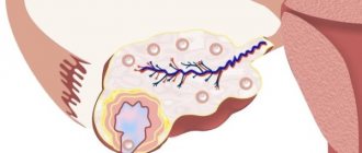
The corpus luteum is a temporary gland that produces hormones. It has an irregular shape, heterogeneous structure and jagged edges on ultrasound images. The formation forms on the left or right ovary during the period of ovulation, that is, after the rupture of the dominant follicle, which occurs on days 10-16 of the cycle. Subsequently, a blood clot filled with granules appears from the follicle, from which a temporary tumor lines the epithelium.
Corpus luteum and its functions
The corpus luteum (hereinafter - VT) is a gland in one of the ovaries (right or left), which is formed for a time after the release of the oocyte (egg) from the follicle, that is, after ovulation itself - an important process without which conception is impossible.
When the dominant follicle develops in the ovary in the first half of the menstrual cycle, it is responsible for the synthesis of hormones such as progesterone and estrogen.
Ovulation has occurred: the follicle ruptures to release the egg. However, the above hormones must continue to be produced, since after the expected fertilization, the body needs to strengthen the endometrium in the uterus so that the egg can attach to it. Subsequently, hormones nourish the fetus during the first trimester, until the placenta becomes responsible for this function.
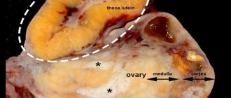
Thus, to synthesize the necessary hormones, a corpus luteum is formed in the same place where the follicle was . Moreover, the larger its size, the greater the amount of hormone synthesized. The iron received its name due to the yellow tint of the parenchyma of which it consists, as well as the cream color of the lutein.
Important ! If ovulation occurs in two ovaries in one month, one corpus luteum appears in each of them.
Why is it called that?
Iron got its name due to the yellowish color of the internal structure, as well as lutein, which itself has such a creamy tint.
A similar clinical picture is observed throughout the luteal phase of the cycle, and the size of the formation changes every day. Ultimately, it gradually decreases towards the end of the cycle and then disappears once menstruation begins. The reason here is simple: fertilization has not occurred.
When a corpus luteum is detected on an ultrasound, nothing terrible happens, but this only indicates that the woman ovulated, but it is not known exactly when it was. For these purposes, the doctor asks the patient the day of her menstrual cycle, checking the data with the results of ultrasound diagnostics.
IMPORTANT! The temporary gland can only be seen on ultrasound images or CT scans. A routine examination on a gynecological chair will not provide any reliable information.
Pathology of the corpus luteum during pregnancy
There are not many pathological conditions of VT, however, due to the disruption of progesterone synthesis, they have a significant impact on the ability to conceive a child and maintain a pregnancy.
Absence of corpus luteum
One of these conditions is the absence of the corpus luteum, which is manifested by the inability to get pregnant.
In the case when pregnancy is observed, but the luteal body cannot be visualized during ultrasound examination, several reasons can be assumed:
- the temporary gland is less than 10 mm in size, which indicates a threat of interruption and the need for hormone replacement therapy;
- anatomical feature of the location of the ovary;
- consequences of pathological processes suffered previously (for example, adhesions after an inflammatory disease);
- physiological characteristics of a woman’s body during pregnancy (many women indicate increased gas formation);
- impossibility of performing transvaginal ultrasound;
- insufficient qualification of a specialist.
Hypofunction
This is a pathological condition characterized by insufficient production of progesterone. This deficiency leads to miscarriage in early pregnancy.
Functional deficiency can be suspected if the corpus luteum on ultrasound is determined to be smaller in size than it should be normally. When an ultrasound doctor suspects hypofunction of the corpus luteum, the patient is sent for a blood test to determine the level of progesterone. Thanks to the achievements of modern medicine and pharmacology, insufficient amounts of the hormone can be replenished by taking medications prescribed by the gynecologist.
Corpus luteum cyst
A cystic formation is a small “bubble” with liquid contents, which on ultrasound is identified as a black, often round, inclusion.
If a cyst is suspected on ultrasound, the corpus luteum will be larger than 35-40 mm in size. The cyst does not affect the ability of the gland to secrete a sufficient amount of progesterone. But if it is large enough, it can rupture, which can lead to bleeding, and this is a surgical pathology. Emergency conditions requiring surgical treatment also include torsion of the cyst stalk.
The conclusion “corpus luteum cyst” on ultrasound without clinical manifestations is not an indication for any treatment. By the time the placenta is formed and the corpus luteum undergoes reverse development, the cyst will also go away.
You should immediately consult a doctor if a cyst has previously been identified and the following suddenly arise:
- lower abdominal pain;
- decreased blood pressure;
- loss of consciousness.
Such complications can be avoided if you follow your doctor's recommendations to avoid physical activity and limit sexual contact.
What is the role?
The corpus luteum does not form in a woman’s body just like that. It performs an important function in synthesizing the hormone progesterone. The larger the size of the formation, the more hormone is secreted.
Progesterone is necessary to prepare the body for pregnancy, as it strengthens the endometrium for implantation of a fertilized egg into the uterus. During the period of bearing a child, the hormone provides nutrition to the fetus in the first trimester, and then this function is taken over by the placenta.
It is known for certain that the corpus luteum produces not only progesterone, but also other vital hormones in small quantities:
- Estradiol.
- Androgens.
Speaking in a general sense, the functions of this neoplasm come down to the following biological processes:
- Production of significant amounts of progesterone, as well as other hormones.
- Nutrition of the fertilized egg in the first 12 weeks of pregnancy.
- Preventing the development of new follicles.
- Strengthening the endometrium and its transformation for fertilization.
- Decreased contractile function of the uterus.
When does the corpus luteum form in the ovary?
The corpus luteum becomes visible in the ovary after ovulation is complete. After the egg is released, the follicular fluid is poured out and the follicle cavity closes. A glandular endocrine structure is immediately formed in it; in the first days it is small in size.
This temporary formation increases gradually. Its diameter is directly related to hormonal activity. The larger the size, the more progesterone it produces.
Stages of formation
The process of formation of the corpus luteum can be divided into several stages:
- First (proliferation). This is the formation of a neoplasm after the follicle bursts and the egg is released. Cell division begins, during which lutein, which has a yellow pigment, is formed. The gland takes on a certain shape, uneven edges and heterogeneous walls.
- Second (vascularization). This stage is characterized by the growth of a neoplasm, which begins to wrap around blood vessels, penetrating into the epithelial layer. A similar picture is recorded on ultrasound images in the form of a small tumor with moderate blood flow. This process is usually observed on days 13-17 of the menstrual cycle.
- Third (flourishing). The gland reaches its maximum size, and its outlines become brighter and more visible, and blood flow increases. The color of the new growth acquires a purple tint, and its diameter reaches 25-27 mm. The blossoming stage is fixed on days 19-25 of the menstrual cycle.
- Fourth (regression). This final stage appears only if fertilization has not occurred and the egg has died. The gland begins to decrease in size, and then completely disappears by the beginning of menstruation. Scars or hylian formations (“white body”) remain on the ovary due to degenerative tissue changes.
All these stages occur sequentially one after another according to the days of the menstrual cycle. If pregnancy occurs, the corpus luteum does not disappear until 12-16 weeks. In size it should not exceed 30 mm, but by the end of the first trimester the iron gradually decreases to 10-15 mm, and then completely disappears.
IMPORTANT! Detection of a temporary neoplasm on ultrasound images is not the main qualifying sign of pregnancy. The endocrine gland always forms after ovulation and can persist for a long time in the presence of a cyst.
Ultrasound diagnostic methods of the corpus luteum
Ultrasound of the female reproductive system, including the ovaries, is performed in two ways.
Transabdominal ultrasound
. The sensor is applied to the surface of the abdomen in the pubic area. This procedure requires that the bladder be full. To do this, a woman should drink 0.5 liters of water 1 hour before the procedure. This allows you to get a more accurate image.
Transvaginal ultrasound
. It is carried out in the middle of the cycle (on days 14-15). The examination is carried out using a vaginal sensor. With such a study, the bladder, on the contrary, should be empty. This method is not used during pregnancy for more than 12 weeks, as well as when examining girls who are not sexually active.
Video: How a corpus luteum cyst is formed
The corpus luteum is formed in the second phase of the female cycle, immediately after the ovulation process. The function of the corpus luteum is to produce progesterone. This hormone reduces contractions of the uterine walls, which is necessary for the preservation of the embryo.
When the third week of the menstrual cycle arrives, the main follicle ruptures, releasing the egg. And at the site of its rupture, this yellow gland is formed. By the way, this shade is given to it by the luteinizing hormone of the pituitary gland.
If fertilization has not occurred by this time, the woman begins menstruation, along with which the endometrium is rejected.
The formation of the gland occurs in several stages. The first is called proliferation and begins when an egg is released from the follicle. Follicle cells actively divide, forming the corpus luteum.
The second stage is called vascularization, in which blood vessels grow into epithelial cells. It is believed that it is the temporary gland that is most actively supplied with blood, which determines the high-quality production of progesterone.
The bloom of the temporary gland occurs in the third stage, during which the production of hormones necessary for pregnancy occurs. If the egg has been fertilized, the gland produces hormones until the placenta is formed.
This takes about 24 weeks. If there has been no fertilization, the activity of the gland lasts only one to two weeks.
The fourth stage is the final one. This is a regression stage that ends with menstruation if there has been no fertilization. Hormones stop being produced, and luteinizing hormone decreases in quantity. Due to the fact that progesterone is not produced by the corpus luteum, the uterus begins to contract, which gives rise to monthly bleeding in the female body.
If pregnancy occurs, the embryo is implanted into the walls of the uterus. The membranes of the embryo begin to produce hCG, which further stimulates the corpus luteum to produce progesterone. From this time on, the gland begins to play a huge role in the proper development and course of pregnancy until the formation of the placenta. Subsequently, it is she who produces progesterone. During pregnancy, the fourth stage of the corpus luteum occurs after the placenta takes over its functions.
It is worth noting that gland plays a very important role in the development of the female body and in its cyclical processes. The function of the corpus luteum is reduced in adolescents, as well as during menopause. And in a woman 18-35 years old, the iron lasts longer. It is the correct production of progesterone and estrogen that affects how cyclic processes will occur in the female body, and how the embryo is implanted into the walls of the uterus.
Size of the corpus luteum on ultrasound after ovulation
The normal size of the corpus luteum depends on the day of the menstrual cycle. Ideally, the gland appears only after ovulation, and before this moment the doctor detects the follicles.
In the first three days of the cycle they do not exceed 4 mm, and then before ovulation they increase to 20-24 mm.
It is quite easy to distinguish them from the corpus luteum. The follicle has smooth edges and a uniform structure. After its rupture (days 11-16 of the menstrual cycle), a temporary gland is formed, the size of which is at first difficult to determine (proliferation).
In the future, its diameter will look like this:
- Days 13-18 of the menstrual cycle – 15-20 mm.
- 18-21 days of the menstrual cycle – 18-20 mm.
- Days 21-24 of the menstrual cycle – 20-27 mm.
- Days 25-29 of the menstrual cycle – 10-15 mm.
Such data is relevant if a woman’s menstrual cycle is 28-29 days, so the above values are averaged.
In general, the size of the tumor should not exceed 30 mm in any phase of the menstrual cycle. Most often, the diameter of the gland 7-8 days after ovulation is 17-24 mm. This is the optimal and desirable norm, which indicates the absence of pathologies.
IMPORTANT! If the size of the gland after follicle rupture is less than 10 mm, then this clinical picture indicates insufficiency of the corpus luteum, which is the main cause of infertility.
Corpus luteum in the ovary: dimensions
The process of formation of a glandular structure from granulosa cells of a ruptured follicle starts after ovulation. When the follicle ruptures and the mature egg is released, blood is released from the damaged blood vessels. It accumulates in the center of the developing temporary gland. In the same place, a stigma is formed - a connective tissue scar, around which the cells of the granular layer begin to multiply. They are covered with a dense network of capillaries. This stage is called proliferation and vascularization. The size of the hormonal formation during this period is 12–15 mm.
Then the granulosa cells begin to accumulate lutein (a pigment that gives the characteristic yellow color) and degenerate into glandular cells - granular luteocytes. Theca luteocytes are formed from cells of the internal folder. This stage is called ferruginous metamorphosis.
Progesterone production begins at the 3rd stage of flowering. At this time, the temporary organ reaches a diameter of 24 mm. During this period, doctors call the formed glandular structure cyclic or menstrual.
2-3 days before menstruation, it decreases to 12-15 mm, and scar tissue forms from it. This leads to a decrease in progesterone production and the onset of menstruation.
If fertilization has occurred and an embryo has formed, then the flowering stage lasts 10-16 weeks. The hormone produced by the corpus luteum of the ovary is required for the maintenance and normal development of the fetus.
During pregnancy, the size of the corpus luteum in the ovary reaches 30 mm. Over time, its functions are transferred to the placenta, and it begins to regress.
Reasons for deviation from the norm
Often the size of the corpus luteum does not correspond to normal values. In most cases, this condition is preceded by one or another reason. Deviations from average values are possible in the presence of:
- Cysts.
- Insufficiency of the corpus luteum.
If ultrasound images show an increased diameter of the gland, the doctor suspects a cyst. Doctors attribute this pathology to functional neoplasms that arise as a result of hormonal imbalance and resolve on their own after 2-3 menstrual cycles.
Only cysts larger than 40 mm are dangerous. They require serious treatment in the form of hormonal therapy or surgical laparoscopy. Formations larger than 60 mm require surgical intervention, as there is a high risk of rupture.
The small size of the temporary gland is a lack of progesterone production, which is the cause of infertility. This condition is not life-threatening, but requires serious treatment in the form of hormonal therapy.
How long does the corpus luteum live in the ovary?
If there are no problems with reproductive health, then a temporary hormonal formation appears immediately after the release of the egg and continues to develop for 12-14 days. If during this period the egg is fertilized and begins to implant into the wall of the uterus, then it will continue to develop. The lifespan of the corpus luteum in the left ovary during pregnancy can last 16 weeks.
When should you start worrying?
Concern should be shown in the following cases:
- The diameter of the temporary gland is more than 40 mm.
- The patient has pronounced gynecological symptoms.
- A woman cannot get pregnant.
The situation when the size of the tumor remains enlarged deserves special mention. Often it begins to progress, and then does not respond to drug and hormonal therapy. This is a serious cause for concern and usually ends with surgery in the form of laparoscopy.
REFERENCE! Small cysts with a diameter of 30 to 40 mm in most cases disappear on their own after 2-3 months. The patient does not need to do anything to treat them, but a control ultrasound examination after 3 menstrual cycles must be done as a control.
Gynecological symptoms
Menstrual irregularities and pain do not always mean a serious problem, but in any case this is an important reason to consult a doctor and have an ultrasound scan.
The small size of the tumor is a significant anomaly, especially when planning pregnancy, where increased production of progesterone is so necessary. In this situation, the best treatment option would be hormonal therapy followed by follow-up with a doctor.
Which hormone promotes regression of the corpus luteum of the ovary
The process of formation and regression of the temporary luteal gland is under the control of the pituitary gland, growth factors and the immune system. Luteolytic action is characterized by O2 radicals, cytokines, prostaglandin F2α, prolactin, oxytocin.
The corpus luteum in the right (left) ovary during pregnancy is necessary for the production of peptides and steroids necessary for the preservation and development of the embryo. When an egg is fertilized and implanted, hCG synthesis begins in the body. This hormone affects the formed structure, provoking an increase in the process of progesterone production.
In the absence of pregnancy, the stage of reverse development begins. The production of progesterone, inhibin A and estradiol decreases. By reducing the concentration of inhibin A, its blocking effect on the pituitary gland is eliminated. As a result, the secretion of follicle-stimulating hormone is resumed. This is also facilitated by a decrease in the concentration of progesterone and estradiol.
The corpus luteum forms in the ovary immediately after the release of the egg. Thanks to its presence, it is possible to confirm the occurrence of ovulation. During fertilization and the formation of the embryo in the current menstrual cycle, the luteal gland is preserved; you can understand the peculiarities of its effect on the body by watching the video:
https://youtu.be/4CxTxw539CQ
If fertilization does not occur, then it regresses.
What do gynecologists and uzologists mean when they say that there is a corpus luteum in the ovary? What is it, what should it be (and should it be at all), what function does it perform in the female body?
When to see a doctor?
With minor pathologies of the temporary gland, symptoms manifest themselves very sparingly. A woman usually does not notice its clinical manifestations.
The problem is discovered during preventive diagnostics, which is usually ignored by patients. However, a significant increase in the size of the tumor is accompanied by the following symptoms:
- Pain in the right or left ovary of varying nature and intensity.
- Delay of menstruation by more than 5 days.
- Severe pain in the mammary glands that has not previously been observed.
- Increase in basal temperature.
- Pain, discomfort in the lower back, groin.
If the above clinical manifestations are observed in the second phase of the cycle, then the problem probably lies in the presence of a cyst.
This is the main reason to consult a doctor, as the consequences can be unpredictable.
IMPORTANT! In some situations, it is necessary to call an ambulance, especially if there is acute abdominal pain, vomiting, nausea, or a rapid rise in temperature. These are indirect signs of cyst rupture, which requires emergency surgery.
Signs of the corpus luteum in the left ovary
Only with the help of ultrasound can one determine the presence of a corpus luteum in the ovary. It is impossible to determine on your own whether it appeared in the 2nd half of the cycle. The temporary structure responsible for the production of progesterone forms and disappears asymptomatically, no visible changes occur.
If during ovulation a woman felt pain or tingling in the left lower abdomen, then we can assume that a temporary hormonal gland will appear on the left.
If an ultrasound shows that the corpus luteum in the left ovary is 18 mm in diameter, this means that the egg has emerged from the left female reproductive gland. This size corresponds to phase 2 of the cycle. When planning pregnancy, it is better that its diameter is more than 20 mm. This is an indirect confirmation that progesterone is produced in sufficient quantities. When the size of the hormonal formation is less than 16 mm, they speak of phase 2 insufficiency.
Can the corpus luteum in the ovary hurt?
The endocrine gland formed at the site of the ruptured follicle should not provoke pain. Although some gynecologist patients report the appearance of pulsation and discomfort in the lower abdomen, which are concentrated on one side.
Severe pain usually occurs when a cyst forms. This condition is characterized by the fact that by the time menstruation begins, the temporary organ does not decrease in diameter, but continues to grow. Intracellular fluid begins to accumulate in it. Cysts can reach 8 cm in diameter. In 90% of cases, they resolve on their own within a few months.
Sudden sharp pain may indicate that the corpus luteum in the ovary has burst. This is a pathology that doctors call apoplexy. The condition requires immediate hospitalization and surgery during which resection or complete removal of the problem organ is performed. Due to the rupture, blood begins to accumulate in the abdominal cavity, and the lack of timely medical care provokes the development of serious consequences.
Corpus luteum in the ovary and delay
If a delay in menstruation occurs during an ultrasound, the doctor must evaluate the size and structure of the formation that appears in phase 2. The absence of menstruation on time in the presence of the luteal gland may be due to pregnancy. Normally, its size should be 20-30 mm. On ultrasound it looks like a hyperechoic formation with heterogeneous content.
If menstruation is delayed, on ultrasound the doctor may see an anechogenic round formation with a homogeneous structure - a cyst. Its diameter varies from 30 to 80 mm. To normalize the condition, patients are prescribed progesterone drugs in a short course. A few days after their cancellation, menstruation should begin.
A situation is possible in which the delay is caused by the persistence of the corpus luteum in the right or left ovary. Persistence of an immature hormonal gland is characterized by a decreased level of luteinizing hormone. The temporary organ cannot fully mature, because of this, phase 2 of the cycle is lengthened.
When a mature gland persists, it does not decrease in size by the beginning of menstruation, but moves into the next cycle. In this case, a full follicular phase and an extended luteal phase are observed. It lasts 20-25 days and is accompanied by hyperproduction of progesterone.