For the musculoskeletal system, a spinal fracture is the most serious injury. These types of fractures have a wide range of classifications and several different outcomes. In some cases, destruction of the bone of one of the segments can only result in long treatment and an uncomfortable corset. In another situation, it will take several years to heal, and there will still be consequences. And some of the victims die even before the ambulance arrives.
What is a spinal fracture? This is a dangerous pathology that occurs due to impaired integrity of the bones in the spinal column and threatens with serious complications for later life.
Causes
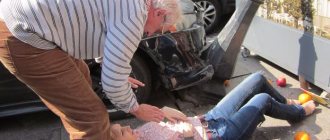
A spinal fracture occurs with a variety of injuries. The bone tissue of the vertebrae cannot withstand the pressure and is destroyed as a result of an unsuccessful fall on the back, straightened legs or buttocks. Rupture of discs and ligaments often occurs due to sudden braking of the car and throwing back of the head as a result of an accident.
A fall from a height, a direct strong blow to the back during a fight or sports activities are another category of common causes. In addition, any vertebra can break due to long-term osteomyelitis and osteoporosis. Especially in old age. Rickets, congenital genetic pathologies or an oncological process in the bones are also a potential threat of fracture.
Diagnosis of fractures
Naturally, the basis for an accurate diagnosis is a qualified X-ray examination. In any case, irrefutable evidence of a fracture will be the detection of both the fracture line and individual displaced fragments. In children, a fracture with the beautiful name “green twig” often occurs when the young and flexible periosteum remains intact.
In this case, as well as in impacted fractures, identifying the fracture line presents certain difficulties. But, “fortunately”, for a beam injury in a typical place, an impacted mechanism is not typical, and yet variants are very rare.
The radiologist then determines the position of the fragments. Sometimes the distal fragment is not whole, but fragmented. In some cases, a fracture of the styloid process of the ulna is detected. This “surprise” is noted in 70% of all cases.
It is very important to determine the type of fracture - what mechanism caused the injury, and specifically on a lateral X-ray. When repositioning, you need to put the fragment in place so that it does not stand anteriorly or posteriorly. If this is not done, then after the bone heals, either flexion or extension of the hand may be limited.
Classification
The classification of vertebral fractures includes categories based on several characteristics. According to the mechanism of occurrence:
Flexion - the spinal column suddenly bends when falling on the buttocks, when falling on the shoulders of a strong weight, and when a person jumps from a great height on straight legs.
Extension - due to strong extension of the spine, the longitudinal ligament, intervertebral cartilage is torn, and the vertebral body is destroyed. This type of fracture is mainly characteristic of whiplash injuries in the cervical spine in car accidents.
Rotational - the spine in the cervical or lumbar region bends and rotates along the axis. A fracture-dislocation or dislocation of the vertebra occurs. In most cases, such injury is accompanied by damage to the spinal cord and is characterized by subsequent vertebral instability.
Compression - occurs when there is excessive vertical pressure on the vertebral body and intervertebral discs. The vertebrae are deformed - flattened. The spinal cord is often affected.
In some cases, the victim is diagnosed with not only isolated types of fractures, but also their combinations.
Fractures are distinguished separately by the number of broken vertebrae:
- Multiple;
- Isolated.
Depending on the threat of displacement:
- Stable;
- Unstable.
If the vertebral body is crushed into several fragments, this is a burst or comminuted type of fracture.
With secondary damage to the spinal cord, rupture of discs and ligaments, the fracture is considered complicated. If only the bone is damaged, the fracture occurs without complications.
Depending on the specific location of the injury, injuries are classified into fractures of the cervical, thoracic, lumbar, sacral and coccygeal spine.
Cervical fractures
The seven vertebrae in the cervical region are the most fragile and flexible. The integrity of the bone in this segment can be disrupted even by a sharp turn of the head. The most life-threatening fracture is considered to be from the 1st to 3rd vertebrae. Such injuries mainly occur in road accidents, diving in shallow water, a heavy object falling on the head, and during extreme sports.
Clear signs of a fracture include:
- Neck pain of varying intensity;
- Nausea, dizziness;
- Sometimes - urinary incontinence;
- Hypertonicity of the neck muscles;
- Dysfunction of the respiratory and cardiovascular systems up to clinical death - in cases of severe complicated fracture;
- Impaired sensitivity from mild numbness to complete loss of tactile sensation in both the neck and limbs.
Multiple fractures of two or more vertebrae are characterized by difficulty breathing, severe headache with dizziness, and often painful shock. In other cases, a vertebral fracture may only be felt as minor discomfort. And such a situation is similarly dangerous due to developing consequences in the short term if diagnosis is not carried out in a timely manner.
Fractures of the spine in the cervical region are compression, comminuted and compression-comminuted. The last type of fracture is the most dangerous and complex.
Fractures of the thoracic and lumbar spine
Compared to injuries in the cervical spine, these types of spinal fractures are more common. The high degree of susceptibility to thoracic injuries is explained by limited mobility and, therefore, less depreciation of the intervertebral discs.
The largest vertebrae, the 10th, 11th, and 12th, which function as supports for the chest, are the most commonly fractured. A vertebral body fracture can be either isolated or combined with a head injury, rib fractures, abdominal injuries, and fractures in the upper or lower extremities. This mainly occurs during falls from great heights, road accidents, work injuries, and bone pathologies. For example, everyone who suffers from bone tuberculosis, osteomyelitis, osteoporosis and oncology with metastases in the spine is at risk.
In addition to severe pain with the slightest movements, a fracture in the thoracic region can be determined by the following signs:
- Numbness and loss of sensation in the legs up to paralysis;
- Asphyxia;
- Difficulty urinating;
- Irradiation of pain to the heart, shoulder blades or abdominal cavity;
- Traumatic shock.
The thoracic region is characterized by a compression type of fracture, when the vertebral body is flattened under strong pressure. Due to the close proximity of the heart and lungs, severe complicated fractures for the victim are sometimes fatal.
There are 5 vertebrae in the lumbar region. Of these, the most vulnerable is the first, and if it is fractured, the vertebral body is completely destroyed. A broken second vertebra is less common and usually results in injury to the first and third.
The fourth vertebra usually fractures due to compression of the second and third. This happens as a result of severe subsidence of the intervertebral disc. Destruction of the 5th vertebra often occurs after a sudden fall onto the buttocks from a height, including the height of a person’s own height.
Of the most typical symptoms of a spinal fracture in this case, the following are noted:
- Severe muscle weakness in the legs;
- Severe lower back pain;
- Urinary incontinence or vice versa - difficulty urinating;
- Paresis or complete paralysis of the lower limbs.
Only after 3 or 4 months of active rehabilitation does the period begin when you can sit after a compression fracture of the spine.
As with fractures in other vertebral regions, prognosis depends directly on whether the spinal cord is affected and to what extent. For example, its complete transverse rupture completely immobilizes the victim and negatively affects the functioning of the rectum and bladder of a person for many years or for life.
Fractures of the sacral and coccygeal regions
A fracture of the sphenoid bone at the base of the spine is a fairly rare injury, but very serious. An isolated sacral fracture occurs in no more than 20% of all cases of injuries in this segment. In all other situations, the sacrum is damaged along with the bones of the pelvic ring.
Among the most common causes of a fracture are a fall on the buttocks from a height, compression of the pelvic bones as a result of a domestic or work injury, and sometimes difficult childbirth.
In this case, vertebral fractures are manifested by pain in the lower back and groin area. Especially in a sitting position. Victims also complain of problems with urination, limited movement and numbness of the skin in the lumbar region.
A fracture of the coccyx occurs for similar reasons as a fracture of the sacrum. In addition, additional predisposing factors for this include systematic strong shaking while driving and downhill skating on smooth plastic ice skates.
A fracture can be suspected by severe, prolonged pain, swelling and hematoma in the tailbone area, and difficult and painful defecation. In some cases, the consequences of only conservative treatment without surgery lead to chronic constipation, frequent migraines due to bone marrow displacement, gout and inflammation of the nerve plexus in the coccyx.
What types of fractures are there? Classification of bone fractures
Classification of fractures:
- Based on their origin, fractures are divided into congenital and acquired. Congenital occurs when there are malformations of the fetus. Acquired ones are divided into traumatic and pathological. Pathological fractures occur with metastases of malignant tumors, tuberculosis, osteomyelitis, etc. To establish the correct diagnosis, the international classification of diseases is used.
- Based on the presence of soft tissue damage, fractures are divided into open, closed and gunshot.
- Depending on the nature of the bone damage, fractures can be complete or incomplete. Incomplete fractures include fissures, subperiosteal greenstick, marginal, and perforated fractures.
- According to the direction of the fracture line, transverse, oblique, longitudinal, comminuted, helical, impacted, and compression fractures are distinguished.
- Based on the presence of displacement of bone fragments relative to each other, fractures can be either displaced or without displacement. Displacement is possible: in width, length, at an angle and rotationally.
- Based on the damage to the part of the bone, fractures are divided into diaphyseal, metaphyseal and epiphyseal.
- The number of fractures can be single or multiple.
- According to the development of complications, complicated and uncomplicated fractures are distinguished. The following complications may occur: traumatic shock, fat embolism, damage to internal organs, bleeding, nerve damage, and the development of surgical wound infection.
Signs of a fracture
Signs of a spinal fracture manifest themselves in different ways. Each symptom always depends on the location of the damage, whether the spinal cord is affected or not, whether the ligaments and neighboring organs are affected.
When one or more vertebrae is destroyed, victims experience:
- Local pain from minor discomfort to unbearable;
- Weakness in the limbs up to paresis and paralysis;
- Severely limited movement activity in the damaged segment;
- Irradiation of pain to the back, head and abdominal cavity;
- State of traumatic shock;
- Swelling and hematomas in the damaged area;
- Dizziness and severe weakness;
- Labored breathing;
- Difficulty with full urination;
- Involuntary muscle contractions.
The most striking manifestations of symptoms occur with comminuted fractures of the spine and complicated compression injuries. In such situations, the spinal cord, nerve endings, and blood vessels are often damaged, and there is no doubt about the presence of a fracture.
How to identify an injury - symptoms:
How to detect a fracture?
- formation of a tumor around the site of injury;
- bruising;
- limited mobility (if we are talking about limb fractures);
- feeling of severe pain when trying to move;
- deformation at the site of injury;
- change in the length and width of the damaged area;
- crunching sound when injured;
- bone mobility outside the joints.
Please note that the signs of a fracture may vary significantly depending on the location of the injury. So, for example, when receiving a spinal injury, the patient may not feel pain at all at the site of the injury, but feel discomfort in the legs. Deformity, as a rule, does not occur with joint fractures. A clear sign of damage in this case is acute pain.
First aid
For the victim, a spinal fracture is sometimes not only a threat to health, but also a great danger to life in general. First aid must be provided in a short time, but each action must be extremely competent. Otherwise, there is a high probability of significantly worsening the person’s condition. Especially if the injury is a displaced fracture of the spine. Each mistake can lead to either lifelong disability or instant death.
So, what should you do if someone in front of you breaks their spine? First of all, a person must be limited in movements as much as possible. To do this, carefully lay him on his back on a flat surface. After this, moving it in any way until medical help arrives is strictly prohibited.
If the fracture is in the cervical region, secure the victim’s neck to the shoulder with a collar made of thick fabric or cardboard. Regardless of which segment the fracture occurs, it is strictly prohibited to adjust the vertebrae yourself.
To prevent painful shock when the victim experiences unbearable pain, he needs to be given any strong analgesic in the form of an injection or tablets. But tablets are categorically excluded due to the risk of getting into the respiratory tract if a person is on the verge of losing consciousness.
At least three people must move the victim for transportation. One holds the neck and head, the second is responsible for the chest area, and the third is responsible for the legs and pelvis. If doctors have a soft fabric stretcher, the person is placed on his stomach. All vertebral sections, including the legs, are securely fixed with special belts or tourniquets.
Where can the beam break, and what does it look like?
The radius is a rather long formation. It connects the elbow and wrist joints, and can break in the following places:
- the head and neck of the radius near the elbow joint.
Most often, this is the result of a sharp hyperextension of the elbow, or a jerk of the forearm outward or inward around the elbow joint. Swelling occurs in the elbow area, along the front and outer surface of the forearm. After this, movement of the elbow, especially rotation and extension, causes severe pain;
- diaphyseal fracture of the radius (in its middle part).
Quite often, a fracture of the diaphysis is combined with a fracture of the ulna. A single fracture of the radius is more hidden, since there is no deformation of the forearm, and there are no signs of gross dysfunction.
However, there is pain and swelling at the fracture site. The range of motion (rotation of the forearm) is reduced, and when moving, you can hear the crunching of fragments, or crepitus. A characteristic symptom of a radial fracture is a “silent” and non-rotating head of the radial bone when the forearm is rotated.
- fracture-dislocations of Monteggia and Galeazzi.
This is the name for combined injuries, in which one bone breaks and the second is dislocated. With a Montage injury, the ulna bone breaks (in the upper third, closer to the elbow), and the head of the radius is dislocated, but remains intact. But Galeazzi’s fracture-dislocation leads to the radius breaking in the lower third, and the ulna dislocating its head.
A Monteggia injury occurs when the upper third of the forearm receives a blow. There is a sharp disturbance in movement in the elbow, the forearm becomes slightly shortened, and it swells near the elbow.
With a Galeazzi injury, swelling and pain occur in the area of the wrist joint, and deformation of the radial bone contours occurs at a certain angle.
All these types of injuries can be treated conservatively or surgically, depending on the severity of the injury, the presence of displacement, tissue interposition and other factors.
Diagnostics
The traumatologist finally confirms the signs of a fracture with the result of an x-ray in the lateral and direct projections. If there is a suspicion of damage to the spinal canal, CT or MRI results of the spine will be needed. An examination and conclusion by a neurologist, as well as a study of cerebrospinal fluid, are often required. Without such an examination, it is impossible to make an accurate diagnosis of a spinal fracture.
Treatment of open fractures
Open injuries are very serious injuries that are treated in a hospital. If the wound is small in area and there are no small crushed bones, then the wound and broken bones are disinfected.
In the presence of extremely severe fracture, surgical intervention is necessary. Before the operation, instrumental studies are carried out in order to accurately identify the nature of the damage: the depth of displacement of bone fragments, the severity of the fracture and damage to soft tissues. To do this, the following studies are carried out:
- computed tomography (CT);
- X-ray;
- magnetic resonance imaging (MRI).
The next step in the trauma recovery process is:
- disinfection of the wound and removal of dead tissue and skin;
- manual comparison of bone fragments;
- suturing and installing drainage;
- bone fixation using special orthopedic structures (Ilizarov apparatus).
For injuries with the formation of open wounds, external fixators are used, which imply access to the wound for monitoring it. If a plaster cast is applied, be sure to leave a window for treating the wound.
Treatment
How to treat a spinal fracture? Treatment for a spinal fracture is difficult and lengthy. Closed fracture reduction and the traction method are now used quite rarely. The most effective methods are conservative therapy and surgery, depending on the complexity of the fracture.
Conservative technique
It is used in cases of closed, stable and uncomplicated fracture without spinal cord injury or vertebral displacement. Treatment involves wearing a brace or collar if the neck is broken. An important part of therapy is prolonged bed rest with a roller in the area of injury. The exact period directly depends on the severity of the fracture and the presence of complications: the period ranges from one to several months. The same goes for the use of corsets. In some cases, it must be worn for up to six months.
The patient must be prescribed several groups of drugs.
- Preparations containing calcium and multivitamin complexes to speed up the fusion process - Calcemin, Calcium D3;
- Drugs that support the full structure of the intervertebral disc - Alflutop, Teraflex, Dona and their analogues;
- Meloxicam, Diclofenac, Nimesulide - as an analgesic and anti-inflammatory agent;
- External ointments and gels for relieving swelling and eliminating hematomas - Fastum gel, Ultrafastin, Ketoprofen and Voltaren.
Physiotherapy and feasible therapeutic exercises with a gradually increasing load are of great importance in the treatment complex.
Vertebral fixation corset
The treatment uses a supporting rigid corset made of durable plastic or an alloy of light metals. What is the essence of a corset? It stably fixes all vertebrae in the damaged segment. Thus, the therapeutic effect is as follows:
- The vertebrae, for example, of the thoracic region are almost completely immobilized for a long time. Because of this, they get used to a full-fledged stable position and become entrenched in it;
- The back muscles stop spasming, displaced bones do not touch the nerve endings, which significantly reduces pain;
- The corset takes on the entire weight of the back, relieving the muscles;
- Blood circulates well in bone and muscle tissue, supplying them with oxygen and essential nutrients. Thanks to this, they recover faster;
- Active blood flow warms up the back well. In this case, the corset performs the functions of a radiculitis belt.
After 4 or 5 months, it is advisable to replace the rigid corset with an elastic one with the possibility of semi-free fixation. This option allows you to bend, taking on a significant part of the load and at the same time reliably fixing the vertebrae.
Operation
Injuries complicated by spinal cord damage and compression of nerve endings, open, unstable and comminuted fractures require surgical intervention. In this case, the specific type of operation depends not only on the nature of the fracture, but also on the age of the patient and his general condition.
In the case of an uncomplicated compression fracture, vertebroplasty is used. The destroyed vertebral body is restored by injecting bone cement, a special plastic material. Bone cement is injected transpedicularly through a needle, each action being monitored by radiography. The length of hospitalization after surgery is minimal. Most patients feel significant relief within a few hours.
Vertebroplasty is relevant only for those patients in whom the height of the vertebral body as a result of compression has decreased by less than 70%.
Kyphoplasty is similar to vertebroplasty. Kyphoplasty is also used to restore the volume of a damaged vertebra. In this case, the restorative material is supplied to the spinal bone using a special oxygen cylinder. A balloon with continuous air supply is first inserted into the vertebral body, and only then a puncture needle with fixing material is inserted.
Unlike vertebroplasty, kyphoplasty requires more time and mechanically causes more trauma to the skin.
For burst and comminuted fractures, operations are performed, during which the fragments are first removed, the height of the vertebral body is restored, and its deformity is corrected as necessary. The surgeon creates a fixed block between the broken and adjacent vertebrae using anterior spinal fusion. The vertebral body is completely or partially replaced by an implant.
Treatment at home
The results of home treatment depend entirely on strict adherence to all recommendations received. Any loads and active household mobility are excluded. Bed rest is extremely important.
If the patient wears a corset, several rules must be followed:
- It should not be worn on a naked body. For clothing you need a natural cotton T-shirt or T-shirt;
- At night, the retainer must be removed;
- The corset should not interfere with free breathing and blood circulation. If this happens, it indicates that it was selected incorrectly.
The patient should sleep on a fairly hard surface. To do this, you need to put strong, hard plywood of the appropriate size under the mattress. In some cases, the bed must be tilted so that the patient's head is higher than his feet.
To prevent bedsores, it is extremely important to wipe the patient’s skin with camphor alcohol and antiseptics. You can place soft cushions under your knees and feet. If a bedridden patient suffers from constipation, he needs systematic siphon enemas.
What to do with a closed fracture
What to do with a closed fracture - this question interests everyone who has at least once witnessed such injuries. Sometimes those around them become confused when they see the victim in such a situation, or, due to panic, they do something completely different from what is required. Anxiety is transmitted to the victim, which worsens his already serious condition.
All measures should be taken to transport the victim to the clinic. First aid before the ambulance arrives and the victim is transported to the trauma center is as follows:
- Give painkillers to prevent painful shock;
- Stop the bleeding (if there are bleeding wounds in the area of the fracture, it is necessary to treat them - disinfect them, try to stop the bleeding, apply a sterile bandage).
- Ensure immobility of the affected limb. If possible, apply a bandage (splint) to secure the limb.
Correctly provided first aid reduces the likelihood of further complications and increases the effectiveness of treatment. The emergency team can also apply a splint. If you don’t have a tire, you can make one from boards. First aid should also include stopping bleeding, preventing the migration of bone fragments and damage to adjacent tissues. All this must be done carefully and without panic.
Diagnosis and treatment of bone injuries
To diagnose traumatic bone injury, radiography is necessary. The broken area is examined in two projections. Additionally, a computed tomography or MRI may be prescribed. Treatment of limb injury is carried out conservatively or surgically. With conservative treatment, bone fragments are compared and immobilized for several weeks, depending on the nature of the damage. Most often, this treatment is carried out if a person has a closed fracture without displacement.
The likelihood of normal bone fusion depends on bone mass, age, and body condition. The older the patient, the slower his bones heal. The time of fusion is influenced by the presence or absence of complications in the patient.
In case of an injury complicated by displacement, the bone is compared and fixed through surgery. For this, screws, knitting needles, and staples are used. In some cases, wearing an Ilizarov device is indicated. During the recovery and rehabilitation period, limb massage, physiotherapy, and exercise therapy may be indicated.
Performing exercises is of great importance for restoring a person’s normal performance and improving his overall physical and emotional condition.
About rehabilitation
What rehabilitation measures are necessary to fully restore the mobility of the limbs and return a person to an active life? These may include special exercises, massage, physiotherapy, a special diet, and, if necessary, wearing orthoses. The goal of rehabilitation is to minimize any manifestations of injury, restore blood circulation, muscle strength and quickly return the patient to a healthy state.
When a callus forms, the patient can benefit from sunbathing and electrophoresis. As soon as the plaster is removed, the patient may be shown mud therapy, therapy with paraffin or ozokerite. Any type of bone injury requires responsibility on the part of the doctor and the patient.
During treatment, it is very important to quit smoking and drinking alcohol. High-quality nutrition, positive emotions, and physical exercise help to cope with the consequences of a fracture and prevent its complications.
A closed fracture is a serious injury that is treated conservatively. In some cases, surgery may be required. Provided that first aid was carried out correctly and the patient followed all the doctor’s instructions, the injured bones heal and the rehabilitation time is relatively reduced.
Rehabilitation
The inevitability of prolonged immobilization is atrophy of the adjacent muscles. During this period, the muscles weaken, and after a certain time after the fracture, the victim needs functional restoration and strengthening of the spine.
Physical therapy exercises and massage
Physical therapy is prescribed in accordance with the severity of the fracture and the individual characteristics of its treatment. Equally important are the patient’s age and the time interval since the injury.
Full recovery without exercise therapy is almost impossible, especially after surgery and wearing a corset. But in each case, the gymnastics complex is selected purely individually, and for each stage of rehabilitation it changes in increasing complexity.
Breathing exercises are important for the cardiovascular system, and are sometimes prescribed in the very near future after the fracture has occurred. This could be simple diaphragm breathing, blowing up balloons and other similar exercises.
In addition, at the first stage, you can make rotational movements with your hands. During any movements of the legs, in order to prevent possible pressure on the damaged segment, the foot should not be lifted off the bed. The duration of the stage is, as a rule, no more than 10–14 days, after which the patient is allowed to smoothly and carefully roll over onto his stomach.
For the second stage of rehabilitation, exercises are selected to strengthen the back muscles.
- From a lying position on your stomach, raise your shoulders and head;
- Lying on your back - alternately lifting your legs at an angle;
- Circular movements with legs bent at the knees - “Bicycle” exercise.
The third stage of recovery occurs 1.5 - 2 months after the injury. The gymnastics complex includes:
- Alternately raising your legs up from a lying position on your stomach;
- Lying on your back - simultaneous lifting and spreading of straightened legs to the sides;
- Slow walking with your toes raised and your elbows out to the sides.
At the final stage of rehabilitation, exercises in a standing position are shown. These are swings of the legs straight, back and to the sides, rolls from toe to heel and vice versa, as well as smooth squats.
To improve blood flow and restore muscles, massage is equally important. The optimal number of sessions for one course is from 10 to 15. But for different ages of patients, the technique for performing it is also different. For example, for young patients after injury, it is important to form a muscle frame. At the same time, elderly patients require a soft and gentle technique.
Each session begins with relaxing stroking movements, after which they begin kneading. However, some techniques can cause harm to bone tissue that has not yet strengthened. Therefore, the procedure should only be carried out by a qualified specialist who knows how to break muscle knots without the slightest impact on the bones.
Features of open fractures
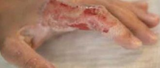
The main characteristic of such fractures is damage to the skin. Open fractures cannot be confused with other fractures. They occur when sharp bone fragments cut through the skin. Bone fragments can be seen with the naked eye, since they come out through the wound and communicate with the external environment. A closed fracture is characterized by the integrity of the skin, and the presence of a fracture can only be determined by secondary signs (pain, swelling, impaired mobility).
Injury to bone structures in closed and open ruptures is of the same nature, but a violation of the integrity of soft tissues is observed in the second case and can occur in several ways:
- rupture of fibers due to their significant tension;
- a cut in the skin with a specific object that caused the injury itself;
- skin damage from sharp fragments of broken bone.
An open fracture is dangerous because the open wound can become infected, extensive bleeding can also occur, and bone fragments can cause damage to the vascular system and rupture of nerve fibers.
Rehabilitation and consequences of a fracture in children
In the first 3–5 days after a fracture, the child needs to reliably block the pain and relieve the damaged part of the spine. Strict bed rest with spinal traction and a reclining roller is prescribed. The bed must be raised 30 degrees and a firm bed surface must be provided.
The most commonly prescribed physiotherapeutic procedure is electrophoresis. Just like for adults, breathing and therapeutic exercises are very important for proper functioning of the lungs and normal blood circulation. But the child performs all exercises exclusively lying down, without raising either his legs or his head. 2 weeks after the injury, magnetic therapy is added to the procedures.
To form a full-fledged muscle corset, symmetrical massage and appropriate exercises from the therapeutic complex are prescribed. To do this, the child can lie on his stomach with support on his elbows. Over time, you need to turn from your back to your stomach 6 to 8 times daily. The total amount of time in this position should be at least one and a half hours. After another 3 weeks, exercises on all fours are included in the complex.
Grade I compression fractures are best treated. But, unfortunately, this is not a guarantee that in adolescence or even in adulthood the consequences of a spinal fracture will not remind of themselves. Even after several years, a child may periodically experience pain in a previously broken back due to osteochondrosis, spondylitis, intervertebral hernia or kyphosis. Such diseases develop as a result of instability of the spinal segments. Therefore, parents should patiently go through all stages of treatment consistently and without deviations.
How to treat a fracture of the radius?
It is important to remember that healing of a radius fracture requires the following conditions to be met:
- Accurately bring the fragments together along the fracture line;
- Squeeze them tightly until the gap disappears;
- Immobilize fragments as much as possible for at least 2/3 of the immobilization period.
Of course, these are ideal conditions, and both the quality and the period of fusion depend on them. What do doctors usually do for uncomplicated types of radial fractures?
With an extension fracture
First, the traumatologist anesthetizes the fracture site. For this, 20 ml of a 1% novocaine solution is sufficient, and manual closed reposition of the fragments is performed. To do this, the forearm is bent and a counter-thrust is created towards the elbow along the longitudinal axis of the hand. This position must be maintained for 10 - 15 minutes. This is necessary so that the desired muscle group relaxes and does not interfere with reposition. After this, the fragment usually easily moves towards the palm and elbow.
In order for the angular deformity to disappear, the hand and the fragment are bent in the palmar direction, usually at the edge of the table. After this, with palmar flexion and slight abduction to the elbow, a plaster splint is applied to the back of the hand. It should cover the space from the upper third of the forearm to the metacarpophalangeal joints, leaving only the fingers free.
If the injury is flexion
The difference is that here the force and direction are created by moving the fragment to the back side, and not to the palmar side. To prevent angular displacement, everything is done in the opposite way, that is, the hand is extended at an angle of 30 °, and a plaster splint is also applied.
After the reposition is carried out, you need to make sure that everything is aligned correctly. To do this, an x-ray is taken, and in complex cases (for example, with a helical fracture line), the reposition itself is carried out under x-ray control.
Possible complications
Fortunately, an infrequent but unpleasant complication is injury or rupture of the radial nerve. This is an indication for urgent surgery. Symptoms of damage are:
- dorsal numbness of the hand and the first three fingers (from the thumb);
- causalgia (burning pain on the back of the hand).
Then surgical treatment should be carried out, sometimes with the involvement of a neurosurgeon, if the use of operating microscopes and microsurgical intervention is planned.
When is surgery needed?
Most often, it is possible to restore the integrity of the radius without any incisions, osteosynthesis or other types of operations. But there are cases when surgical care is indispensable, and then urgent hospitalization in the trauma department is necessary. After all, if you miss several weeks, then the ability of the ends of the bone to consolidate sharply decreases, and either improper fusion or the formation of a false joint is possible. There are the following absolute indications for surgery for any type of fracture:
- Open fracture. Naturally, primary surgical treatment is required, removal of necrotic tissue, fragments, and prevention of secondary infection;
- Interposition of soft tissues. This is the name for the situation when soft tissues get between bone fragments, along the line of future fusion: muscles, fascia, fatty tissue. Under such conditions, no fusion will occur, but a false joint will form. It is necessary to get rid of any traces of foreign tissue in the fusion zone;
- Injuries to the vascular and nerve bundles;
- Difficulties during reposition, a significant number of fragments;
- “Uncontrollable” fragments. This is the name given to pieces of bone to which nothing is attached and which can move freely.
How to treat a radial fracture while wearing a splint and after removal?
It is important to follow a diet with plenty of proteins, microelements and vitamins. The patient must receive cottage cheese, fish, eggs, and meat. It is possible to take vitamin and mineral complexes. From 10 to 15 days after injury, the use of calcium supplements is recommended - in the form of chloride or gluconate.
Recovery prognosis
At the initial and subsequent stages of treatment, it is impossible to predict an unambiguous result, because much depends on the degree of the fracture. To predict a complete recovery, the severity of the injury is determined not by the number of destroyed vertebrae, but by the percentage of longitudinal and transverse damage to the spinal cord.
How soon after a spinal fracture can a patient walk fully? Based on the results of MRI and neurological examination, a specialist can only make an approximate prognosis. But several months after rehabilitation, it can diverge greatly from the actual result if the patient, for example, suffered a grade 3 fracture and we are talking about a rupture in the spinal cord.
As for fractures in which neither the spinal cord nor ligaments with blood vessels were damaged, here the chances of completely curing the spine are as high as possible. In any case, much depends not only on the type of fracture and the professionalism of the rehabilitator, but also on the patience and perseverance of the patient himself.
Help with fractures
Providing first aid for fractures is a very important step. It must be provided quickly and accurately. However, it is worth remembering that different types of injuries require different manipulations. What types of fractures are there and how to behave correctly so as not to harm the victim?
Closed limb fracture
First of all, it is necessary to fix the injured limb using a splint, which you can make yourself. If there are no suitable materials at hand, then one lower limb can be tightly tied to the other, and the upper limb can be hung using a scarf, scarf or headscarf.
Thanks to these actions, the injured limb will be immobilized. This will avoid deterioration of the victim’s condition during transportation. In addition, it is advisable to apply ice cubes or another cold object to the site of injury to relieve swelling and give the patient an anesthetic.
Open limb fracture
The open type of fracture is very dangerous.
The victim's limb is severely deformed, and severe bleeding often occurs. The wound surface must be treated with an antiseptic as quickly as possible and covered with a sterile bandage. Of course, as with a closed fracture, the limb must be fixed. Under no circumstances should you try to straighten the damaged area yourself. This procedure is performed only by a qualified specialist, after an x-ray. With such injuries, the patient may experience traumatic shock. To avoid this, the person must be given a medication that will help relieve pain and taken to the emergency room as quickly as possible.
Jaw bone fracture
The main sign of a jaw fracture is deformation of the oval of the face. It also becomes difficult for a person to swallow, and his speech becomes slurred.
The victim must take a horizontal position. In this form, he needs to be taken to the hospital. During transportation, you can gently hold the broken jaw with your hands or tie it up in advance.
Spinal column fracture
The most dangerous are injuries to the spine. After this injury, a person may develop partial or complete paralysis. In some cases, the spinal cord may also be damaged, leading to the development of severe complications. The first symptoms of a spinal fracture are the inability to perform certain movements and severe pain.
The victim must be immobilized as much as possible and placed on a hard surface in a horizontal position. If there is no stretcher, you can use boards, doors, etc. Under no circumstances should you pull him by the arms or legs - this can lead to severe damage to the spinal cord. After this, the patient must be transported to a medical facility as quickly and carefully as possible.
Rib fractures
One of the most common types of fractures. If a person has a broken rib, they will experience pain when taking deep breaths, coughing, sneezing, and sudden movements. If the victim produces blood and foam when breathing, attacks of suffocation and severe thirst appear, then his internal organs are injured. The most common injury is to the lungs.
After an injury, the victim must be brought to a lying or semi-sitting position and given an anesthetic. Then the patient must exhale, in this position his chest is bandaged.
Consequences
Ankle fractures are quite common. What complications can follow after receiving such an injury? How is it dangerous? This injury, if you pay attention to all the complications, can be called quite mild. If good treatment is not provided, the bone may not heal properly and the victim will experience extremely unpleasant consequences. Such unpleasant consequences include ankle dislocation, formation of a false joint, chronic pain, problems with motor activity, and secondary arthrosis of the deforming type.
How long to walk in a cast if you have a broken ankle
In any case, the plaster cast or other fixation device is removed only after a control x-ray, which confirms the complete and correct fusion of bones and fragments, as well as the normal condition of the ligaments and tendons.
Wearing time
First of all, the timing of wearing the fixation device depends on:
- Timeliness and correctness of first aid.
- Type and complexity of the fracture.
- Individual characteristics of the patient’s body.
A balanced diet and compliance with the recommendations of your doctor will help speed up recovery.
With offset
In this case, the determining factor is the correct preliminary fixation of the joint during first aid and the rapid delivery of the victim to the emergency room. Otherwise, the displacement may become difficult to correct using closed reduction and surgical intervention will be required.
No offset
In most cases, with such fractures, immobilization lasts from one to two months. The time for complete recovery depends on the intensity of rehabilitation measures and the individual characteristics of the patient.
If the outer part is damaged
Such fractures are treated with surgery, so wearing a fixing bandage will take two months or more. As after any surgical operation, in this case the recovery period is also determined by the speed of healing of the postoperative wound.
For a fracture of the lateral malleolus without displacement
This is the mildest case of destruction of the integrity of the ankle, and fixation of the joint is required for a period of one to one and a half months. Within a week, gradual normalized load on the leg is allowed.
Causes of fractures
The bone structure of the skeleton is constantly subject to various loads, so it is not surprising that 80% of the world's population faces the problem of bone fractures. There are two types of reasons:
- Injuries include fractures that damage healthy bones that are not subject to pathological influences. This type of injury is caused by strong impacts, falls from a significant height and other mechanical impacts.
- When bones break, affected by any internal processes, they speak of pathology. Under the influence of such factors, the bone structure loses strength, and even the slightest bruise, muscle tension or uncomfortable position during sleep can lead to a fracture.
In order to carry out proper treatment, the doctor must clearly know what the nature of the fracture is. If pathology is present, then part of the complex therapy must be aimed at eliminating it.
Physiotherapy
If you do it only under the supervision of a doctor, a broken leg will remind you of itself for a very long time
You need to develop the injured limb yourself, using simple exercises and actions - and being careful not to overstrain it. Recommended:
- A lot of walking, but leisurely.
- Rotate your foot (start a week after removing the cast).
- Holding on to something, raise your legs forward one by one and keep them in this position.
- Carefully, but it is possible to swing sharply to the side.
- Slowly rise onto your toes, and just as slowly lower onto your heels.
In about four weeks you will be able to start exercising on an exercise bike.
Painful sensations
Often, pain from a non-displaced fracture of the inner ankle makes itself felt immediately after the person is injured, however, there are exceptions; due to some psycho-emotional states, they may not appear immediately (for example, if a person participated in a sports competition and “ on adrenaline" finished it). The pain is acute, and because of it, a person cannot even fully step on the leg, because the pain intensifies due to an increase in the load on the leg or even when trying to move. All this causes a person great discomfort. If you palpate the area of injury during a displaced fracture of the inner ankle, the pain will become sharp. When a person receives a large number of injuries (for example, after an accident), he may experience a phenomenon called painful shock.
Possible complications
If you visit a doctor late, self-medicate, or violate the rules and timing of wearing a fixation device, the bones and their fragments can grow together in an unnatural position, which will interfere with the normal functioning of the joint and provoke dislocations and the development of flat feet.
An improperly formed callus can impinge on nerve fibers and impede or block the innervation of the adductor muscles of the foot and the sensitivity of the skin. Untimely treatment of a postoperative wound can cause the development of an inflammatory process or infectious disease of muscle tissue, bones and blood vessels.
Complications of fractures
The immediate complication of fractures is trauma to the neurovascular trunks in combination with damage to soft tissues or internal organs.
All patients presenting to the emergency department with suspected fractures should have the peripheral vasculature and nerves above and below the fracture site identified in the medical records assessed. An intermediate consequence of fractures is fat embolism. Long-term effects include nonunion, avascular necrosis, angular deformation of the bone, shortening of the bone, excessive callus growth, joint stiffness, post-traumatic ossification, and arthritis.
Surgical treatment
Indications for this type of treatment for a fracture of the medial malleolus (photo not included for ethical reasons) are: open fracture of the medial malleolus, closed fracture of the medial malleolus with displacement for which closed manual reduction cannot be performed, chronic injuries, severe ruptures of the ankle ligaments. Before performing a surgical operation, the doctor communicates with the patient and collects anamnesis to determine the presence of contraindications.
With the help of surgical intervention, very important goals are achieved: in case of open fractures, bleeding is prevented and the wound is cleaned of contaminants, bone parts are compared (reposition), they are fixed in the correct position (osteosynthesis), ankle ligaments are restored, and the functionality of the joint is restored.
In most cases, surgical intervention involves open reposition of bone fragments (restoration of its anatomically correct shape) and their fixation - osteosynthesis (with special bolts, nails, screws).
There are several types of operations for fractures of the inner ankle: osteosynthesis of the tibiofibular joint - a special bolt is inserted at an angle from the outer ankle through the tibia and fibula. Additionally, the tibiofibular joint is fixed with a nail. Osteosynthesis of the medial malleolus - at a right angle to the inner malleolus, a two-bladed nail is inserted to fix it.
An additional pin is used to secure the outer ankle. If there are fragments, special screws are used to fix them. An additional method is the use of Kirschner wires, a wire loop and an anti-slip plate for oblique fractures. After the operation, an immobilizing bandage must be applied with access for wound treatment. It is also necessary to perform a control x-ray. As a result of surgical operations for fractures of the medial malleolus, an excellent result and restoration of joint function are observed in 90% of cases.
Diagnosis
Open fractures are easy to diagnose. The fracture should be palpated to identify any damage. Next, you should take an x-ray, which allows you to determine the exact duration of the damage, the type of fracture, the nature of the displacement, and the number of bone fragments. Open fractures of the extremities, fractures of tubular bones and the spine require a minimum of two radiographs taken in two mutually perpendicular planes. In some cases, assessment of the condition of soft tissues requires an MRI. With an open fracture, there is a risk of damage to the integrity of nerves and blood vessels. If there are any, or there are suspicions, you should consult with a neurosurgeon and vascular surgeon.
Consequences of injuries
His further recovery depends on how correct the assistance to the victim is. Trauma is a serious intervention in the skeletal structure. This is why various consequences of fractures can occur:
- Bleeding that is not stopped in time will cause significant blood loss. This will lead to oxygen depletion of cells and tissues, which will slow down bone fusion.
- Injured bones can cause damage to other nearby organs. For example, the lungs - with a rib injury, or the brain - with a skull injury.
- With an open form of injury, infection penetrates into the wound, and there is a risk of infection of the blood and bone marrow.
Any of the consequences will cause the bones to heal poorly (or incorrectly). Especially if the fracture is located near the area where the so-called growth point is located.
Further treatment
After providing first aid, the victim should be hospitalized in the Traumatology department. Moscow has many specialized centers operating around the clock, where the patient will be provided with all the necessary assistance. Providing qualified assistance, doctors will determine the severity of the injury, evaluate hemodynamic parameters, and conduct a primary diagnosis of the fracture, which includes examination and treatment of the wound, detection of clinical signs of injury, and radiographs. The patient will be given novocaine blockades and tetanus injections, and broad-spectrum antibiotics will be prescribed to avoid infection.
Next, the patient is transferred to the operating room, where the wound will be cleaned of foreign bodies and contamination, bone fragments lying separately will be removed, severely damaged, non-viable tissue will be excised, the wound will be closed and turned into a closed fracture. The edges of the wound should be sutured without tension; if this is not possible, then skin grafting is performed.
The stage of primary surgical treatment is very important, as it neutralizes flora favorable for the development of pathogenic microorganisms and creates conditions for favorable wound healing. Moreover, excision of “poor” tissue is a good biological factor, because healthy living tissue fights infections and heals better.
It is better if primary surgical care is performed in the first 8 hours after injury. During this period, microorganisms will not have time to penetrate deep into the wound to the tissues and spread throughout the body through the blood and lymphatic pathways.
PSO can be: early, carried out in the first 24 hours after injury; delayed up to 48 hours; late. The reasons for the delay may be traumatic shock, severe bleeding, or surgery associated with damage to important organs.


