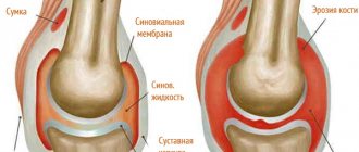
Injury to the medial meniscus of the knee, the treatment of which will depend on the severity, is a common injury. The cartilage layer that is located inside the knee is called the meniscus, there are 2 types - medial (internal) and lateral (external). They perform shock-absorbing and stabilizing functions.
The knee joint is one of the most complex and bears the greatest load. Therefore, meniscus damage is a very common occurrence. According to statistics, more than 70% of damage occurs precisely there. Athletes involved in athletics, skiing and speed skating are at risk. However, a similar injury can be obtained at home by performing simple exercises.
Meniscus damage
In the cavity of the knee joint there are 2 menisci: internal (medial) and external (lateral). The medial cartilage pad is firmly fixed and practically immobile, therefore it is damaged in 85% of cases of knee injury.
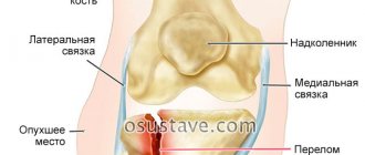
Medial and lateral menisci
The outer lateral cartilage is more mobile and moves when injured. Its rupture is rare; it is more often subjected to compression and compression. Any injury to the knee meniscus is accompanied by a thorough diagnosis and a long rehabilitation period.
Nature of damage:
- traumatic
- degenerative
Symptoms
In the acute period, symptoms are usually pronounced at the beginning and subside after some time (subacute period after 15-20 days).
Three possible phases of a rupture:
- Acute: pain, swelling, problems with bending the limb and stepping on it.
- Subacute: pain is tolerable, swelling subsides. But fluid collects in the joint cavity, and a blockade develops (some actions become impossible or very painful).
- If the rupture is not treated, it will become chronic. This form occurs without pronounced symptoms; sometimes a sign of such a lesion is pain during physical activity after a long rest (usually in the morning), which goes away as soon as the person “disperses” a little.
Traumatic injuries
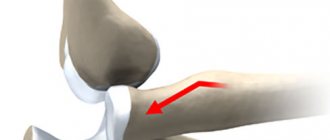
They arise as a result of closed combined knee injuries. When the tibia is rotated outward, burdened by a high load on the joint, damage to the internal meniscus occurs. Internal rotation of the tibia injures the outer cartilage. Rupture of the lateral meniscus at a young age occurs as a result of strong impacts and falls.
In older people with degenerative changes, an unsuccessful movement is enough to injure the cartilage pad. Repeated and untreated injuries to the knee joint lead to chronic damage to the menisci (meniscopathy).
Causes of injuries:
- a fall
- hit
- injury
- bad turn of the shin
- heavy load on the joint
- sudden movement of the knee
- degenerative changes in cartilage tissue
Types of damage
Meniscus cartilage is a shock-absorbing cushion, so there is no fracture-type injury to the meniscus.
It is not elastic enough and is not subject to stretching. When injured, tears and cracks form, parts of the cartilage are completely torn off, and the meniscus may be pinched or compressed.
Some of the most common types of ruptures
The most common meniscus injuries are:
- tear - violation of the integrity of the cartilage;
- avulsion - complete separation of part of it from the cartilage;
- pinching - the cartilage remains intact, but is displaced in the articulation cavity.
The rupture of the cartilage pad can be complete or partial, transverse or longitudinal in the form of a “watering can handle,” flap-like or fragmented, with or without displacement of the torn part. A “watering can handle” rupture of the medial meniscus of the knee joint occurs with extensive disruption of the integrity of the middle part of its body. The anterior and posterior horns remain intact. Such an injury causes severe pain, joint blockade and requires immediate surgical attention.
Severe forms of damage
A tear can form in different parts of the cartilage: central, horns, internal, but mainly the body is damaged. A rupture of the posterior horn of the internal meniscus is diagnosed more often than the anterior one. The most complex injuries are considered to be combined injuries, in which the rupture of the cartilage pad is accompanied by a knee bruise and damage to the elements of the articulation. Displacement of the meniscus or entry of its torn part into the articular cavity causes blockade of the joint, manifested in pain and limitation of the range of passive and active movements.
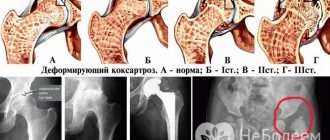
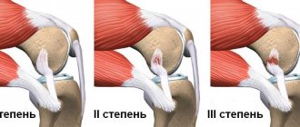
Degree of meniscus damage
There are different lesions, which correspond to three degrees of severity according to stoller:
- First degree. It is diagnosed by internal changes in the cartilage. The mobility of the knee is preserved, there are clicks and discomfort during physical activity. Symptoms go away on their own within 1-2 weeks. May be asymptomatic.
- Second degree. It is characterized by moderate pain in the knee, aggravated by movement. There may be slight swelling. In the second degree, there is no rupture of the cartilage pad, but there is a violation of the structure. After a week, symptoms may disappear, but periodically return with movements resembling the mechanism of injury.
- Third degree. It is diagnosed when the cartilage ruptures. May be accompanied by swelling, hemarthrosis and joint blockade. Symptoms intensify and treatment is required.
3 degrees of meniscus damage according to stoller
Main Causes of Injury
Damage to the internal meniscus of the right knee joint and other similar injuries can be caused by a variety of reasons, which are usually grouped into two large groups:
- injuries;
- degenerative damage.
Young, active people mostly deal with injuries. The main causes of injury are the following:
- sudden extension movements;
- high loads on the legs and knees;
- static position of the joint, maintained for a long time;
- strong unnatural circular movements in the joint, caused, for example, by walking on toes;
- individual ligament weakness;
- direct injury caused by a strong blow, fall, etc.
Older people predominantly face chronic and degenerative problems. Damage to the lateral meniscus of the knee joint can be caused by a blow, a fall from a height, strong vertical loads, lifting heavy things, jumping, diseases of the musculoskeletal system, degenerative processes in the bones, diseases that cause metabolic and blood supply disorders, arthrosis, gout, arthritis, oncology, diabetes mellitus and age-related changes occurring in the human body.
It is necessary to take into account that any injuries require surgical intervention; chronic damage to the medial meniscus of the knee joint can lead to the development of chronic meniscitis, as well as repeated damage of the same type.
Additional factors that increase the chances of injury under any conditions are:
- excess body weight;
- sudden movements;
- high loads on the knees;
- unnatural movements in the knees;
- weak ligamentous apparatus, the cause of which may be congenital or acquired characteristics.
Degenerative damage
This section includes pathologies caused by degenerative changes in cartilage tissue.
Causes of degenerative meniscus tear:
- repeated injuries
- meniscopathy
- age over 40 years
- chronic joint diseases (gout, rheumatism, arthritis)
Repeated injury to the meniscus can cause degenerative changes in cartilage tissue, accompanied by the formation of cysts and decreased elasticity. In addition, frequent microtraumas resulting from heavy loads on the knee joint can cause degeneration.
Degenerative changes are an integral part of aging, which involves age-related breakdown of collagen fibers and loss of structural support. These processes start after the age of 30 and progress with age. The internal structure of the cartilage pad becomes weak and less elastic, so the rupture of the internal meniscus of the knee joint occurs with minimal physical activity.
Degenerative cartilage tears are also the result of chronic joint diseases (gout, rheumatism, arthritis) and are common in people over 40 years of age. For their appearance, the presence of a traumatic factor is not necessary - a rupture of the body or damage to the posterior horn of the medial meniscus can form during habitual actions (squatting, getting out of bed, etc.) or during an unsuccessful movement.
Symptoms of degenerative damage:
- pain is intermittent
- the pain is moderate and increases gradually
- pain worsens with exertion
- swelling and swelling of the knee are mild
- Joint blockade occurs extremely rarely
Surgery
Surgical therapy for a medial meniscus tear is used to eliminate the blockade of the joint, cleanse its cavities from accumulated blood, and restore the integrity of cartilaginous structures if they are damaged.
Before the operation, the doctor conducts a diagnostic examination and analyzes the extent of the damage. Surgical intervention is permissible if the patient is under 45 years of age and does not suffer from degenerative pathologies of cartilage tissue.
Main types of operation:
- Meniscectomy. The essence of the procedure is the removal of part or all of the meniscus. It is painful and can cause arthritis.
- Cartilage restoration. Refers to gentle procedures. This is done to maintain knee mobility. Surgery techniques include the use of clamps to connect destroyed tissues, transplantation of new structures in case of complete destruction of the meniscus, and arthroscopy.
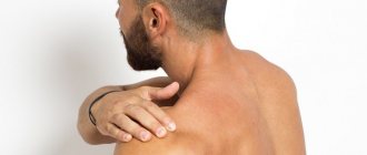
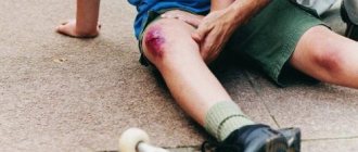
Symptoms of meniscus damage
If the knee is injured, it swells and pain of varying intensity occurs. The pain can be moderate, occurring when repeating certain movements (squats, going up or down stairs) or acute, excluding even minimal load. A small injury to the medial meniscus causes discomfort and painful clicking when moving. Large ruptures are accompanied by severe pain, cause limited movement in the knee and lead to blockage of the joint.
Symptoms of an injured meniscus
A rupture of the posterior horn of the meniscus makes it impossible to bend the knee, while injuries to the anterior horn lead to difficulties in straightening it. Internal rotation accompanied by pain indicates damage to the medial cartilage; pain when rotating the tibia outward indicates injuries to the lateral cartilage. Symptoms of damage to the meniscus of the knee joint gradually gain intensity and most fully manifest themselves 2-4 days after the injury.
Signs of meniscus damage:
- pain
- joint swelling
- clicks when moving
- limited range of motion in the knee, especially extension
- hemarthrosis
- effusion
- joint block
In case of a primary uncomplicated injury, a bruise, a tear of the meniscus, pinching, and compression of the cartilage without its separation from the capsule and tibial plateau usually occur. The fundamental factors for severe rupture are degenerative and inflammatory processes.
Knee meniscus tear
The most common knee injury is a torn internal meniscus. There are traumatic and degenerative meniscus tears. Traumatic ones occur mainly in athletes and young people aged 20-40 years; if left untreated, they transform into degenerative ruptures, which are more pronounced in older people.
Based on the location of the tear, there are several main types of meniscal tears: a tear shaped like a watering can handle, a transverse tear, a longitudinal tear, a flap tear, a horizontal tear, damage to the anterior or posterior horn of the meniscus, paracapsuar injuries. Meniscal tears are also classified by shape. There are longitudinal (horizontal and vertical), oblique, transverse and combined, as well as degenerative. Traumatic ruptures occur mainly at a young age and run vertically in an oblique or longitudinal direction; degenerative and combined - more often occur in older people. Longitudinal vertical tears, or watering can handle tears, can be complete or incomplete and often begin with a tear in the posterior horn of the meniscus.
Consider a tear in the posterior horn of the medial meniscus. This type of tear occurs most often, as most longitudinal, vertical, and watering can handle tears begin with a tear in the posterior horn of the meniscus. With long tears, there is a high probability that part of the torn meniscus will interfere with the movement of the joint and cause pain, even blocking the joint. The combined type of meniscal tears occurs, covering several planes, and is most often localized in the posterior horn of the meniscus of the knee joint and mostly occurs in older people with degenerative changes in the menisci. With injuries to the posterior horn of the medial meniscus that do not lead to longitudinal detachment and displacement of the cartilage, the patient constantly feels the threat of joint blockade, but it never occurs. It is not common for the anterior horn of the medial meniscus to rupture.
A rupture of the posterior horn of the lateral meniscus occurs 6-8 times less frequently than the medial one, but carries no less negative consequences. Adduction and internal rotation of the tibia are the main causes of rupture of the lateral meniscus. The main sensitivity for this type of damage occurs on the outer side of the posterior horn of the meniscus. Displaced rupture of the lateral meniscus in most cases leads to limitation of movements in the final stage of extension, and sometimes causes blockade of the joint. A tear of the lateral meniscus is recognized by a characteristic clicking sound during inward rotation of the joint.
If the meniscus is damaged, you cannot do without a doctor.
Symptoms of a rupture
With injuries such as a torn meniscus of the knee joint, the symptoms can be quite different. There are acute and chronic, long-standing meniscus tears. The main sign of a rupture is joint blockade, in the absence of which it is quite difficult to determine a rupture of the medial or lateral meniscus in the acute period. After some time, in the subacute period, the rupture can be identified by infiltration in the area of the joint space, local pain, as well as using pain tests suitable for any type of damage to the meniscus of the knee joint.
The main symptom of a meniscus tear is pain when palpating the joint space line. Special diagnostic tests have been developed, such as the Epley test and the McMurry test. The McMurry test is produced in two types.
In the first option, the patient is placed on his back, the leg is bent to an angle of about 90° at the knee and hip joints. After that, they clasp the knee with one hand, and with the other hand they perform rotational movements of the lower leg, first outward and then inward. If you hear clicks or crackles, you can talk about pinching of the damaged meniscus between the articular surfaces; such a test is considered positive.
The second version of the McMurry test is called the bending test. It is done like this: clasp the knee with one hand as in the first test, then bend the leg at the knee to the maximum level; after which, the tibia is externally rotated to identify tears of the internal meniscus. If the knee joint is slowly extended to approximately 90° and the shin rotates, if the meniscus is torn, the patient will experience pain on the surface of the joint on the back inner side.
When performing the Epley test, the patient is placed on his stomach and the leg is bent at the knee, forming an angle of 90°. With one hand you need to press on the patient’s heel, and with the other at the same time rotate the foot and lower leg. If pain occurs in the joint space, the test can be considered positive.
Treatment of rupture
A meniscus tear is treated both conservatively and surgically (resection of the meniscus, both complete and partial, and its restoration). With the development of innovative technologies, meniscus transplantation is becoming increasingly popular.
Conservative treatment is mainly used to heal small tears of the posterior horn of the meniscus. Such injuries are often accompanied by pain, but do not result in pinching of the cartilage tissue between the articular surfaces and do not cause clicking or rolling sensations. This type of rupture is characteristic of stable joints. Treatment consists of getting rid of such types of sports activities, where it is impossible to do without quick jerks from the defender and movements that leave one leg in place; such activities worsen the condition. In older people, such treatment leads to a more positive result, since the cause of their symptoms is often degenerative tears and arthritis. A small longitudinal tear of the medial meniscus (less than 10 mm), a tear of the lower or upper surface that does not penetrate the entire thickness of the cartilage, transverse tears of no more than 3 mm often heal on their own or do not manifest themselves at all.
There is also another way to treat a meniscus tear. Stitching from inside to outside. This type of treatment uses long needles that are passed perpendicular to the line of injury from the joint cavity to the outside of the strong capsular area. In this case, the seams are applied one after another quite tightly. This is one of the main advantages of the method, although it increases the risk of damage to blood vessels and nerves when removing the needle from the joint cavity. This method is ideal for treating a tear in the posterior horn of the meniscus and a tear extending from the body of the cartilage to the posterior horn. If the anterior horn is ruptured, it may be difficult to pass the needles.
In cases where damage to the anterior horn of the medial meniscus occurs, it is more correct to use the suturing method from the outside to the inside. This method is safer for nerves and blood vessels; in this case, the needle is passed through the meniscus tear on the outside of the knee joint and further into the joint cavity.
Seamless fastening of the meniscus inside the joint is gaining more and more popularity with the development of technology. The procedure takes little time and takes place without the participation of such complex devices as an arthroscope, but today it does not provide even an 80% chance of meniscus healing.
The first indications for surgery are effusion and pain that cannot be eliminated by conservative treatment. Friction during movements or blockade of a joint are also indicators for surgery. Resection of the meniscus (meniscectomy) was previously considered a safe procedure. Thanks to recent studies, it has become known that in most cases, meniscectomy leads to arthritis. This fact has influenced the main methods of treating injuries such as tears of the posterior horn of the internal meniscus. Nowadays, partial removal of the meniscus and grinding of the deformed parts have become more popular.
Damage diagnostics
Symptoms accompanying a tear of the meniscus of the knee joint are individual and depend on the severity and nature of the damage, and the age of the patient. Due to the lack of specific symptoms, self-diagnosis is impossible.
The diagnosis is established after interviewing the patient and conducting an examination. To clarify the diagnosis, diagnostic studies and special pain tests are used (Roche, Chaklin, Baikov, McMurray, Landa, Shteiman - Bragarda, etc.).
What do meniscus tears look like on MRI images?
Conventional radiography of the knee joint is not informative, since the cartilage is not visible on the X-ray. Contrast X-rays are more visual, but with the advent of modern diagnostic methods (ultrasound, CT, MRI), they have lost their relevance.
Treatment without surgery
Treatment of a meniscus tear with conservative methods includes the following procedures:
- Maintain bed rest, minimize the load on the sore leg;
- Using cold compresses to prevent swelling;
- The use of ointments, tablets and injections with an analgesic effect (Movalis, Nimesulide, Ibuprofen are suitable);
- Puncture to remove accumulated fluid from joint structures;
- Using a bandage to fix the injured limb;
- The use of a plaster splint, which is applied for 3 weeks;
- The use of rehabilitation measures in order to completely restore the motor function of the joint.
Recovery after primary therapy or surgery is also carried out conservatively. The doctor prescribes therapeutic exercises, physiotherapeutic procedures and special massage.
To restore the cartilaginous structures of the meniscus, chondroprotective agents and hyaluronic acid are used. Therapy is carried out annually and lasts from 3 to 6 months.
Diagnostic methods
- Ultrasound (ultrasound examination)
- CT (computed tomography)
- MRI (magnetic resonance imaging)
- specific pain tests
Ultrasound examination makes it possible to diagnose cartilage tears, for example, a horizontal tear of the posterior horn of the medial meniscus, the presence of cysts and degenerative changes in the structure. Computed tomography is widely used to diagnose various pathologies of the knee joint, but shows a low percentage of reliability in the study of cartilage and ligamentous apparatus.
They are better visible during magnetic resonance imaging, therefore, if a tear or pinched meniscus is suspected, it is more advisable to prescribe an MRI of the knee joint, the accuracy of which reaches 95%.
Classification of meniscal injuries visualized on MRI according to Stoller:
0 - healthy meniscus;
1 - there are minor structural changes inside the cartilage tissue, which can only be detected using MRI. There are often no symptoms. The patient is recommended to limit physical activity, take vitamins and chondroprotectors.
2 - a lesion is visualized in the tissues, which does not reach the surface. Sometimes the condition of the tissues is borderline in nature and in the conclusion they indicate grade 1-2 according to Stoller. The second degree is considered a condition preceding the rupture. There are often no significant symptoms, but slight pain and swelling are possible. In addition to the previous recommendations, massage, physiotherapy and exercise therapy are prescribed.
3 - the signal from the source of damage reaches the surface, which corresponds to a cartilage rupture. The rupture can be complete or partial and requires mandatory treatment. Depending on the severity of the condition, conservative or surgical treatment is indicated. Pathologies of the 3rd degree are accompanied by severe pain, swelling, hemarthrosis and joint blockade.
Rehabilitation period
An important stage in the treatment of such injuries is the restoration of normal functioning of the joint. The rehabilitation process should be supervised by an orthopedist or rehabilitation specialist. During the recovery process, the victim is shown a set of the following procedures:
- physiotherapy;
- physiotherapeutic procedures;
- massage;
- hardware methods for joint development.
Rehabilitation activities can be carried out both at home and in a hospital. However, being in a hospital would be preferable. The duration of the rehabilitation course is determined by the degree of damage and the type of treatment performed. Typically complete recovery occurs after 3 months.
During the rehabilitation process, it is important to relieve the swelling that forms inside the joint as a result of surgery. Swelling can persist for a long time and interfere with the complete recovery of the joint. To eliminate it, the use of lymphatic drainage massage will be effective.
A tear of the posterior horn of the medial meniscus, despite its severity, has a favorable prognosis if the main condition is met - timely treatment.
The prognosis becomes less favorable if a horizontal tear of the medial meniscus is accompanied by concomitant severe injuries.
First aid for meniscus injuries
In case of knee injuries, the first step is immobilization (immobilization). If a joint blockade occurs, immobilization is carried out in the existing position.
It is strictly forbidden to independently reduce the pinched meniscus and try to remove the blockage of the joint.
Unprofessional actions can cause broken bones and sprained ligaments. This procedure should only be performed by a doctor. Next, you need to apply a cold compress for 15-25 minutes. It will help reduce pain, narrow blood vessels, reduce swelling and accumulation of exudate in the joint cavity.
Treatment of a torn meniscus
The symptoms of a meniscus tear and the degree of injury determine the method and methods of treatment. Therapy can be conservative or surgical. Surgical intervention is resorted to if there is no other way to restore mobility to the joint or the pathology becomes chronic.
The duration of therapy and rehabilitation varies under the influence of many factors: the age of the victim, the degree and cause of the injury, and the development of complications.
Treatment must be correct and timely. It is important to properly help the victim immediately after an injury:
- It is necessary to immobilize the injured knee by fixing the leg;
- To eliminate swelling, you can use an ice-based compress;
- If the spasms of a meniscus tear are severe, the patient needs to take an anesthetic medication (Diclofenac or Indomethacin).
Prevention
The treatment of meniscus injuries must be taken with full responsibility. Chronic injury causes many complications. In some cases, a person is unable to fully restore motor activity. To avoid unpleasant consequences, preventive measures are necessary. They include physical exercises aimed at strengthening and increasing the elasticity of cartilage tissue. It is recommended to visit the gym several times a week, do morning exercises, and go swimming or jogging. During physical activity, you should remember to be careful and not overload your joints.
It is important to prevent the appearance of extra pounds, as they increase the load on the musculoskeletal system. It is necessary to adjust the diet by creating a low-calorie menu and enriching it with foods containing fiber, collagen and chondroitin.
Causes, symptoms and complications of medial meniscus
- Tear of the medial and internal knee meniscus, causes and symptoms
- Treatment of knee meniscus with folk remedies
- Knee meniscus cyst
- Removal of the meniscus of the knee joint
- Other articles
The main function of the medial meniscus of the knee joint is to properly redistribute the load on the bones of the lower extremities and maintain stability in the knee joint.
Causes of damage to the medial meniscus of the knee joint
Despite the fact that the medial meniscus of the knee joint is less mobile than the lateral one, it is injured much more often. A sudden change in body position with a relatively stationary ankle can lead to damage, as can unsuccessful turns, too much load with excess body weight.
Injuries to the medial meniscus often occur in athletes, especially football players, basketball players, and tennis players. Damage to the meniscus can occur completely accidentally in the most ordinary people; according to statistics, men suffer from this disease 2.5 times more often than women.
Symptoms of damage to the medial meniscus of the knee joint
Injury to the medial meniscus is characterized by the immediate appearance of acute pain immediately after the injury; the knee joint noticeably increases in size due to swelling. In severe cases, there may be a blockage of the joint, that is, the joint may simply jam in one position.
The patient can rarely move independently; as a rule, he requires outside help. If the leg is in a bent position, the person feels almost no or mild pain, but when trying to straighten the leg, the pain instantly intensifies. 2-3 weeks after the injury, in the absence of proper treatment, you can independently palpate the joint cavity, enlarged due to effusion and infiltration.
When the limb is extended, a characteristic clicking sound can be heard.
For chronic damage to the medial meniscus, the main symptom is a dull pain in the knee that gets worse as you go down. If the patient complains of repeated constant blockades of the joint, then they speak of old damage to the meniscus, which is usually accompanied by inflammation of the periarticular bursa.
Diagnosis of damage to the medial meniscus of the knee joint
To diagnose damage to the medial meniscus of the knee joint, the X-ray method, as well as ultrasound, are ineffective due to the transparency of the meniscus for X-rays and ultrasound. They are carried out mainly to ensure that there are no fractures.
Damage to the meniscus is well identified by magnetic resonance imaging or endoscopic arthroscopy - examination of the inner surface of the joint using modern endoscopic equipment, combined with a thorough questioning of the patient and physical examination of the site of damage.
Treatment of damage to the medial meniscus of the knee joint
Emergency care consists of pain relief, fixing the damaged joint in a motionless state and immediate hospitalization.
In the absence of a fracture, puncture of the knee joint removes the hematoma (accumulated blood in the cavity) and removes the resulting blockage. It is necessary to apply a tight bandage with an elastic bandage and local application of cold in the first hours after the injury. It is recommended to keep the limb elevated and, in case of severe damage, immobilization is prescribed for 10 days.
Read also: Meniscus what is it
If the damage is severe, then upon examination there is muscle wasting on the injured limb, sharp pain when palpating the joint space, and positive symptoms inherent in this disease (symptom of clicking and rolling).
As a rule, this is a direct indication for surgical intervention; the meniscus is surgically sutured in case of a tear or resection of the damaged part is performed. In the postoperative period, the patient is given a plaster splint or the limb is fixed with a tight bandage.
Physiotherapy and regular exercise therapy bring good results.
Complications of damage to the medial meniscus of the knee joint
If all of the above symptoms are typical for the clinic of acute damage to the medial meniscus, then over time, when the person returns to normal life, including sports, symptoms of a chronic nature appear.
As a consequence of an acute injury, severe pain occurs in the damaged area; there may be sudden blockades without clinical manifestations of hemoarthrosis. During physical activity, these manifestations become pronounced and create certain inconveniences for a person, that is, they negatively affect the quality of life.
A section of the meniscus damaged as a result of injury contributes to rapid wear of the articular cartilage, which often ends in osteoporosis with the inevitable progression of the disease.
Expert editor: Mochalov Pavel Alexandrovich | Doctor of Medical Sciences general practitioner
Education: Moscow Medical Institute named after. I. M. Sechenov, specialty - “General Medicine” in 1991, in 1993 “Occupational diseases”, in 1996 “Therapy”.
Functions of the menisci
The menisci are a buffer between the bony surfaces of the thighs and legs. When moving, they do not come into contact with each other and are not micro-traumatized. The menisci contribute to the optimal displacement of the femur and tibia relative to each other, but at the same time they limit the range of motion. As a result, the resulting loads are correctly distributed, and premature wear of the knee structures does not occur.
| Function of the knee meniscus | Characteristics |
| Stabilization | Menisci stabilize all structures of the knee joint, promote their displacement only within a certain range, and prevent their damage |
| Protection | Menisci reduce the likelihood of premature destruction of cartilage and bone tissue by reducing friction. They are smooth and elastic, so the bone surfaces slide easily and do not cling to each other. |
| Depreciation | The menisci have a fibrous structure, due to which they can compress and unclench, eliminating increased stress on the muscles and ligamentous-tendon apparatus |
Preventive actions
To prevent the occurrence of a pathological process, experts recommend that patients exercise maximum caution when making any movements. The main importance is attached to shoes - poorly made, incorrectly selected or uncomfortable ones can become a provocateur for displacement or damage to the meniscus.
During training, all athletes must wear protective knee pads as a preventive measure against accidents.
Other patients should be careful about their own body weight - every extra kilogram of weight negatively affects the condition of all joints. And with careless movements it can cause accidental injuries.
Damage to the meniscus of the knee joint is a common problem in sports and at the everyday level. Following the rules of prevention will help avoid damage and the need for long-term treatment.
Rehabilitation
The rehabilitation program after a meniscus injury is compiled individually for each patient, since its volume depends on the complexity and type of injury. The start date is also set by the doctor for each patient. To restore lost functions of the knee joint, such a program includes therapeutic exercises, massage and physiotherapy.
Damage to the meniscus of the knee joint is accompanied by a violation of the integrity of these cartilage “shock absorbers”. Such injuries can vary in severity, and the tactics of their treatment depend on the type and complexity of the injury. Both conservative and surgical techniques can be used to treat meniscus injuries.
General signs
Signs of a meniscus tear:
- a specific sound (crackling, clicking in case of injury), repeated during certain actions (descending stairs), passive flexion and extension of the leg with the hands;
- unbearable pain;
- swelling;
- hemarthrosis;
- blockade;
- the appearance of a painful cushion in the area of the joint space;
- decreased tone of the thigh muscles.
Blockades can be quite long, painful, and difficult to treat.
In the future, deforming arthrosis may develop.
Treatment methods
Meniscus injury is completely treatable, especially those under 40 with red zone tears.
Mild tears with minor symptoms are easier to heal than serious injuries. Therefore, methods are chosen based on the severity of the injury:
- For mild disorders, conservative methods are used;
- for moderate and severe cases – surgical (suturing, removal, prosthetics).
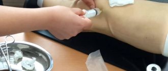
First aid for injury
After a characteristic click and the appearance of acute pain, it is necessary to provide first aid to the patient, providing:
- Complete rest of the limb in the position in which the injury occurred.
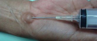
- Apply a cold compress (heating pad or ice pack) to the injured area (for 10–30 minutes).
Cold compress on the affected joint
Under no circumstances should you remove the blockade (if there is one) or try to straighten or straighten the limb, as this can tear ligaments and damage blood vessels and soft tissues.
For unbearable pain, local anesthetic ointments (Diclofenac, Indomethacin) can be used.
Immediately after the grass, any movement causes severe pain, and the victim takes care of his leg. Sometimes home treatment with painkillers is effective and, despite some discomfort, the patient begins to walk.
If the meniscus is injured, this cannot be done; moving before treatment is strictly not recommended. This can be triggered:
- crushing and fiberization of cartilage;
- displacement of the anatomical elements of the joint (torn tissue, muscles, bones).
These injuries can cause lameness and joint deformity in the future.
When can you walk fully after an injury?
- 15-20 days after removing a small part using gentle methods (arthroscopy);
- 4-6 weeks after stitching or removal;
- 6-8 weeks after traditional treatment.
Stepping on the limb and doing therapeutic exercises are allowed a day after the operation and removal of the cast.
Traditional methods
Conservative methods are used for tears, ruptures of the red and transition zones.
First, the doctor removes the blockage by removing fluid from the cavity and administers pain medication. As a result, when the knee straightens without effort, the limb is fixed with a plaster splint (a bandage with a solid base) from the foot to the thigh for a period of 5–6 weeks.
During this period, the patient is prescribed:
- Chondroprotectors (Rumalon, Chondroitin) and the introduction of hyaluronic acid into the joint for rapid tissue regeneration.
- Painkillers to relieve severe pain (Meloxicam, Diclofenac).
To accelerate the restoration of damaged tissues after removing the plaster cast, physiotherapeutic treatment methods are used:
- special therapeutic massage that restores muscle tone;
- UHF therapy, which accelerates tissue regeneration using high-frequency electromagnetic radiation;
- electrophoresis with solutions of Lidocaine and Novocaine (substances penetrate the skin under the influence of a constant electric current);
- a set of exercise therapy exercises to improve mobility.
If there are no complications (repeated blockades do not occur), then treatment lasts 6-8 weeks.
Surgical methods
It is impossible to do without the intervention of a surgeon:
- in the absence of results from traditional treatment;
- for moderate and severe injuries;
- with repeated blockades;
- for chronic degenerative destruction of the menisci (cystic changes in fibers).
| Type of operation | Characteristic |
| Arthroscopy | A gentle restoration method, suturing torn fibers using an arthroscope Not used to treat degenerative changes in old age |
| Bonding | A method of gentle surgical treatment using thin curved clamps (reconstruction) that are inserted through a puncture Bonding is not used to treat degenerative changes in old age |
| Transplantation | Performed after complete removal of serious damage (crushing cartilage) Carried out with implants |
| Removal | In case of serious tissue degeneration or severe damage, the meniscus or part of it is removed A common complication of meniscectomy is arthrosis deformans (in 70%) |
About 3 weeks after the operation, bandaging is carried out; after 5-7 days, physiotherapy is prescribed for recovery. Complete rehabilitation after surgery takes 2 to 3 months, and full restoration of mobility takes up to a year.
Progress of minimally invasive surgery on the knee joint for meniscal damage. Click on photo to enlarge
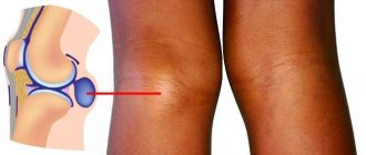
A little anatomy
This is how the knee joint works.
There are two menisci in each knee joint:
- lateral (or external) - its shape resembles the letter C;
- medial (or internal) - has the shape of a regular semicircle.
Each of them is divided into three parts:
- anterior horn;
- body;
- posterior horn.
The menisci are formed from fibrous cartilage tissue and are attached to the tibia (front and back). In addition, the inner meniscus along the outer edge is attached by the coronary ligament to the joint capsule. This triple fastening makes it more stationary (compared to the external one). Because of this, it is the inner meniscus that is more susceptible to injury.
A normal meniscus consists primarily of special collagen fibers. Most of them are located circularly (alongwise), and the smaller part is located radially (from the edge to the center). Such fibers are connected to each other by a small amount of perforating (i.e., random) fibers.
The meniscus consists of:
- collagen – 60-70%;
- extracellular matrix proteins – 8-13%;
- elastin – 0.6%.
In the meniscus there is a red zone - an area with blood vessels.
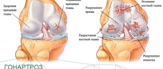
Possible complications
Sometimes, in the absence of unbearable pain, meniscus damage is confused with a regular knee bruise. The victim may not seek help from a specialist for a long time, and the painful sensations may disappear completely over time. Despite this relief, the meniscus remains damaged and ceases to perform its functions.
Subsequently, destruction of the articular surfaces occurs, leading to the development of a severe complication - gonarthrosis (deforming arthrosis). This dangerous disease may in the future become an indication for knee replacement.
If you have a knee injury, the following symptoms are a reason to consult a doctor:
- even mild pain in the knee when walking up stairs;
- the appearance of a crunching or clicking sound when bending the leg;
- episodes of knee locking;
- swelling;
- sensations of interference when moving in the knee joint;
- inability to squat deeply.
If at least one of the above symptoms appears, you should contact an orthopedist or traumatologist.
Causes and types of ruptures
Depending on the causes of meniscus damage, there are:
- traumatic ruptures - appear as a result of traumatic impact (awkward turn or jump, deep squatting, squatting, rotational-flexion or rotational movements during sports, etc.);
- degenerative ruptures - appear as a result of chronic diseases of the joint, which lead to degenerative changes in its structures.
Depending on the location of the damage, meniscal rupture can occur:
- in the anterior horn;
- body;
- posterior horn.
Depending on the shape, a meniscus tear can be:
- horizontal - occurs due to cystic degeneration;
- oblique, radial, longitudinal - occurs at the border of the middle and posterior third of the meniscus;
- combined - occurs in the posterior horn.
After an MRI, specialists can judge the extent of meniscus damage:
- 0 – meniscus without changes;
- I – a focal signal is recorded in the thickness of the meniscus;
- II – a linear signal is recorded in the thickness of the meniscus;
- III – intense signal reaches the surface of the meniscus.
Severity of rupture
When determining therapeutic tactics, the traumatologist must take into account the type of injury and its severity. Minor injury to the meniscus often does not require surgical intervention, but when it is fragmented or displaced fragments, surgery is necessary. There are 3 degrees of severity of rupture of the cartilage lining of the knee:
- 1 degree of severity. Clinically, the injury is manifested by moderate pain, mild swelling in the knee area;
- 2 degrees of severity. The leading symptoms of damage are severe pain, the severity of which gradually decreases, swelling, hematoma;
- 3rd degree of severity. A rupture is characterized by acute pain, extensive swelling, hematoma, and inability to put weight on the foot.
A rupture of any severity requires seeking medical help. Due to improper fusion of cartilage tissue, the knee joint will not be able to fully function.
How to treat?
If your leg is injured in the knee area, you should definitely seek medical help. The doctor, using special diagnostic measures, will establish a diagnosis and prescribe the necessary treatment for the damaged meniscus of the knee joint.
Diagnostic measures
To establish the correct diagnosis, the doctor interviews the victim and then uses diagnostic tests to accurately determine the meniscus injury. The following types of tests are used:
- according to Steinman . The inspection is performed by rotating the shin in different directions (at bends and at right angles). So, if pain bothers you during internal movements of the leg, then this is a medial injury. Pain on the opposite side indicates damage to the outer part of the meniscus;
- according to Baikov . The limb bends at a right angle (with medical assistance), then straightens, and severe pain occurs;
- according to Polyakov . The limb is examined in a supine position. The doctor raises the healthy leg, placing emphasis on the heel of the injured limb;
- according to Chaklin . An injury to the meniscus of the knee joint can be determined by a click when flexing and extending the injured leg.
The listed medical tests are considered basic during the first examination. The next step in diagnostic activities is the use of the following techniques:
- magnetic resonance therapy makes it possible to establish a diagnosis in 95% of limb injuries in the knee area;
- Ultrasound is used less frequently as an additional diagnostic measure;
- X-ray.
After diagnosis, if the meniscus of the knee joint is damaged, treatment is prescribed. Treatment options depend on the type and severity of the injury. The following types of treatment are distinguished:
- conservative;
- surgical intervention;
- restoration activities.
For incomplete meniscus tears, conservative treatment usually helps. In case of joint immobility, surgical intervention is most often necessary. We will tell you in more detail about each treatment method if the meniscus of the knee joint is damaged.
Conservative technique
The main part of conservative treatment is to ensure complete rest of the injured knee. First, the accumulated fluid is pumped out, and at the same time an anesthetic drug is injected.
The next step is to reduce the meniscus. With proper reduction, joint immobility is eliminated. A fixing, elastic bandage is applied to the injured knee. The doctor recommends rest and the following procedures (if necessary):
- UHF (thermal effect on the damaged area);
- painkillers are prescribed, as well as medications that restore cartilage tissue;
- special physical education and massage sessions.
What type of procedure to prescribe depends on the specific indicators and type of damage.
Surgical intervention
Surgery for injuries to the knee joint is indicated in the following circumstances:
- the body of the meniscus is damaged (flattened);
- abdominal bleeding began;
- the horn of the meniscus came off during a rupture;
- complete separation;
- tissue deformation with meniscal displacement;
- Conservative treatment had no effect;
- re-injury.
During surgical intervention, the following techniques are used:
- removal of the meniscus (complete or in parts);
- restoration of the meniscus structure;
- arthroscopy is performed through holes made in the tissue. This technique allows you to sew torn parts of the meniscus;
- fastening of torn elements;
- transplantation technique.
Although knee joint injury is considered a common problem, it is a very dangerous injury. When at least one sign of damage appears, there is no need to hope that the pain will disappear. It is necessary to seek medical help as soon as possible. Only a doctor can make a diagnosis and prescribe the correct treatment. If you self-medicate, you will only get worse and the traumatic condition will develop into a chronic disease.
Diagnostics
The diagnosis is made on the basis of anamnesis (fact of injury), examination data (including surgical tests), patient complaints and the results of instrumental research methods.
You can confirm the diagnosis using:
- X-ray to identify damage (the study can be carried out with contrast); the value of the study in excluding possible fractures of bone structures;
- MRI, characterized by significantly higher accuracy compared to radiography;
- CT, which is less informative than MRI, is used when the latter is not possible;
- Ultrasound, which provides the opportunity to identify and assess the degree of damage to connective tissue structures;
- arthroscopy, which provides the opportunity to: visualize the injury;
- remove damaged cartilage fragments;
- administer medications.
Treatment, methods, what and how
In some cases, it is necessary to provide assistance immediately after injury before preliminary diagnosis. The set of measures includes:
- Providing rest and a comfortable position to the victim.
- Applying local cold pressure to the painful point (the same compress).
- If possible, administer an anesthetic mixture.
- Applying the final hardening dressing.
If the injury does not require surgery, then medical specialists may recommend conservative treatment , which includes the combined use of painkillers and anti-inflammatory medications. Subsequently, you will have to turn to physiotherapy procedures to return the limbs to their previous motor functions.
If the resulting injury is severe, the defective medial meniscus is treated through direct surgery . Depending on the situation, damaged cartilage can be “restored” or completely removed based on the following operations:
- Removal of damaged cartilage formation in case of extensive rupture.
- Removal of the damaged part of the gasket with restoration of the healthy area.
- Replacement of a damaged meniscus with a donor implant.
In some cases, the operations performed may be complex.
Etiology
The most common cause of a meniscus tear is an injury in which the tibia is sharply rotated inward, in which case the lateral meniscus is damaged, or outwardly, the medial meniscus is torn. Other predisposing factors are:
- excessive bending of the knee under the influence of gravity;
- sharp abduction of the lower leg;
- rheumatism and gout are the main causes of degenerative rupture, in which the formation of cysts is observed;
- secondary injuries, bruises or sprains;
- strong physical activity with high body weight;
- prolonged twisting on one leg;
- running on uneven surfaces;
- congenital abnormality of functioning in the form of weakness of joints and ligaments;
- inflammation in the knee of a chronic nature.