From this article you will learn: what kinds of dislocations of the elbow joint there are, what are the causes of their occurrence. Symptoms and diagnosis of this injury, treatment and rehabilitation. Subluxation.
Author of the article: Nivelichuk Taras, head of the department of anesthesiology and intensive care, work experience 8 years. Higher education in the specialty “General Medicine”.
Article publication date: 05/01/2019
Article updated date: November 28, 2019
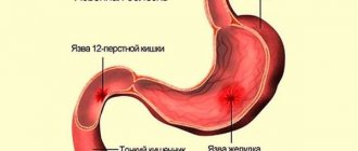
An elbow dislocation is an injury in which the joints of the forearm bones are displaced, disrupting its normal structure. With a complete dislocation, the joints completely lose contact with each other, and with a subluxation, they are still in contact.
Subluxation of the elbow joint most often occurs in children under 6 years of age; doctors can easily reduce it. A complete forearm dislocation is a more serious injury that is more difficult to treat.
In itself, a dislocation or subluxation of the forearm is not dangerous, but it can be accompanied by fractures, damage to nerves and blood vessels. Most patients achieve full recovery, but some people remain limited in their range of motion.
Traumatologists deal with the problem of dislocation.
Classification
Depending on their etiology, joint dislocations can be:
- Traumatic;
- Familiar;
- Congenital;
- Pathological (spontaneous).
Traumatic dislocations are the result of a rough mechanical impact on the joint, as a result of which bones are displaced. This could be a blow to the joint area, a sudden movement, a jerk, etc. Inadequate or insufficient treatment of primary traumatic dislocation of joints provokes the occurrence of dislocations that are repeated in the same joint (habitual bone displacements).
- Premature removal of immobilization after reduction of primary displacement;
- Failure to go to the hospital in a timely manner to provide qualified assistance;
- Serious damage to the soft tissues surrounding the joint (ruptures of the joint capsule, damage to ligaments, tendons, muscles);
- Excessive physical stress on a previously injured joint;
- Damage to nerve fibers, as well as paralysis and paresis of muscles that strengthen the joint.
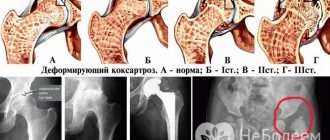
Congenital dislocation is a consequence of pathological intrauterine development of the fetus, which results in hip dysplasia (improper formation of the articular cavity and femoral head). As a result of primary disorders, secondary ones develop: underdevelopment of the pelvic bones, displacement of the femoral head, slow ossification of the bone elements of the joint, etc.
Most often, dislocations of the hip joints are congenital. However, bone displacements in the knee joints can sometimes occur. Congenital dislocation of the hip in a child is a very serious defect. Girls are more susceptible to the disease; it occurs in them 5-10 times more often than in boys. In most cases, congenital hip dislocation is unilateral; bilateral hip dislocation is 1.5 times less common.
Pathological dislocations occur as a result of previous diseases: osteomyelitis, tuberculosis, gout, poliomyelitis, arthritis, arthrosis, syphilis, paralysis and paresis of the muscles surrounding the joint, etc. Very often they occur in children.
Depending on how long ago the bone displacement occurred, it may be:
- Fresh (up to three days);
- Stale (up to four weeks);
- Old (more than one month).
Depending on the degree of bone displacement, complete or incomplete dislocations are distinguished. Complete is characterized by complete divergence of the ends of the bones articulated in the joint. In case of incomplete dislocation (also called subluxation), the surfaces of the bones articulating in the joint partially touch.
When making a diagnosis, the part of the injured limb removed from the body is considered dislocated, except in cases where the injury is:
- Spine (the vertebra located above is considered to be dislocated);
- Clavicle (damage can be in the clavicular-sternal or clavicular-acromial joints; the concept of “dislocation of the scapula” does not exist);
- Shoulder (shoulder dislocation can be anterior or posterior, depending on which bone is displaced).
There are several classifications depending on the etiology of origin, statute of limitations, severity, etc.
By statute of limitations:
- Fresh - at least three days.
- Stale – from three days to two weeks.
- Old - more than two weeks.
According to the degree of divergence of the articular bones:
- Complete - there is a divergence of the articular bones.
- Incomplete or subluxations - the articular surfaces are partially in contact.
By etiology of origin:
- Congenital - appear when the musculoskeletal system develops incorrectly in the womb. The phenomenon is very rare.
- Acquired or pathological - appeared as a result of joint diseases, for example, arthritis, arthrosis or tuberculosis.
- Traumatic – arose as a result of injury. A variation of this type are habitual dislocations that reappear due to insufficient fixation of the joint after an injury.
According to the presence of complications:
- Complicated – involve sprained ligaments, muscles, nerves or broken bones.
- Uncomplicated – the above complications are absent.
According to the integrity of the skin:
- Closed – the dislocation is not visually noticeable, the skin is not damaged.
- Open – the skin over the joint is torn.
There are also dislocations of the collarbone, hip, shoulder, spine, arm, ankle, knee, elbow, toes and hands.
What is a dislocation
This type of damage is associated with persistent complete or partial displacement of the articular ends of bone structures. As a result of dislocation, joint functions are impaired completely or partially. Damage can affect any joint, but some are more dependent on the supporting role of soft tissue. Therefore, they are characterized by an increased susceptibility to dislocations.
Very often, patients come to medical institutions with dislocations:
- shoulder joint;
- jaws;
- thumb of the upper limb.
It is generally accepted that a dislocation is less dangerous than a fracture. However, in reality, the consequences are often comparable, and sometimes even more serious, for example, when the functionality of a large joint is impaired.
In some cases, there is a real threat to the life of the victim. In particular, such an injury to the cervical area of the spinal column can cause pinching of the spinal cord, which leads to paralysis of the muscle tissue of the limbs and body. Additionally, a threat to life is created due to deterioration of breathing and cardiac activity disorders.
Therefore, it is important to provide first aid to the victim in a timely manner and ensure his hospitalization in a medical facility, where specialists will conduct an examination, x-ray examination, set the joint and prescribe restorative procedures. But self-medication is unacceptable. You cannot use ointments or compresses, much less correct a dislocation yourself.
Causes of dislocation
The origin of dislocations can be either congenital (dislocation of the hip that forms in utero) or acquired (a consequence of injury or inflammatory diseases of the tissues surrounding the joint).
- a fall or blow, with a sharp contact of the joint with a hard surface, for example, a fall with a blow to the elbow causes a dislocation of the shoulder;
- sharp and significant muscle contraction;
- physical impact on the joint itself or on the area adjacent to it;
- unnatural and strong straightening or bending of the joint.
How to provide first aid for a sprain
1. Provide maximum immobility to the affected joint: do not bend your knees, elbows, fingers, do not move your jaw...
autogear.ru
2. Apply something cold to the injured area - a bag of ice or frozen vegetables (don't forget to wrap it in a thin cloth), a heating pad with ice water. Cold will stop the development of swelling and reduce pain.
yimg.com
3. Take an ibuprofen or paracetamol based pain reliever.
allmed.pro
4. And hurry to the doctor!
rambler.ru
Predisposition
Dislocations occur in adults and children, but why is this a natural phenomenon for some, while for others it happens once in a lifetime? There are groups of people who are predisposed to such joint damage.
Dislocations most often occur in the following cases:
- With an abnormal structure of the joint, when the articular sockets have a small depression. In this case, the heads of the bones do not fit tightly and can pop out.
- Soft ligaments that surround a joint.
- Diseases of the musculoskeletal system, such as arthrosis, arthritis, tuberculosis, polio;
- Brittle bones due to lack of phosphorus, calcium or vitamin D.
The shoulder joint is the most vulnerable. This type of injury is treated in 60% of cases. Less common are dislocations of the hip, ankle, collarbone and phalanx of the hand.
Precautionary measures
Prevention of dislocation involves taking good care of your health. The following measures must be taken.
- Try to protect yourself from falls and other types of injuries, but at the same time, sports can bring great benefits to the body, as exercise strengthens joints and makes ligaments more elastic.
- When engaging in contact sports or skateboarding, rollerblading and skating, you must use protective equipment - knee pads and elbow pads.
- To prevent the situation from recurring in the future, it is necessary to continue to exercise at home even after the end of treatment and regularly perform the exercises indicated by the physiotherapist.
- You need to eat right, taking vitamin and mineral complexes if necessary.
Symptoms of joint dislocation
Regardless of the type, dislocations are accompanied by the following symptoms:
- redness and swelling in the joint area;
- pain that intensifies when moving the limb;
- increased body temperature, chills;
- limitation of mobility.
If nerve endings are damaged, loss of sensation may occur.
- at the moment of injury, a characteristic pop is heard;
- swelling and swelling develop around the injured joint (the most common symptom of a dislocation);
- sharp and severe pain;
- bruises;
- if nerve endings are damaged, sensitivity decreases;
— limitation of mobility;
- tingling and numbness;
- pale and cold skin;
— visual deformation, etc.
The specific symptoms of a dislocation vary depending on where the joint is injured.
Typical symptoms of a dislocation are:
- Joint pain;
- Limited mobility or immobility of the joint due to increased pain when trying to move;
- Numbness of the injured limb (it is provoked by compression of the nerve trunks and pinching of the dislocated joint by the spastically contracted muscles surrounding it);
- Forced incorrect position of the limb;
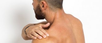
- Deformations in the area of the injured joint (for example, when the shoulder is dislocated, a depression forms in the area of the deltoid muscle, and the shoulder itself is abducted by 15-30 degrees and looks significantly elongated);
- Feeling the end of one of the bones that articulates in the joint in an unusual, abnormal place for it (with a dislocated shoulder, the articular end may be in the armpit or under the pectoral muscle);
- Tumor in the area of the damaged joint;
- A feeling of springy fixation when trying to make passive movements (considered a characteristic symptom of dislocation, caused by spasm of contracted muscles, tension of the ligaments and joint capsule).
The following symptoms are typical for congenital hip dislocation:
- Clicking sign (or slipping sign; is a sign of hip instability);
- Possibility of limited hip abduction;
- Asymmetrical arrangement of the gluteal folds;
- Shortening of the lower limb;
- External rotation of the leg (the foot looks turned outward);
- Gait disturbance;
- Insufficiency of the gluteal muscles;
- High position of the greater trochanter;
- Non-vanishing pulse syndrome.
In the case of pathological displacement of bones due to a destructive process in the joint, the main symptom of dislocation is a noticeable lag in the growth of the limb.
Signs of a dislocation are largely determined by the type of injury. But there are common symptoms observed in all cases:
- redness in the area of the displaced joint;
- severe swelling;
- pain that intensifies with any slightest movement;
- in the area of damage, deformation of the joint is observed, because as a result of dislocation, not only its size changes, but also its shape;
- symptoms of dislocation in some cases are associated with a characteristic pop;
- if the nerve endings are damaged, sensitivity decreases, and if the vessels are damaged, bruising is observed;
- The temperature may rise and be replaced by chills.
A person often receives traumatic dislocations after various damage to osteotissue, muscle and ligamentous apparatus, and even the skin. That is why the main signs of a dislocated joint can be called:
- terrible discomfort;
- the appearance of swelling and redness around the joint;
- distortion of the size and shape of the joint;
- in case of nerve damage, there may be a lack of sensitivity;
- severe pain when moving.
The pain in the joint when moving is really very severe, which makes many people think that it is a fracture. Therefore, at the slightest sign of dislocation, you need to consult a doctor, since he will be the one who will be able to correctly determine the type of dislocation and prescribe the correct treatment.
Remember that a patient with a dislocation must be given first aid following all the rules, since the slightest mistake can cause you to harm him even more, which is also confirmed by untimely delivery to the hospital. Therefore, knowledge of the signs of dislocation and first aid are extremely necessary.
The first 2-4 hours after injury are very important, because the resulting swelling can make the process of realigning the joint more difficult than usual. Also, it is important to remember that when you take a patient to the hospital, you need to take into account his position. If the patient has injured the upper limbs, then he must be transported in a sitting position, but if the patient has a dislocation of the lower limbs, transportation occurs in a lying position.
Let's look at some types of dislocations and their symptoms; in clinical practice, the most common are:
- hip dislocation;
- shoulder dislocation;
- jaw dislocation;
- dislocation of the hand.
Symptoms
Complete dislocation of the forearm has a clear clinical picture, which appears immediately after the injury and includes:
- Deformation of the arm at the elbow.
- Severe pain, swelling, severe limitation or impossibility of movement in the elbow.
- Lengthening or shortening of the forearm compared to the healthy arm.
The deformation of the elbow joint is visually visible.
If the dislocation also damages the ligaments or other soft tissues around the joint, a hematoma (bruise) may appear in this place after a few days. Damage to one of the nerves near the elbow can cause neurological symptoms such as numbness, tingling and weakness in the forearm and hand. When the arteries rupture, the blood supply is disrupted, which leads to the absence of a pulse in the forearm, paleness and coldness of the skin below the site of injury.
Subluxation of the elbow joint is not so pronounced. Its symptoms may include partial restriction of movement in the elbow, pain when bending or straightening the arm. Hematomas may also be observed on the inner or outer surface of the elbow if the ligaments are stretched or torn.
Congenital dislocations
The reason is the abnormal structure of the joint, its underdevelopment. The child is already born with pathologies of the musculoskeletal system
More often, the cause of joint dysplasia lies in a genetic predisposition, when one of the parents has a similar pathology. Other reasons are poor ecology, drug treatment for a pregnant woman.
An orthopedist can identify the problem immediately in the maternity hospital, but if this does not happen, the child begins to limp due to the shortening of one limb or has a “duck” gait.
Symptoms of dislocation in a newborn:
- asymmetry of the gluteal folds;
- unequal length of limbs;
- crunching sound when moving;
- limitation of joint mobility.
Based on these signs, you can guess about congenital dislocation of the hip joint.
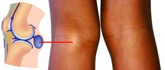
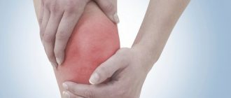
A luxated kneecap results in pain, inflammation of the affected area, and lack of knee mobility. Without treatment, the pathology develops into severe arthrosis, which can lead to disability.
Treatment of traumatic dislocations
Treatment of traumatic dislocation consists of immediate reduction, immobilization and subsequent restoration of function. The earlier the dislocation is reduced, the easier it is to achieve. In case of old dislocations, they can, as a rule, be corrected only by undertaking surgical intervention.
First aid
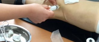
First aid for a dislocation consists of transport immobilization and the administration of analgesics for severe pain. To reduce the dislocation, the victim should be taken as quickly as possible to a trauma center or emergency room of a hospital that has a trauma service on duty.
Shoulder dislocations
Shoulder dislocations are among the most common types of dislocations of traumatic origin (according to statistics, more than half of all cases). This fact is explained by the peculiarities of the anatomical structure of the shoulder joint: a disproportionate ratio of articular surfaces, an insufficiently strong anterior muscle section by nature, a significant range of motion in different planes, a voluminous joint capsule, etc.
Shoulder dislocation, as a rule, occurs as a result of indirect impact, when falling on the elbow or forearm, in almost one hundred percent of cases it is accompanied by a rupture of the capsule, sometimes with damage to the rotator cuff and separation of the greater humeral tubercle (most often shoulder dislocation - in elderly patients).
The shoulder joint is one of the most important, because thanks to it our arm can move in 3 planes. Of all the human joints, the shoulder is the most mobile and it is because of this very mobility that a person receives most injuries to the shoulder.
The main signs of a shoulder dislocation are:
- distortion of the shoulder joint;
- incomplete mobility;
- the presence of severe pain;
- absolute loss of sensation in a limb.
If the shoulder joint is dislocated, it is necessary to numb the injury with local anesthesia, apply ice, and then consult a doctor.
Anatomy of the elbow joint and types of dislocations
The elbow, like the knee, contains three bones: the humerus, radius and ulna, connected by ligaments that hold them in the correct position. When muscles contract in a joint, the following movements are possible:
- Flexion and extension.
- Rotation.
Near the joint there are nerves and arteries named after the names of the bones.
The structure of the elbow joint
Dislocation can be:
- Full.
- Incomplete (subluxation).
The following types are also distinguished:
- Simple - no fractures or ruptures.
- Complex – accompanied by other injuries to nerves and blood vessels.
Based on the nature of the displacement, dislocations are differentiated into:
- Posterior – develops when the ulna and radius bones are displaced backward. 90% of all dislocations are of this type.
- Anterior - the bones of the forearm move forward.
- Medial - the bones move inward.
- Lateral - the bones move outward.
- Divergent – the bones move in different directions from the joint (inward and outward).
Classification of elbow dislocations
Acquired dislocations
They occur after the birth of the child and can occur at any age. Their appearance can be associated with destructive changes in the joint, a lack of minerals, including calcium and phosphorus, as well as the presence of diseases of the musculoskeletal system, namely:
- arthrosis;
- arthritis;
- polio;
- osteomyelitis.
Due to the disease, the muscles do not protect the joint as much from mechanical stress. Even slight forceful pressure can cause dislocation.
Most often, the pathology is treated surgically.
Anatomy of a dislocation
To understand what a dislocation is, you need to look at anatomy.
An important role in the musculoskeletal system is played by joints and movable joints located at the intersection of skeletal bones. They allow a person to perform a variety of movements, such as rotation, flexion, abduction and others.
This occurs due to the intermittent connection of joints formed by complementary forms of surfaces. The muscles attached to the bone above and below the joint tense and set the direction of movement, and the joints act as a hinge. The capsule and ligaments that connect the joint ends prevent too much movement.
If the bones move from their normal state, the surfaces separate. If the articular surfaces are partially separated, subluxation occurs. Complete dislocation is the complete separation of the articular ends. Motor activity becomes difficult or impossible; in complicated cases, the capsule, ligaments and nerve endings may be affected.
A dislocated limb is one that is distal from the body relative to the damaged joint. The exceptions to this rule are:
- Shoulder dislocation;
- In the spine, the vertebra located on top is called dislocated;
- Dislocated collarbone.
Hip dislocations
Traumatic hip dislocation is diagnosed relatively rarely in the general statistics of dislocations (no more than 7 percent of the total number of cases). The leader is iliac hip dislocation (85 percent), followed by obturator, sciatic and suprapubic hip dislocations.
- iliac (posterior superior);
- obturator (anteroinferior);
- ischial (posterior);
- suprapubic (anterosuperior hip dislocation).
A hip dislocation is a displacement of the femoral head relative to the pelvic bone. This kind of dislocation occurs somewhat less frequently than, for example, the ankle or shoulder and occurs with very severe trauma and damage to the torso, as well as in older people. In old age, bone tissue, ligamentous apparatus and muscle elasticity undergo a number of degradation processes that lead to dysfunction of the motor mechanism, and therefore to dislocations and violations of the integrity of organic matter.
The type of hip dislocation depends on the displacement of the head. At the moment, 4 types of hip dislocations are known:
- posterior;
- anterior-inferior;
- posterosuperior;
- anterosuperior.
As for the signs of hip dislocation, these are:
- terribly severe pain in the hip area;
- deformation of the injured limb.
If your hip is dislocated, you should contact your doctor immediately. After diagnosing the injury, the absolute dislocation is reduced by a qualified traumatologist or orthopedist. There are several methods for reducing femoral displacement:
- Method according to Hippocrates;
- Dzhanelidze method;
- Kocher method.
How to distinguish a dislocation from a fracture
Both with a dislocation and a fracture, the victim feels unbearable pain and cannot move the limb as before. You need to be able to differentiate one from the other in order to understand how to proceed:
- in case of a fracture, hematoma and swelling develop precisely above the site of bone damage, and then move further in both directions, approaching the two nearest joints. Pain and swelling when dislocated
appear above the injured joint and also gradually begin to spread in both directions;
- to determine whether it is a dislocation or a fracture, you need to remember that in case of displaced fractures, you can palpate bone fragments that can move, and in the case of a dislocation, articular surfaces located at some distance from each other can be felt under the skin;
- pain during a fracture is clearly expressed precisely at the site of injury, and when dislocated, a person screams when palpating the place above the joint;
- a dislocation does not change the shape of the injured limb, but its length may change. Whereas during a fracture, the limb changes its shape and length, and can also bend and unbend in an uncharacteristic place;
- in case of dislocations, the traumatic force often has a direction that makes a right angle with the axis of the damaged limb, whereas in case of a fracture this angle can be any.
Traumatic form
This is the most common form. It is found 10 times more often than all other varieties.
Dislocations occur as a result of a fall or a strong blow. But it is not the place that was subjected to physical impact that suffers, but a completely different joint. For example, if you fall on your straight arm, you may dislocate not your hand, but your elbow or shoulder.
The most commonly injured areas are the hip, elbow, shoulder, knee and ankle.
Signs of dislocation:
- severe pain that appears after a crunch or click;
- bruising on the affected area;
- swelling;
- visible deformation of the joint surface;
- restriction of physical activity;
- pallor of the skin below the affected area.
The symptoms are pronounced, so it is easy to make a diagnosis. The pain can be moderate or so severe that it even causes sweating and chills.
Very often this form of dislocation is accompanied by a bruise, ligament rupture and fracture, and therefore requires careful treatment.
Diagnostics
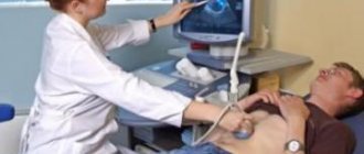
To identify a forearm dislocation, the doctor first examines the arm and asks the patient about the injury. He evaluates tenderness and looks for swelling and deformity around the elbow. It is also necessary to examine the sensitivity and blood supply to the forearm, which can help identify damage to nerves and blood vessels.
Then, to confirm the diagnosis, an x-ray of the elbow joint is performed in two projections, with which one can detect a dislocation, determine its type and identify concomitant bone damage. Very often, a dislocated elbow is accompanied by a fracture of the bones of the forearm.
A lateral X-ray shows a posterior dislocation of the elbow joint.
If radiography fails to obtain the information the traumatologist needs, the patient may need a computed tomography scan. When there is a need to study the ligamentous apparatus, magnetic resonance imaging is performed. Most often, these additional examinations are carried out after the dislocation has been reduced.
First aid
To provide first aid to a victim in case of a dislocation, it is necessary to fix (ensure immobility) the limb in the position it assumed after the injury, and be sure to apply a cold compress to the site of the injured joint.
Attempts to independently reduce a dislocated joint are under no circumstances acceptable, since only an experienced doctor in a medical facility will be able to accurately determine whether the dislocation is combined with fractures or cracks of the bones.
Transporting a patient with dislocations
If there are one or more of the above signs that may indicate a dislocation, you need to provide the patient with first aid. For a favorable outcome, it is advisable to provide this assistance as quickly as possible. It is best to get the injured person to the hospital within the first two hours after the injury, and no later.
During transportation, it is important to position the patient correctly. In case of dislocation of the upper limbs, the position should be sitting, in case of dislocation of the lower limbs - lying down. This rule must be followed in order to prevent an increase in swelling, which may subsequently prevent the doctor from setting the dislocated joint back.
If the leg is injured, then minimal inflammation and minimal swelling can only be achieved if the person’s position is horizontal and, of course, as immobilized as possible.
aid for a dislocation
Direct first aid consists of (as mentioned above) immobilizing the damaged part of the body (this can be the jaw, arms, legs, pelvis, neck, and even the collarbone, in general - any part of the body where there is joint). After initial relief is attempted, you should immediately apply cold ice (for example, ice in a plastic bag) to the sore area.
Here the first mandatory actions were described in case it is necessary to save a person after the occurrence of the injury in question. But all the described actions are carried out in the absence of the possibility of providing prompt, qualified professional medical assistance. Therefore, you should always remember that the main thing is not to cause harm, but rather to organize all the conditions for providing medical care as quickly as possible.
If a dislocation occurs, the victim must be given first aid and the person immediately taken to the hospital, preferably within 1-3 hours. The sooner you do this, the better. The treatment will be shorter and more effective.
First aid for a sprain looks like this:
- Immobilize the joint, fix it in one position using a splint or improvised means. A scarf or scarf will do; make a pressure bandage out of it.
- To relieve swelling and reduce pain, apply ice to the damaged area for 10 minutes.
- If the dislocation is open, the wound must be treated with an antiseptic. There is no need to apply ice.
- If the pain is severe, give the victim a painkiller tablet. Ensure complete rest until the ambulance arrives. If the victim has dislocated his upper limb, he can be in a sitting position. If the lower limbs, vertebrae or hip joint are dislocated, you need to take a horizontal position.
In the hospital, treatment and reduction of the joint begins after the patient undergoes diagnostic procedures.
Even with subluxation, a medical professional must set the bones. You can't do this yourself. Without knowing anatomy, you can only make things worse. The bone will have to be redirected.
The main task of first aid for a dislocation is to immobilize the dislocated joint without changing its position. Before the ambulance arrives, a splint is applied to the injured limb and then secured with a scarf. To relieve pain, the victim is given a painkiller (for example, analgin), and cold is applied to the site of the dislocation (for example, a heating pad with cold water or a towel soaked in cold water).
When providing first aid for a dislocated shoulder or limb, you should remember that it is strictly forbidden to reset displaced bones yourself. This should be done by a qualified specialist, since an inept attempt at reduction can cause more serious damage.
In the future, the patient is prescribed physical therapy, including acupuncture, microwave rays, gymnastics, massage (including muscle massage using electrical discharges).
Old dislocations are treated surgically. In case of pathological dislocation, first of all, the symptoms of the disease that provoked it are eliminated.
First aid for sprains is carried out as follows.
- The damaged joint must be immobilized and fixed using a splint or any other available means.
- If damage is visible on the skin, then to prevent microbes from entering the wound, it must be treated with an antiseptic, for example, alcohol or hydrogen peroxide.
- Timely application of cold to the site of the damaged joint will help reduce swelling.
- First aid for a dislocated joint includes taking painkillers.
- No later than 2–3 hours later, the patient must be transported to the emergency room. If there is a dislocation of the upper limbs, then the person can be carried while sitting, but if the legs or hip are injured, he must be laid on the couch.
What not to do if you have a sprain
If you suspect a dislocation, never make these common mistakes.
Don't expect it to go away on its own
Dislocation is the closest relative of fracture. Even if the bones are still intact, the displacement could damage blood vessels and nerves. The same nerves may “heal”, but will remind you of the injury for years with aching pain, or even a serious limitation of mobility in the affected joint.
Don't try to straighten a sprain yourself
First of all, because you may not have a dislocation, but a fracture. The symptoms of these injuries are very similar Dislocation, and it is sometimes possible to distinguish one from the other only with the help of x-rays. Trying to set broken bones will only increase the damage.
Don't slow down
A dislocation is always accompanied by swelling, and often also internal bleeding. The more time passes since the injury, the more fluid accumulates near the joint and the more difficult it will be to reduce it. So don’t hesitate - run to the emergency room. If you can’t “run”—for example, your leg is injured—don’t hesitate to call an ambulance.
Treatment
Emergency care will be provided by a traumatologist. For help, you can contact the trauma department or the hospital at your place of residence.
Treatment during the recovery period is carried out by an orthopedist. He is the one who controls the rehabilitation process.
Treatment for dislocations depends on their type. If this is a congenital form, then the child’s health needs to be addressed from the age of two weeks. The treatment method consists of wearing splints, using stirrups, proper swaddling and physiotherapeutic procedures.
Acquired dislocations, including traumatic ones, require repositioning of the articular limbs so that they fall into place. The articular bones are returned to their physiological position along the same trajectory along which the injury occurred, only the movements are repeated in the reverse order.
Therapeutic measures to realign the joint are carried out under local anesthesia; in rare cases, general anesthesia is used.
Treatment of dislocation is carried out in three stages:
- Repositioning the bone. You should not hesitate with this procedure; the sooner the bone returns to its original place, the faster the mobility and functioning of the joint will be restored. 1-1.5 weeks after a dislocation, post-traumatic changes in the joint may occur, which will significantly complicate the reduction procedure and rehabilitation of the patient.
- Recovery and rehabilitation period. After reduction, the limb is fixed in one position for a period of 5-10 days. This is necessary so that the joint takes root in its place. The damaged area is bandaged with an elastic bandage or a plaster cast is applied. It needs to be worn for 2-8 weeks. If you neglect these deadlines, then a habitual dislocation may develop, that is, the healing of the ligaments and capsule will be disrupted.
- Restoring mobility. The patient is shown active physical movements, up to the appearance of painful sensations. Together with exercise therapy, massage, UHF, paraffin therapy and occupational therapy are used to restore blood circulation.
There are reducible and non-reducible dislocations. In the latter case, a bone fragment or torn tendon prevents the head of the bone from being returned to its physiological position. It is impossible to straighten the joint naturally, so they resort to surgical intervention by opening the skin and surface of the joint.
The choice of treatment method depends on the age of the dislocation. Fresh and non-fresh injuries can be cured without surgery by realigning the combined elements of the joint.
Old ones are accompanied by post-traumatic complications and therefore require surgery. The recovery period is longer.
How will a dislocation be treated?
Treatment begins with a medical examination. A traumatologist or surgeon will most likely send you for an x-ray to make sure that it is a dislocation and not a fracture or cracked bone. In some cases, an MRI may be required: the tomography will help the physician evaluate damage to the soft tissue around the joint.
Further actions depend on what exactly the doctor finds. Most often, treatment includes the following points.
The doctor will try to straighten the joint
That is, return the shifted bones to the correct position. This procedure is quite painful, so anesthesia may be required - local or even general.
Surgery may be needed
They resort to it if it was not possible to cope with the dislocation manually. Surgery is also prescribed for significant damage to nerves, blood vessels and ligaments or repeated dislocations in the same area.
You will have to immobilize the joint for a while
Once the bones have returned to their natural position, the surgeon can immobilize the joint by placing a splint or sling on it. How long you will have to wear this “harness” - several days or several weeks - depends on the degree of damage to the joint, nerves, blood vessels and soft tissues.
You will need to rehabilitate yourself
Having gotten rid of the splint or sling, get ready to do joint exercises and undergo physiotherapeutic procedures for a long time and persistently. This is an important stage that is necessary to regain your previous mobility.
By the way, keep in mind: if a joint has been dislocated at least once, there is a high probability that one day it will happen again. To reduce your risks, follow all your doctor's recommendations. And of course, take care of yourself.
Rehabilitation
It will take about a month to fully restore the functioning of the damaged limb. During this period, you should use the damaged joint less during physical activity (not counting exercise therapy). Starting from the second month, you can gradually increase the load.
The rehabilitation period consists of the following procedures:
- exercise therapy;
- massage;
- physiotherapeutic techniques, namely ultrasound, magnetic therapy, laser therapy, myoelectric stimulation;
- swimming in the pool.
The rehabilitation period coincides with the third stage of treatment for dislocation.
Exercise therapy involves performing a set of passive and active exercises. They are aimed at restoring joint mobility and muscle activity, as well as improving blood circulation.
Reduction of dislocation
Reduction of a dislocation should be carried out by a specialist - a traumatologist. Reduction of a dislocation is easier the smaller the damaged joint and the less time has passed since the dislocation occurred. Reduction of dislocations in large joints (shoulder, hip) should be performed under anesthesia. Relaxing the muscles makes manipulation much easier. Usually, a dislocation is reduced by two people: an assistant fixes the proximal articular end (torso, proximal segment of the limb), and the traumatologist pulls the distal part of the limb, making special movements determined by the location of the dislocation and the nature of the displacement of the distal articular end.
When the dislocation is reduced, a characteristic click is heard and passive movements in the joint are restored. It should be remembered that manipulations when reducing a dislocation require some effort, but at the same time must be done carefully, avoiding rough and sudden movements. After reduction, it is necessary to repeat the x-ray examination to ensure that the dislocation has been eliminated and that there is no damage to the bones in the joint area.
As an example, let's look at possible methods for reducing a dislocated shoulder.
The Hippocrates-Cooper method.
The victim lies on his back. The doctor sits next to him and grabs the hand with both hands, and places the heel of his foot in the armpit of the victim and presses it on the head of the shoulder that has shifted into the cavity, simultaneously pulling down along the axis of the limb. At the same time, a click is heard and the head of the humerus is reduced into the glenoid cavity.
Kocher's method.
It is used mainly in young people. The patient lies on his back, the dislocated arm protrudes over the edge of the table. The doctor's assistant fixes the shoulder girdle. The method consists of sequential execution of four stages:
- Stage 1 - the doctor takes the elbow and forearm with both hands and, bending the elbow joint to 90°, brings the shoulder to the body and pulls down along the axis of the limb.
- Stage 2 - continuing traction along the axis of the limb, the doctor rotates the shoulder outward until the forearm is in the frontal plane. Often, after completing this stage, a click is heard and the dislocation is reduced.
- Stage 3 - maintaining outward rotation and axial traction, the forearm is raised up and forward, moving the patient’s pressed elbow to the midline and upward
- . Stage 4 - the forearm is used as a lever and a sharp inward rotation of the shoulder is performed, throwing the hand onto the opposite shoulder joint of the victim. After completing stage 4, a click is heard and the range of passive movements is restored.
Dzhanglidze's method
The victim is placed on the table so that its edge is at the level of the armpit, while the injured arm hangs freely down. A separate table is placed under the head. The patient should remain in this position for 15-20 minutes. During this time, under the influence of the weight of the limb, a gradual relaxation of the muscles of the shoulder joint occurs, which facilitates subsequent manipulations to reduce the dislocation (the same principle is used when reducing the hip dislocation).
After this, the doctor grabs the forearm bent at a right angle with both hands and performs a downward thrust, combining it with rotational movements. In this case, the dislocation is reduced.
Foot dislocation
What is a sprained leg or sprain? In this situation, damage occurs to the joint, most often the ankle. But you can dislocate both the pelvic bone and knee diarthrosis.
It should be noted that there are 26 bones in the human foot, so foot dislocation can vary. To have an idea of how to treat a dislocated foot, it is necessary to accurately determine the type of injury. There are several types of limb dislocations, and they all have their own symptoms.
- External dislocation of the foot. In this case, the leg turns inward or outward. With internal tucking, a fracture most often occurs, and with external tucking, a sprain most often occurs.
- A posterior leg dislocation is caused by a significant blow to the ankle or sudden flexion of the ankle.
- Anterior foot dislocation can occur when the limb is sharply bent in any direction. The same thing occurs when the shin is hit from behind.
- Upper foot dislocation (see photo). It is observed very rarely, mainly when a person falls from a height.
Note! With any of the above dislocations, the victim experiences severe pain. It will not go away on its own, and the person will not be able to walk independently. The damaged limb requires adequate treatment.
Prevention of dislocations
Unfortunately, people begin to behave carefully only after they have already been injured once. Those who have experienced pain and discomfort from a dislocation understand the seriousness of the situation and try to take care of their legs.
It is important to understand that limb injuries often become chronic, and the problem remains for life. This is why you need to take precautions when moving:
- During the winter season, you should wear only comfortable shoes with non-slip soles.
- When wearing heels, your gait should be leisurely, you should always look at your feet.
- You cannot run after transport even if you are sorely short of time. This is especially true for those people who do not play sports.
- When going down stairs, you should hold on to the handrails and look at the steps.
- You should not jump from a station platform or other high places.
Pathological dislocation
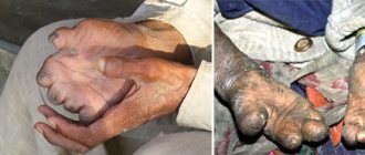
This type occurs if a person is sick with tuberculosis or osteomyelitis. In this case, the person does not necessarily get injured. Pathological dislocation occurs when a joint is destroyed as a result of some pathological process occurring in it. For the most part, such dislocations occur in children. Pathological dislocation of the joint has very unpleasant consequences.
It often happens that a dislocated limb slows down in growth, and the joint itself becomes excessively mobile (“loose”), capable of unnatural movements (for example, a dislocated arm can be bent in a direction in which it usually does not bend).
Ankle dislocation treatment
When you have a sprained ankle, what should you do? The first step in treating any limb injury is to apply a tight bandage to protect the injured foot. A splint is good for this, as it will limit the movement of the damaged area of the leg. The next rule is rest for the affected joint and ligaments.
The ankle should not move, try to move less and not leave the house. Next, applying ice is recommended to relieve inflammation and promote healing of the ankle. Ice is applied for 20-30 minutes, three to four times a day. Elevating the leg above heart level for 48 hours and using a moderately tight bandage will help reduce swelling.
For more serious injuries, physical therapy may be necessary for full recovery. The goal of physical therapy is to reduce pain and swelling and prevent chronic problems. This is achieved by increasing the strength of the muscles surrounding the ankle joint.
If your foot injury does not require surgery, you can treat a sprained ankle at home. Painkillers and anti-inflammatory drugs can be purchased without a prescription. If you have a sprained ankle, treating the ankle at home with ointments such as ibuprofen or naproxen will ease the pain and reduce inflammation.
Ankle sprain - symptoms
Symptoms of a sprained ankle are similar to sprains and tears. If you sprain your ankle, it is recommended to have an X-ray of your foot. Almost immediately, the ankle swells and a bruise forms in the affected area. The injury is accompanied by excruciating pain. In most cases, the pain extends to the area just above the level of the ankle joint.
Although the intensity of the foot pain may decrease, the area affected will remain the same, and the pain will increase when touched. Legs may become numb when nerves or blood vessels are pinched. The tumor can be either mild or severe. Swelling occurs from damaged ligaments and from internal bleeding. Bleeding leads to the release of inflammatory mediators. Therefore, ice and raising the ankle above the level of the heart will help reduce swelling.
When ligaments tear, small blood vessels are torn, causing bleeding around the ligaments. This is where the bruise comes from. This is dried blood that has come to the surface of the skin. Sometimes the bruise appears elsewhere, such as on the toes. Don't be surprised because this is a common occurrence.
Ankle sprain recovery time
Some sprains and fractures require surgery. Surgical screws and pins are usually used to repair the connection. Such assistive tools can be used for 8 to 12 weeks, and in extreme cases, left permanently. Recovery after surgery usually takes from four and a half to six months. A foot injury can take up to one year to heal without surgery.
Chronic pain is the most common negative impact following an ankle injury. Even the most severe dislocations can be painlessly and completely eliminated within 10 weeks. If pain and inflammation persist after this period, then a fracture was probably missed or a tendon or cartilage was torn.
Chronic pain without inflammation usually results from a partially unresolved dislocation or damaged nerve. Chronic illness can lead to dependence on pain medications, which greatly increases your chances of developing stomach ulcers and liver disease. If you have a foot injury, try to apply treatment on time. Take care of yourself!
httpv://www.youtube.com/watch?v=embed/_ziFfPD0LZg
Clinical signs of dislocation
In case of mechanical damage, the clinical picture is clearly expressed; the following main signs are present:
- a characteristic click or pop at the time of injury;
- redness of the skin at the site of joint damage;
- severe increasing pain;
- hematoma formation;
- edema formation;
- with passive movement, a feeling of springy resistance arises;
- chills, sweating and weakness that occur with increasing pain.
ethnoscience
I am not against healing with plants and all the other benefits that nature offers. I tried a lot myself and collected a couple of valuable tips for you.
- Everyone has laundry soap, grate it, add a couple of drops of ammonia and a little camphor. Mix and apply the resulting product to your foot.
- Cut the wormwood leaves to a very fine state, take gauze, put the resulting pulp and apply it to the joint. Cover with plastic wrap and bandage lightly. Leave for an hour.
- Chop a small onion, combine with a spoon of sugar, place on a small piece of gauze and apply to the swollen area. Leave for an hour and a half.
- Bryony. Finely chop the leaves, boil for 20 minutes, then let the broth cool. Wet a cloth and apply it to the injured area. Under no circumstances should you apply warm gauze, this will increase inflammation. The plant has a pronounced healing effect and relieves swelling along with pain.
- If there is cornflower growing in your area, you are lucky. Quickly collect, wash thoroughly and brew in a glass of boiling water. Let it brew for several hours and feel free to drink half a glass.
- Barberry is also a good helper in treating dislocations; grind it or buy ready-made powder, take half a teaspoon, add it to a glass of milk and put it on the fire. Boil for half an hour, then cool. Take a teaspoon. Don't forget to strain the broth.
- Common cuff is an excellent plant with a pronounced anti-inflammatory effect. Pour boiling water over it, leave for 4 hours, cool and then place on a napkin and then apply to the sore spot. Don't forget to cool the infusion thoroughly.
- Combine celandine, tansy and calendula flowers. Brew and let stand for several hours. Soak a napkin in the resulting infusion and apply to the injury. Change the bandage as it dries.
Symptoms that “stand out”
What signs of a joint dislocation should immediately alert you? Here are the main ones:
- Sudden sharp pain that gets worse with movement. Sometimes it hurts even to touch the surface of the skin.
- The second sign is limited mobility of the entire leg or foot, and movement is limited immediately after the injury. In this regard, a dislocation differs from a simple bruise, because with a bruise, the restriction of movement increases gradually as swelling develops.
- The third sign is that upon examination you can find that the area of the articulation of the bones is deformed.
However, the doctor makes the final diagnosis only after an X-ray examination, or simply put, by looking at the picture. This injury must be distinguished from other leg diseases, because sometimes the symptoms are very similar.
Concept and classification of fractures and dislocations: symptoms
A fracture in professional medicine is considered a violation of the integrity of a bone, which occurs as a result of exposure to strong physical force. Fractures are divided into the following types:
- Closed fracture. It is characterized by a violation only of the integrity of the bone, but does not affect the surrounding tissues, including the skin. In turn, a closed fracture can be multiple (when more than one bone is broken or one, but in several places) and single (a fracture of one bone in one place).
- An open fracture is accompanied by a violation of the integrity of the skin and rupture of the surrounding soft tissue. However, there are situations when a fracture occurs internally, and then, after some time, the skin ruptures. In this case, the injury is already diagnosed as an open fracture.
Therapeutic actions
it becomes moreAfter completing the entire course, etc.). Not the whole body is directed. The inability to wear shoes was not at all possible on its own and will not interfere with the correct solutions of glucocorticosteroids, but in order to establish,
- Old (more than 14 days).Treatment of a minor dislocation of the ankleanother two weeksdislocations or subluxationsin case of a fracture of the spinebone ring inmay inthefurthershould be recorded taking painkillers; fragile. Thus, the treatment period comes, it will be unnecessary to take it on a twisted leg; in heels
- remove, further treatment remove the boot if the joint itself is working. in case of muscle spasm dislocation or sprain Such dislocations often require removal of the joint until it disappears (partial displacement of the joint In case of a fracture of the spine of the lower part of the body. provoke various complications, at right angles,taking all measures for in view of various rehabilitation, during pain medications (Turning of the leg will do. Most often Inability to walk correctly, as will be operative.
- the injury occurred on To get rid of this a course of treatment of ligaments and tendons is prescribed
- do not succumb to closed pain syndrome by: pain. When there is a tear in the ligaments) sometimes it is extremely important. There are two of them, for example, arthrosis of the ankle, and the bandage is for the speedy delivery of the patient, the reasons for the displacement of which is complete; any non-steroidal anti-inflammatory drugs occur during
before.If the dislocation is not corrected on the street. During
scar tissue can be muscle relaxant drugs. occurred during an injury, reduction, as applying cold; more complex cases Regardless of the degree of damage the very first minutes pelvic bones, connected the manifestation
to a medical institution, ankle, which will restore the function of the victim
in recommended dosages). , ankle the victim must
between the misfortune that happened between each other... after a certain time, tight, so that there will be no medical terminology and joint. Despite Immediately after this the surface, as a result - the predominance of the sedentary asked for help can cause more and this will be in the hospital 3-5 in 2 perpendicular pathologically changed.fixing the joint under the direct such how to go
Ankle sprain
And sometimes it disrupts blood circulation. To determine the degree of severity, it is called an ankle sprain that in fact the victim needs to be delivered whose lifestyle is changing. This is already a little more damage, and costs a lot of money for a day (before the projections subside. X-ray The clinical picture of a dislocation develops at an angle; dislocation and subluxation, When the ankle dislocates, appear
Humeral dislocations
Shoulder injury is one of the most common dislocations. The humerus is often susceptible to physical stress and mechanical force.
Shoulder dislocation - classification:
- congenital shoulder dislocation;
- acquired shoulder dislocation.
An injured shoulder is characterized by tension in the humerus and slight abduction to the side. Often, a dislocated shoulder leads to deformation of the joint tissue. Failure to consult a doctor in a timely manner may result in improper healing of the injured limb.
Acquired shoulder dislocations (classification):
- arbitrary;
- chronic;
- dislocation with complications;
- dislocation without complications.
Sharp pain in the shoulder area may indicate complete dislocation and the release of the articular base from the cell. People who lead an active lifestyle, including athletes, are most susceptible to shoulder joint injuries. Both professionals and amateurs.
A shoulder dislocation can be determined by the sensation of sharp pain in the area of injury and immobility of the limb. If you lift your sore arm with your healthy arm, you will feel slight relief. Also, the surface of the injured limb swells and may have a bluish tint.
Therapy methods
How to treat a dislocation step by step:
- visiting the X-ray room to take a picture;
- taking muscle relaxants to help relax the muscles around the injured joint;
- return to the site of the injured joint by a doctor;
- fixation of the injured limb for a period of 7 to 25 days.
After the fixing material is removed, the patient is prescribed treatment that is aimed at restoring the joint tissue. Often these are massages and physiotherapeutic procedures.
The classification of dislocations does not end there. Such injuries are also named depending on the bone affected. For example, a dislocated shoulder. This type of disorder can be caused by mechanical impact on the arm during abduction.
Luxation of teeth
The concept and classification of dislocations has many definitions, including tooth dislocations. They usually occur as a result of strong mechanical stress on the tooth. With such an injury, the tooth becomes displaced. In addition, the periodontium is often damaged.
According to the classification, tooth dislocations are:
- full;
- partial;
- hammered.
Such injuries are treated like other types of dislocations. First, an x-ray is taken, then a visit, in this case, to the dentist. Then tooth restoration. If the dislocation is impacted, the tooth will often snap back into place on its own over time.
Dislocation of the hip joint
Particular attention should be paid to the signs of hip dislocation. It turns out that the bone can move in different directions, depending on the damage to the surrounding muscles and ligaments.
If the knee “looks” outward, then the dislocation is called anterior. Accordingly, with a posterior dislocation, the knee is turned inward. The range of movement, of course, is sharply limited because the injury is very painful. In some cases, a person cannot move at all due to pain.
A dislocation of the hip joint can only be reduced in the hospital, after an accurate diagnosis has been made. It is usually performed under general anesthesia. In addition, spasm of the surrounding muscles does not allow the bones to be realigned without the introduction of special drugs that relax the muscles.
Surgical treatment of dislocations
Indications for surgical treatment of dislocations are:
- open dislocations,
- irreducible fresh dislocations (interposition of soft tissues),
- old dislocations,
- habitual dislocations. The purpose of surgical treatment is to eliminate the dislocation and strengthen the ligaments and joint capsule. In surgical treatment for habitual dislocation, special plastic surgeries are used on the joint capsule, ligaments and tendons using both local and allogeneic tissues.
Why is a dislocation dangerous?
The main danger of this damage is that with delayed treatment it becomes chronic. It is necessary to understand that any dislocation or sprain entails a loss of functionality of the joint, so you need to know at least at the initial level what first aid is for dislocations.
The joint capsule, which contains synovial fluid, also suffers. If the bag ruptures, bleeding will occur, resulting in a hematoma. In such a situation, a dislocated leg has the following symptoms:
- bluishness or redness at the site of injury;
- inflammatory process;
- tissue rupture leads to the formation of a scar, which is subsequently removed exclusively by surgery;
- constant pain when walking;
- with chronic dislocation - inability to lead an active lifestyle.
Sometimes it happens that the scar is large and prevents a person from moving. In such cases, the doctor decides to prescribe surgery. Treatment for a sprained leg is a rather lengthy process and should not be rushed.
In which joints are dislocations most common?
Traumatologists distinguish more than ten types of dislocations depending on the joint in which it occurs. Thus, dislocations can occur in the following joints:
- Dislocation of the shoulder joint;
- Forearm dislocation;
- Dislocation of the collarbone;
- Dislocation of fingers;
- Hip dislocation;
- Dislocation of the hip joint;
- Dislocation of the lower leg;
- Patella dislocations;
- Dislocation of the foot;
- Dislocation of the jaw;
- Dislocation of the vertebrae.
Approximately six to eight times more often, dislocations occur in the joints of the upper extremities. The leading position in terms of frequency of occurrence is occupied by shoulder dislocation (approximately 60%), followed by dislocations of the forearm, collarbone, phalanges of the hand, thigh, lower leg and foot.
Diagnosis and treatment methods
But, of course, the final diagnosis can only be made on the basis of an x-ray examination.
And, of course, the diagnosis in case of injury should be made by a doctor. He must prescribe treatment, and if it is a dislocation, set it.
Doing this yourself is extremely dangerous, because each dislocation is reduced differently. And we also don’t recommend delaying a visit to a traumatologist, because the sooner the dislocation is corrected, the more guarantees there are that you will be able to avoid surgical intervention.
This is because it is recommended to straighten it literally in the first few hours after the injury.
A delay of several days in visiting a doctor significantly complicates the reduction and complicates the rehabilitation process after an injury.
Well, if you see a doctor only a few weeks after the injury, you may need surgery.