© Glebstock — stock.adobe.com
Share:
The shoulder joint is the most mobile in the human body. All types of movements are possible in it: flexion-extension, abduction-adduction, supination-pronation, rotation. The price for such freedom of movement is the significant “fragility” of this joint. This article will talk about the most common injury that awaits athletes who systematically overload their shoulder joints. This is a dislocated shoulder. In addition to the injury itself, we will touch upon issues of anatomy, biomechanics, first aid and, most importantly, preventive measures.
Description of the joint and the mechanism of its dislocation
The shoulder joint itself consists of three parts:
- articular head of the humerus;
- articular cavity of the clavicle;
- glenoid cavity of the scapula.
The cavity of the clavicle has no connection with the humerus, but has an impact on its functioning. Between the head of the humerus and the socket of the scapula there is an articular lip, which additionally holds the joint and maintains high mobility. The shoulder joint contains several bundles of articular ligaments, groups of muscles that provide great stability.
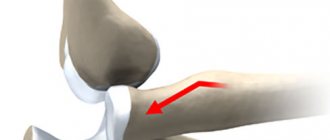
The mechanism of injury is excess of physiological amplitude due to indirect trauma. The joint capsule is destroyed, and the head of the humerus falls out. Sometimes there are fractures, damage to muscles and tendons.
Characteristics of the injury
The shoulder is the most mobile articulation of two bones in the entire musculoskeletal system. The joint is formed by the flat-concave articular surface of the scapula, in which the spherical humeral bone head is located. It is stabilized, preventing displacement, by the rotator cuff, consisting of an articular capsule, muscle fibers, and ligaments. This structure of the shoulder makes it possible for a person to perform a variety of movements with a large amplitude - circular rotations, lifting, abduction and adduction of the upper limb. When a dislocation occurs, the head of the humerus moves relative to the scapula, which leads to complete immobilization of the arm. This type of injury occurs due to the following predisposing factors:
- high mobility of the shoulder joint;
- relatively large joint capsule;
- a small area of contact between the scapula and the bone of the shoulder;
- a person's attempts to soften a fall with his arm extended forward.
Due to the large range of motion in the shoulder joint, traumatologists identify various options for displacement of the articular surfaces of the bones of the shoulder and scapula. The danger of dislocation lies in the high probability of its recurrence. Often this injury becomes habitual, so a person deliberately avoids performing certain movements that provoke displacement of the head of the humerus. The cause of habitual dislocation is primary damage to the ligamentous-tendon apparatus and bursa.
Sometimes a pathological situation is provoked by incorrect treatment or neglect of medical recommendations at the rehabilitation stage. A dislocation, in which large blood vessels rupture, threatens not only the health, but also the life of the victim.
Causes of shoulder dislocation
This injury is the most common of all injuries to the shoulder joint. Causes of dislocation include:
- injury (strong blow to the shoulder, fall on the arm);
- frequent sprains of the muscles and tendons of the shoulders (common in athletes);
- identical hand movements that are often repeated (more often observed in athletes);
- congenital hypermobility - “joint hypermobility” (occurs in approximately 12% of people);
- irregular structure of the scapula (small scapula).
A dislocated shoulder in itself does not pose a serious threat to a person's health. But getting a second injury (habitual shoulder dislocation) within six months after the first injury to the shoulder joint is very high. This does not require strong impact on the site of the previous damage. The reason lies in improper reduction of the shoulder dislocation, treatment, or injury associated with severe rupture of the joint box.
Treatment of dislocation
As for how to treat a dislocated shoulder, we will give only a few general tips, since self-medication in this case can be very dangerous. The treatment process includes several stages:
- reduction of the dislocation by a qualified traumatologist. Better - under local anesthesia. Ideally, under anesthesia. Pain relief provides relaxation to muscles that spasm in response to injury. Thus, the reduction will be quick and painless.
- immobilization and ensuring complete immobility of the shoulder joint. The immobilization period is 1-1.5 months. During this period we try to achieve maximum healing of the shoulder capsule. For this purpose, during this period, a variety of physical therapy is prescribed to help improve blood circulation in the affected joint.
- rehabilitation.
We will talk about the rehabilitation stage for a shoulder dislocation in more detail below.
© belahoche - stock.adobe.com. Reduction of dislocation
Characteristics of types of shoulder dislocations
Depending on various factors, there are several classifications of shoulder dislocations. Based on the presence of traumatic effects, a distinction is made between traumatic (caused by injury) and non-traumatic (habitual) dislocation. Non-traumatic shoulder injury can be chronic (pathological) or voluntary. There is a division of shoulder dislocations into congenital (improper structure of the scapular cavity, hypermobility of the joints) and acquired.
Depending on the type of injury, dislocations can be uncomplicated or complicated (dislocation with bone fracture (fracture dislocation), with damage to the skin and tissue around the joint (open dislocation), with damage to tendons, nerves and blood vessels). Based on the time that has passed since the injury, dislocations are divided into fresh (the first three days), stale (up to five days), old (more than 20 days have passed).
Shoulder subluxation is a common injury that occurs in children and the elderly. It has no complications, but can recur if not properly treated. If the injury occurs for the first time, it is called a primary dislocation. After such damage, the tendon and the joint itself lose their original strength, and the risk of re-injury increases.
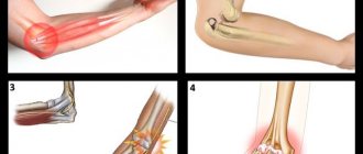
Based on the direction in which the articular head goes and how the articular surfaces diverge, anterior, inferior and posterior dislocation of the shoulder is distinguished.
Anterior dislocation
The most common type of such injury, more than 75% of shoulder dislocations (up to 90%) are anterior dislocations. It has two varieties: subcoracoid and subclavian. In the first case, the head of the bone falls out of the articular capsule and extends beyond the process of the scapula, called the coracoid. In a subclavian dislocation, the articular head moves even further and extends beyond the collarbone. With such an injury, serious complications are possible (rupture of the joint capsule, damage to soft tissues). The shoulder looks to the side.
Lower dislocation
An infrequent type of dislocation (from 8% to 24%). The lower dislocation is called axillary. Here the head of the humerus goes down relative to the glenoid cavity of the scapula. The victim cannot lower his arm; it is moved away from the body.
Posterior dislocation
Posterior shoulder dislocation is very rare (up to 2% of cases). It is observed when a person falls on his outstretched arm. The articular head goes simultaneously to the back and head. Often with a posterior dislocation, ligaments, tendons, and the articular labrum connecting the socket of the scapula and the head of the humerus bone are torn.
Prognosis for recovery
A dislocation of the shoulder joint is not directly life threatening. Their main danger is temporary disability, the development of complications (damage to blood vessels and nerves) and a high risk of relapse. The likelihood of re-dislocation directly depends on the patient’s age:
- Up to 20 years of age, relapse develops in 85% of cases.
- Over 40 years of age, the risk is 10–15%.
Most recurrent dislocations occur within the first 2 years after the first episode.
Symptoms of shoulder dislocation
With the variety of types of dislocations, the symptoms of such an injury are similar:
- sharp and severe pain in the area of injury (shoulder, arm, scapula, collarbone), intensifying when trying to move the arm;
- the appearance of swelling in the shoulder joint;
- restriction of movement (the victim can make a very small number of movements, often springy due to protective muscle contraction and tension of ligaments and tendons; numbness of the hands is possible if the nerve is damaged);
- visible deformation of the shoulder (the shoulders are asymmetrical, the damaged side looks angular).
Signs of a complicated dislocation can be recognized by Bankart damage (increased pain), a characteristic crunch that accompanies a bone fracture, weak palpation of the pulse on the radial artery in case of vascular damage, numbness of the hand in case of nerve damage.
Symptoms
Signs of a dislocation of the shoulder joint, if the injury is primary, are pain, which is mostly caused by rupture of soft tissues. With repeated dislocations, there is much less pain or no pain at all. This is due to the fact that the structures that stabilize the joint have been damaged by a previous injury.
The symptoms of a shoulder dislocation are in many ways similar to those of other joints.
Symptoms of subluxation of the shoulder joint, like dislocation, are characterized by sharp pain in the corresponding place. One of the signs is shoulder deformity and drooping arm. Any movement is impossible due to increased pain and disruption of its functioning. During passive movement, springy resistance is felt.
The asymmetry of the shoulder joints is visually noticeable. The joint itself becomes angular, concave or sunken. Upon palpation, the doctor determines the protruding head of the bone emerging from the bed.
- Anterior dislocation is characterized by downward and forward movement of the head.
- For the anterior inferior - displacement to the anterior part of the axilla or down the coracoid process of the scapula. The victim is forced to keep his arm in a comfortable position: abducted and turned outward or bent.
- In the lower form of the pathology, the head moves into the armpit, the arm goes numb completely or in certain parts.
- With a posterior dislocation, the head moves towards the scapula.
A dislocated shoulder may have other symptoms.
- swelling of the shoulder joint;
- feeling of goosebumps on the arm;
- pain not only in the area of injury, but also along the pinched nerve.
If a shoulder dislocation is severe, complications can develop. The greatest danger is represented by damage to the neurovascular bundle, open damage to the bone structure and soft tissues, and a closed fracture of the humerus.
Habitual shoulder dislocation can be complicated by other pathological situations. Among them is a Bankart injury, when the joint capsule ruptures and a section of the articular labrum is torn off. There are no external manifestations, but there is severe pain and the consequences are unpleasant.
How to identify a shoulder dislocation? Knowing the listed signs of damage, this will not be difficult to do.
Diagnostics
The main symptoms by which a traumatologist determines the type of injury received are described above. A professional examination by a doctor takes place in the form of a thorough and careful palpation to establish the location of parts of the joint, determine its mobility, as well as a conversation with the victim. To clarify the presence/absence of complications, the doctor checks the pulse, palpates the skin, and checks the mobility of the fingers.
To clarify the diagnosis and select the most competent treatment, X-rays and magnetic resonance imaging are used.
Rehabilitation: how to act correctly?
Physiotherapy and massage
Physiotherapeutic treatment of shoulder dislocation and restoration of mobility includes the following procedures:
- magnetic therapy;
- inductothermy;
- ozokerite or paraffin thermal applications;
- electrophoresis;
- exposure to ultrasound;
- laser therapy;
- phonophoresis;
- ultraviolet irradiation.
The massage session should begin with the forearm area.
It is recommended to do a massage, starting the session with the forearm, gradually moving towards the site of the dislocation. Stroking and point pressure are effective. It is useful to develop your arm with hydromassage and swimming, as well as using a spiked rubber ball. The therapeutic effects of procedures on the shoulder joint are as follows:
- help restore mobility and develop the limb;
- promote the production of synovial fluid;
- accelerate tissue regeneration;
- relieve pain and inflammation;
- normalize blood circulation;
- improve lymph outflow;
- saturate tissues with oxygen.
Physiotherapy
To strengthen and develop the arm, the following set of exercises (physical therapy) is used:
- shrugging shoulders - simultaneous and alternate;
- wave your hand;
- raising and lowering limbs;
- putting your hands behind your head;
- circular rotation of the limb;
- bending to the sides while simultaneously stretching the arm;
- push-ups in a standing position with palms resting on the wall;
- exercises with a stick;
- pump up your shoulder muscles using a load weighing no more than 2 kg.
The development of the arm should begin with minimal loads so that the tissues of the joint have time to strengthen and heal.
Treatment with folk remedies
Comfrey roots are used to prepare a medicinal infusion.
Dislocation of the head of the humerus can be cured by preparing a compress of comfrey roots according to the following recipe:
- Take 30 g of the plant and pour 0.5 liters of cool water.
- Place on low heat and simmer for 30 minutes, without bringing to a boil.
- Remove from heat and let sit for 4 hours.
- Filter.
- Soak a cloth in the product and apply it to your shoulder.
If your shoulder is knocked out and hurts, treatment at home includes preparing an ointment. The recipe is as follows:
- Take 5 ml of St. John's wort oil.
- Grind the wheat sprouts into a paste.
- Add 20 g butter.
- Simmer the ingredients in a water bath for 20 minutes.
- Remove from the stove and let cool slightly.
- Remove the top layer - medicine.
- Warm up before applying to the joint and add 2 drops of thuja oil.
- Apply ointment to the sore spot.
Shoulder joint treatment
After receiving an injury, you need to immediately call an ambulance or go to the emergency room yourself. As first aid for someone who has suffered a dislocation, you need to apply cold to the injury site, ensure rest and not move the injured arm. If possible, apply a bandage to your arm to immobilize the injured joint as much as possible.
To reduce pain, you need to give an anesthetic.
You cannot straighten the shoulder on your own until the ambulance arrives.
You can aggravate the situation, damage surrounding tissues, and damage nerves and blood vessels. If there is an open wound, you need to treat it with an antiseptic and apply a bandage.
Next, depending on the situation, the doctor selects a treatment and recovery regimen. All methods are divided into surgical and non-surgical. Only a doctor can determine which one is suitable in a particular case.
Closed reduction of dislocation
You need to move the shoulder joint back into place as quickly as possible. To do this, local anesthesia or general anesthesia must be used: they are used for pain relief and muscle relaxation. There are several methods of reduction:
- according to Dzhanelidze;
- according to Kocher;
- according to Hippocrates;
- according to Mukhin-Mot and others.
After reduction, the pain decreases significantly. Subluxation of the shoulder joint without complications can be reduced without the use of anesthesia. You need to check the success of this manipulation using an x-ray. Then the doctor prescribes painkillers and applies a bandage or special fixation of the shoulder with abduction of the arm.
Even if there is no pain, you need to wear it for at least 3 weeks.
Surgery
This treatment method is often used for repeated habitual dislocations, when surgery is unavoidable. If a second dislocation occurs, it will happen again until the cause of the pathological condition of the shoulder joint is eliminated.
Dislocation of the ACJ (acromio-clavicular joint), often found among athletes, requires only surgical treatment, since such an injury causes ligament rupture.
When eliminating habitual shoulder dislocations, the surgeon pursues such goals as strengthening the ligaments and tendons, and correct alignment of the glenoid cavity and the head of the humerus. There are several types of operations to correct this type of dislocation:
- Turner's operation (removal of an elliptical flap of the articular capsule, suturing of the capsule; advantage - small scar, short recovery period);
- Putti operation (more traumatic, necessary in the presence of complications; the capsule is sutured; does not require a large number of instruments; minus - long recovery period, large T-shaped scar);
- Boychev’s operation (similar to Putti’s operation; a triangular fragment is removed before suturing);
- Bankart operation (not so widespread due to the use of special devices (arthroscope); the goal is to create a new labrum; has a short recovery period; considered the gold standard in the treatment of dislocations).
The doctor’s choice of type of operation depends on the presence/absence of complications, special instruments, and the age of the victim.
The recovery period after such an operation lasts up to six weeks.
After surgery, a brace, a complex device for maximum immobilization and support, is used on the affected shoulder and arm.
Physiotherapy
The use of physiotherapeutic procedures is possible in the presence of a fixing bandage on the shoulder and after its removal. The goal of physical therapy is to reduce tissue swelling, relieve pain in the damaged area, restore good local blood flow and mobility of nearby muscles. They are aimed at restoring the damaged shoulder joint and its functions. Basic physiotherapeutic procedures:
- magnetic therapy (high and low intensity);
- electrophoresis (to accelerate the absorption of drugs);
- diadynamic therapy;
- amplipulse therapy;
- infrared irradiation;
- massotherapy;
- paraffin therapy;
- alcohol compress;
- local cryotherapy (low temperature exposure).
The main contraindications are purulent wounds, kidney and blood diseases, malignant tumors, bleeding, heart disease (heart attack), the presence of pacemakers, infectious diseases, tuberculosis. Some procedures have restrictions such as pregnancy, children under 5 years of age, and a tendency to develop blood clots.
They help shorten the rehabilitation period and reduce the severity of symptoms without medications. But their use must be agreed with the attending physician; you cannot prescribe them to yourself. Physiotherapy procedures do not replace joint realignment or surgical intervention.
Treatment methods
How to treat a patient after an injury? Treatment includes first aid, reduction of the dislocation by a traumatologist with further compliance with the rehabilitation regime and physical therapy exercises. After a full course of therapy, complete restoration of the joint's functionality is possible.
First aid
Immediately after a shoulder injury, when there is a suspicion that a person has dislocated his shoulder, you need to call an ambulance or go to the hospital yourself.
First aid at the scene of the event includes:
- immobilization (immobilization) of the shoulder using a scarf (or any fabric that is at hand);
- applying an ice pack to the damaged area (the ice pack should be wrapped in cloth and applied to the joint for 5 minutes);
- taking painkillers - paracetamol, ibuprofen (dosage is indicated in the instructions for the drug).
Bandage-kerchief for the shoulder joint
The patient should not eat or drink because if anesthesia is needed during the reduction of the dislocation, vomiting may occur.
First aid for subluxation is the same as for complete dislocation of the joint.
Reduction of dislocation
The goal of initial treatment for a shoulder dislocation is to reduce and return the head to the socket.
There are several methods for reducing a dislocated shoulder. The decision about which one to use depends on the type of congenital heart disease and the experience of the doctor.
Typically, reduction of a dislocation is performed on conscious patients. To reduce pain and relax muscles, sedatives and analgesics may be administered during the procedure.
Reduction of dislocation
In complex cases, general anesthesia may be necessary to eliminate congenital heart disease.
In most cases, conservative reduction of the humerus is successful, but sometimes surgery is necessary.
Surgical intervention is performed for:
- If a tendon, ligament or bone fragment gets into the joint cavity, which prevents the head from returning to the glenoid cavity.
- Failure of conservative methods.
- The presence of damage to blood vessels or nerves.
Arthroscopic surgery for shoulder dislocation
Sometimes in young patients (under the age of 25) with a high risk of developing repeated dislocations, traumatologists recommend early arthroscopic surgery.
They are prescribed to restore the labrum and remove soft tissue fragments from the joint cavity.
Reduction is also necessary for subluxation. The technique has no fundamental differences.
Rehabilitation
Immediately after the dislocation is reduced, the traumatologist re-examines the arm to ensure that no arteries or nerves were damaged during the procedure.
To assess the effectiveness of reduction, a control radiography is performed.
Since dislocation causes damage to the tissues that provide stability to the shoulder joint, re-displacement of the bone may develop in the early recovery period.
In order to reduce the load on the injured arm, doctors recommend using a special shoulder orthosis (fixing bandage) or a scarf bandage, the use of which allows the muscles surrounding the joint to relax.
Shoulder brace
In severe cases, traumatologists may apply a plaster cast.
The patient can go home almost immediately after reduction. If the procedure was performed under general anesthesia, the patient must remain in the hospital until clear consciousness is restored.
In this case, it is advisable for the patient to be accompanied home by a relative or loved one.
The duration of immobilization of the injured arm depends on the specific situation. Usually it is 5–7 days, in severe cases it is longer.
Doctors try to strike a balance between providing functional rest for the injured joint and reducing its mobility due to prolonged immobilization.
You should avoid putting stress on the injured arm for 2–6 weeks.
If the patient experiences pain after reduction, doctors may prescribe non-steroidal anti-inflammatory drugs (ibuprofen, naproxen, ketoprofen and others).
An accessible method of pain relief and reducing swelling of damaged tissues is applying ice wrapped in a towel. To do this, wrap the ice in a towel or other cloth and apply it to the joint for 5 minutes.
The principles of rehabilitation for subluxation do not differ from those for complete dislocation of the joint. Typically, with a subluxation, recovery occurs faster: in 3-5 weeks.
Physiotherapy
After the end of the “rest period,” the patient must consult a physical therapist. He will develop an individual physical rehabilitation program aimed at restoring the full range of motion in the damaged joint and strengthening the muscles of the shoulder girdle.
Physical therapy exercises can be started after the pain subsides and immobilization is removed.
First, the patient is recommended to do isometric exercises (they train muscle strength without increasing their mass), also known as static exercises, in which there is no movement in the affected shoulder joint. It is not the strength that matters, but the duration of the load.
Examples of isometric exercises
1. Isometric extension
Stand with your back to the wall, place your arms on the sides of your body. Keeping your arm straight, press firmly against the wall and hold the pressure for 5 seconds. Repeat 5 times.
2. Isometric flexion
Stand facing the wall, place your arms on the sides of your body. Keeping your arm straight, press firmly against the wall and hold the pressure for 5 seconds. Repeat 5 times.
3. Isometric adduction
Place a small pillow or rolled-up newspaper between your arm and body. Press your hand close to your body and hold this position for 5 seconds. Starting with a small object, gradually increase its size. Repeat the exercise 5 times.
4. Isometric abduction
Stand with your side to the wall, place the back of your hand on it. Press down on the wall as if you were trying to lift your arm to the side. Hold the pressure for 5 seconds. Repeat the exercise 5 times.
5. External rotation
Stand facing the door frame. Bend your elbow at a right angle and place the back of your hand on the side of the frame. Press down on it and hold the pressure for 5 seconds. Repeat 5 times.
6. Internal rotation
Stand facing the door frame. Bend your elbow at a right angle and place your palm on the side of the frame. Press down on it and hold the pressure for 5 seconds. Repeat 5 times.
Then they begin to perform dynamic exercises, during which movements are carried out in the affected joint.
Examples of dynamic exercises
1. Internal rotation
The most important exercises for preventing recurrent anterior shoulder dislocation. Attach the expander tape to a stationary object, stand sideways to it. Holding the end of the tape in your hand, bend your elbow at a right angle. Keeping your elbow close to your body, move your hand toward your stomach, then slowly return to the starting position. Repeat 5 times.
2. External rotation
This exercise cannot be performed in the early stages of rehabilitation, as in this case there is a risk of recurrent congenital heart disease. Lie on your side with your knees bent and your affected arm on top. Place a towel under her elbow and take a dumbbell. Bend your elbow at a right angle. Rotate your arm at the shoulder joint, pointing your hand upward from the dumbbells. Slowly return to the starting position. Repeat 5 times.
3. Lead
Standing at one end of the expander band, take the other in your hand. Keeping your arm straight at the elbow, lift it to the side until your hand reaches shoulder level. Slowly return to the starting position and repeat the exercise 5 times.
4. Flexion
Standing at one end of the expander band, take the other in your hand. Keeping your arm straight at the elbow, raise it in front of you, reaching shoulder level with your hand. Slowly return to the starting position and repeat the exercise 5 times.
In addition to exercises aimed at strengthening the muscles of the shoulder girdle, classes are conducted to improve mobility in the affected joint.
Set of classes:
- Stand next to a bench, bend over and lean on it with your healthy arm. One leg should be placed forward, the other should be located slightly behind. Relax your injured arm, allowing it to hang down freely. Shifting your body weight from one leg to the other, make oscillatory movements with your body, during which the injured arm also begins to oscillate from front to back. To make lateral swings of the arm, the feet need to be placed shoulder width apart.
- Take the gymnastics stick by the ends with both hands. Use your unaffected arm to perform flexion, internal and external rotation, adduction, and abduction at the shoulder. The damaged hand will move synchronously with the healthy one.
- Place a rolled towel between the injured arm and body. Bend your elbow at a right angle. With your healthy arm, press the elbow of your injured arm toward your body. This exercise not only improves the mobility of the shoulder joint, but also stretches its capsule.
Exercises after a sprain
Immediately after reduction and application of an immobilizing bandage, as well as approval from the doctor (for uncomplicated dislocations), you can begin a course of exercise therapy. Exercises after a dislocation in the first weeks are passive in nature (performed with the help of a doctor or another healthy hand). Gradually you need to perform the exercises more actively. The first training should begin with flexion/extension and rotation of the hand, clenching the fingers into a fist, and static tension of the shoulder muscles.
A month after the injury and removal of the bandage or fixing bandage, you need to use the joint itself, moving your shoulders forward/backward at a slow pace several times during the day. This exercise helps restore the ligamentous apparatus, the function of the joint itself.
Once the bandage is removed, the importance of exercise increases. You shouldn't start playing sports right away. A properly selected course of exercise therapy helps to quickly strengthen damaged ligaments, strengthen the muscles around the joint, and stabilize the joint itself. The amplitude of movements should be increased gradually, and then include expanders, weights, and rubber bands. In the beginning, you should do the exercises under the guidance of a doctor, and then at home. After exercising, apply a cold compress to the injured area to relieve pain.
By performing simple exercises, you will speed up your recovery after a shoulder injury.
Clinical picture
At the moment of rupture of the joint capsule, acute, piercing pain occurs. Its maximum severity is observed during primary dislocation. If the injury becomes habitual, then the intensity of the pain syndrome gradually decreases, and sometimes it is absent altogether. This is explained by the presence of a large number of nerve endings in the joint capsule. After its rupture, they do not have time to recover, which leads to a significant decrease in tissue sensitivity. A certain role in the gradual reduction in the severity of pain is played by the weakening of the ligamentous-tendon apparatus and the muscular corset of the shoulder.
Immediately after a bursa rupture, movements in the joint are limited. Making any active, purposeful movement becomes impossible. The victim is unable to bend his arm, stretch it forward, or grasp an object with his hand. With outside help, passive movements are possible, but only against the background of so-called “spring resistance”. A person has a feeling of some kind of elastic obstacle. This is how the displacement of the articular surfaces and the complete loss of all functions of the shoulder are clinically manifested. And the symptom of “springy resistance” is provoked by the following pathological conditions:
- contraction of the skeletal muscles of the shoulder in response to pain is a kind of compensatory and protective reaction of the body;
- tension of the tissues of the ligaments and bursa, which remained intact, and the innervation in them was partially preserved.
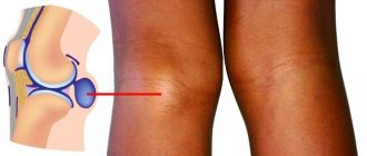
At the time of injury, the joint is deformed, which is clearly noticeable when compared with a healthy joint - the shoulders become asymmetrical. This characteristic sign helps the doctor immediately suspect a dislocation. The damaged joint is somewhat flattened, the protrusion that forms the clavicle and acromion of the scapula is clearly visible. Sometimes, upon palpation, the displaced head of the humerus can be felt.
Another example of a shoulder dislocation on an x-ray.
The pathological situation is aggravated by inflammatory edema, which occurs due to the expansion of small blood vessels in response to displacement of the articular surfaces. Plasma penetrates into the intercellular space from them, which is visualized in severe swelling of the shoulder. Edema develops as a result of compression of any large blood vessel, such as the axillary vein. Pastosity is observed - a mild swelling of the epidermis. The skin turns pale, acquires a bluish tint, and becomes cool and moist to the touch. When you press on it, the depression remains for some time. Sometimes, especially with combined injuries, swelling spreads to the entire arm.
Almost always, with a dislocation, hematomas appear. They form 2-3 hours after injury and persist for several days and sometimes weeks. Their intensity depends on the number of damaged vessels, blood from which has accumulated in the subcutaneous tissue.
Treatment for repeated dislocations
If the dislocation occurs again, the doctor will prescribe surgical restoration of the joint capsule. Other methods will not be able to completely eliminate such injury in the future.
The operation can restore the function of the ligaments and the capsule itself. Therefore, the risk of injury recurrence is minimized. Particular attention should be paid to therapeutic exercises: it will help strengthen the joint, ligaments and muscle frame. Strong muscles reduce the likelihood of recurrent dislocations.
First aid
The speed of recovery and the number of complications that develop depend on whether first aid was provided to the victim. The person should be immediately placed on a hard surface and the injured shoulder should be kept at rest. This can be achieved by positioning the arm in an abducted position - the elbow needs to be slightly bent and a cushion or folded cloth placed under it. A bandage to support the upper limb in the abducted position will allow the joint to be completely fixed. This is a triangular scarf, in the middle of which the forearm is placed, and the ends are tied with a knot at the back of the neck. In addition to immobilizing the hand, the main task of first aid is to improve the well-being of the victim. What can be done to reduce the severity of pain:
- Apply an ice pack wrapped in thick cloth to your shoulder to prevent frostbite. The maximum time for applying a cold compress is 15 minutes every hour. The procedure not only helps eliminate pain, but also prevents the spread of inflammatory swelling to healthy joint structures;
A cold compress and immobilization of the affected arm are first aid measures.
- give the victim a tablet (1.5 for acute pain) of any anti-inflammatory non-steroidal drug - Diclofenac, Nimesulide, Ibuprofen, Nise, Ketorol. You can also dilute a Nimesil sachet in 100 ml of water.
In case of acute pain, you must call an ambulance. This is a characteristic symptom of combined joint damage, so the victim is hospitalized for examination and surgery. If a person complains of mild or moderate pain, then immediately after fixing the arm, he should be taken to the emergency room for realignment of the shoulder joint.
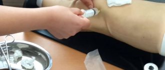
Habitual dislocation of the shoulder joint: injection of anesthetic before reduction.
Hip dysplasia in children, advice from Dr. Komarovsky.
It’s not for nothing that children are sent for routine examinations to specialist doctors every month. Sometimes in the maternity hospital they do not notice, and sometimes such a problem as physiological immaturity of the joints simply does not manifest itself. Parents are very afraid of the diagnosis, but with timely detection and taking certain measures, everything will go back to normal without any difficulties.
Usually, fear appears in front of what we do not know and do not understand. To know what it is and how to help your baby, read this article.
Physiological immaturity.
Immaturity or underdevelopment is called when the development of a joint and its physiological functions do not correspond to the child’s age. This happens under the influence of various natural factors during pregnancy, during or after the birth of the baby.
Most often, problems in newborns arise with the hip joint. It is initially not fully formed and is easily deformed.
Underdevelopment of the hip joint can develop into various forms of dysplasia. What happens in the joint: the head of the femur is displaced from the socket of the pelvis. Depending on the complexity: without displacement, partial or complete.
There are many reasons for a violation of the structure of the hip joint:
- premature birth;
- heredity;
- large fruit;
- breech presentation;
- oligohydramnios;
- gynecological diseases in a woman in labor;
- multiple pregnancy, etc.
The main reason for the appearance of hip dysplasia, according to Dr. Komarovsky, is not a genetic predisposition, but the way children are treated after birth. Indeed, in countries where mothers carry their children on themselves, respectively, the legs are widely spaced, such a disease practically does not occur.
Symptoms and diagnosis of pathology.
The diagnosis can be made in the maternity hospital or during an examination by a pediatrician. You yourself, looking at the baby, may notice some symptoms:
- knees at different heights;
- one leg is longer than the other;
- folds at different heights, or there is (is) one more fold, not a pair;
- when we spread our legs (gymnastic exercise “frog”), different amplitudes.
But not everything manifests itself externally; some signs can only be recognized by a qualified doctor directly upon examination.
Only a pediatric orthopedist can confirm or refute the diagnosis, whom you should contact immediately.
The doctor will examine the baby and listen to the parents. Will refer you to procedures that will help clarify the diagnosis or refute it. These are ultrasound and x-ray.
An ultrasound can be performed at any age; it is not so important if dysplasia is not confirmed. But if a violation occurs, the sooner it is recognized, the sooner measures will be taken to eliminate the defect.
When performing an ultrasound, the doctor carefully moves the sensor near the groin area. It also examines the tissues adjacent to the articular bones, turning from the back to the right or left side.
They are sent for x-rays only based on indicators and at the age of 3 months. During the procedure, the baby must lie still, so it is recommended that it be brought during sleep. This will solve a number of problems and help make your pictures clearer.
Depending on when dysplasia occurred and its course, the name of the pathology differs:
- Congenital dislocation;
- Hip dysplasia;
- Acetabular dysplasia;
- Hip dislocation.
The name “hip dysplasia” is used in infants and young children. And the term “congenital” is used if the joint does not develop or is absent.
Komarovsky can advise you if you do not understand the diagnosis and explain certain features. But also, do not forget to ask your doctor about the peculiarities of the location of your baby’s hip bones, how to treat and handle him during the recovery period.
Treatment.
Treatment of dysplasia in a one-month-old child up to six months old looks more like a set of procedures that do not particularly constrain either the child or the parents. This includes wide swaddling, massages, wearing in a sling (only in those models where the legs hang on both sides), wearing on the hip, wide onesies, a diaper one size larger.
Different types of pillows, pants, stirrups, and tires are also used. Everything is done in order to fix the femoral head in the acetabulum, then the ligamentous apparatus will form correctly and become stronger.
After 6 months, with hip dysplasia, it is necessary to use more rigid fixators: plaster spacers that limit movement. Children are more active than infants, and the load on the hip bones is greater.
Treatment depends on the form of dysplasia.
In severe cases, immediate reduction of the dislocation and a coxite bandage can be used. If no treatment methods help or the disease is detected too late, corrective surgery is performed.
Komarovsky advises not to delay treatment, and even better, if possible, to prevent its development with the help of preventive measures.
You can also view Dr. Komarovsky’s topic: “Hip dysplasia.” In the Video he will clearly show and explain some of the nuances.
Contraindications and dangers of the procedure
X-rays of the shoulder are prescribed for pain and dysfunction of the joint. This condition can be caused by a number of diseases:
- fractures of the bones that form the shoulder joint (humerus, scapula, clavicle);
- damage to the ligamentous apparatus (dislocations and subluxations);
- diseases of inflammatory origin (arthritis);
- degenerative-dystrophic diseases (arthrosis);
- tumors and tumor-like formations of various etiologies.
- congenital pathology.
There are a number of contraindications to x-ray examination:
- Pregnancy. Ionizing radiation has a negative effect on the developing fetus. X-rays are prescribed only for health reasons. In this case, the stomach is covered with lead aprons. It is advisable to carry out the study using a modern device with low radiation exposure.
- Children up to 3 months of age. If it is necessary to examine a newborn, it is best to use other methods, for example, this is done using MRI or ultrasound machines, which do not have a negative effect on the developing body of the child.
- Recent x-ray. There should be a break between studies.
No preparation for the study is required. Before an x-ray is taken, the patient removes all clothing from the upper body, while removing metal objects that could affect the result, such as chains, earrings, and jewelry. The patient is placed on a special table.
Most often, the image is taken in two projections - frontal and lateral. The position of a person will depend on this. The doctor brings the X-ray tube to the area being examined and selects its correct position. A cassette indicating the right or left side of the joint is placed under the selected area.
Then the patient gets up, gets dressed and waits for the result. On average, the procedure, including image decoding, takes about half an hour.
Diagnosis using x-ray is indicated if the following symptoms are present:
- sharp pain in the shoulder on the left or right side;
- limited hand movements;
- swelling.
An increase in the volume of the shoulder joint is one of the indications for an X-ray examination. An x-ray is necessary if shoulder pain is felt after physical activity or sports.
X-ray examination is contraindicated in the following cases:
- During pregnancy.
- Children under 15 years old (relatively).
- During lactation.
- For obesity.
X-ray examination cannot be used more than 2 times a year due to the risk of radiation.
It is known that X-rays negatively affect the body of a patient undergoing X-ray diagnostics. The degree of influence depends on the intensity and time of irradiation. Radiologists wear sievert dosimeters on their coats, which show the dose of radiation absorbed by the body during an X-ray examination.
There are acceptable levels of radiation exposure for various organs of the body. Patients are often reluctant to agree to undergo an X-ray examination due to its negative impact on the body, since even a small dose of radiation is harmful. But if you refuse to conduct an X-ray examination, an incorrect diagnosis will lead to progression of the pathology.
The research method is painless for the patient. No specific preparation is required on the eve of the x-ray examination. The patient's clothing should be easy to remove, without metal elements. Jewelry should be removed.
The patient is placed on a horizontal surface. The groin area is covered with a special lead apron to protect from exposure to radiation. During a forced examination of a child, his body is completely closed, leaving the upper limb area open. The procedure takes place within 5 minutes.
A well-conducted study produces good x-rays, which can be used to accurately determine the condition of the bone tissue.
During the procedure, moving is prohibited to prevent blurry images. The same can happen if the patient is severely obese. In this case, fat cells block the passage of the rays, resulting in unclear images in the pictures.
The results of an X-ray examination can only be described by a radiologist who finds darkened areas, examines the articular structure, and detected foci of pathology. After this, the doctor draws up a conclusion. The description of one x-ray by several doctors may differ slightly, but the diagnosis will be the same.
After examining the results of the x-ray, the radiologist signs the conclusion he has drawn up and passes it on to the attending physician, and the patient discusses the results of the study with him. Sometimes the patient must undergo an x-ray again, which the doctor informs the patient about, indicating the exact reason.
An X-ray of the shoulder joint can be done at any diagnostic clinic. Within a short period of time, specialists conduct x-ray diagnostics, and also describe and interpret the image. In municipal clinics, the patient is given a referral for an x-ray in the institution itself.
In clinics located in the region, there may be no X-ray room, and for examination you need to go to a large city, where you can choose between public clinics and commercial ones. Whether or not an examination in an X-ray room will harm the patient’s health depends on the quality of the equipment.
To confirm the preliminary diagnosis, the patient is sent for X-ray diagnostics
Expensive new X-ray machines emit minimal amounts of harmful rays. And yet, for several days after the procedure, doctors strongly recommend including foods with a high content of vitamin C and B vitamins in your diet.
Dislocation with fracture of the head or neck of the humerus
Dislocation with a fracture of the head or neck of the humerus is rare in middle and old age (Kaplan A.V., 1979). Attempts at closed reduction are fraught with complications, so surgical treatment is indicated. The relationship between bone fragments and the component ends of bones can be quite complex, so in difficult cases, CT should be used to clarify the diagnosis.
X-ray: comminuted fracture of the proximal end of the humerus at the level of the anatomical neck with dislocation of the head
Causes
The most common cause of shoulder dislocation is trauma. Normally, movements of a twisting or everting nature are performed in this joint, and exceeding their amplitude leads to the exit of the articular head from the articular cavity of the scapula. Such an injury can be caused by a fall on the hand, a sudden, intense and unsuccessful movement.
Some additional factors may contribute to the occurrence of shoulder dislocation:
- Frequently recurring sprains of the ligaments and joint capsule. This predisposing factor is especially characteristic of athletes involved in tennis, handball, volleyball, throwing, swimming and similar sports, or people of certain professions whose work activity involves the repetition of a number of excessive movements. Frequent and repeated trauma to the ligaments of the shoulder joint leads to a significant decrease in its stability, and dislocation can occur with any minor traumatic movement.
- Dysplasia of the glenoid cavity of the scapula. In some people, from birth, the glenoid cavity of the scapula is excessively shallow, has a poorly formed lower part (with hypoplasia), or is tilted forward or backward. Such deviations from the norm and some other rarely observed anatomical features of the structure or location lead to an increased risk of shoulder dislocations.
- Generalized joint hypermobility. This deviation from the norm is observed in 10-15% of people and is expressed in an excessive range of motion in the joint.
Contraindications and dangers of the procedure
In order to identify various changes in the joint, the radiologist must know the norms for a healthy shoulder: the joint space is uniform throughout, there should be no additional formations, pathological clearings, or violations of the cortical layer of the bones that form the joint.
Dislocation is characterized by loss of contact between the articular surfaces by half or more. With subluxation, there is a partial disruption of contact, accompanied by a wedge-shaped deformation of the joint space. A shoulder dislocation is shown in the photo below.
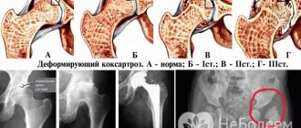
Signs of arthrosis of the shoulder joint at the initial stage are a slight decrease in the height of the x-ray joint space and thickening of the edges of the articular surfaces. There may be bone growths. The second stage is characterized by a pronounced decrease in the height of the joint space, proliferation of osteophytes, subluxations, and deformation of the articular surfaces.
In case of fractures, a violation of the integrity of the cortical layer, areas of linear clearing, bone fragments, displacement of bone fragments, and angular deformations of the bones are revealed.
If the patient has a history of shoulder injury, and the x-ray shows no signs of a fracture, then indirect signs, such as widening of the joint space and uneven areas of darkening, can suggest the presence of fluid in the joint cavity, such as blood, which indicates a ligament or tendon rupture.
In such a situation, it is better to resort to high-precision methods of radiation diagnostics. These include magnetic resonance imaging and spiral computed tomography. Using these methods, ruptures of ligaments and tendons are easily identified and an accurate assessment of the fluid that has accumulated (blood or exudate) is given.
In childhood, x-rays of the shoulder can show various congenital pathologies (shoulder dislocation, deformities). Inflammatory diseases (osteomyelitis) and traumatic injuries involving the growth zone (epiphysiolysis) are common.
Equipment used
An X-ray diagnostic apparatus consists of an X-ray tube, which is suspended above the table where the patient lies. The X-ray film is in a drawer. This may be a photographic plate for developing the image. A portable X-ray machine is used for operational diagnostics. Such a device makes it possible to examine a patient in intensive care or in a hospital bed.
The apparatus for X-ray diagnostics can be stationary or portable
In this case, the X-ray tube is connected to a flexible manipulator located above the patient's body. The photographic plate is located behind the patient. Sometimes, special pillows or sandbags are used to maintain the patient's body in the correct position during the procedure.
While the image is being taken, the patient is asked to hold his breath to obtain a more accurate image. When the X-ray machine is turned on, the doctor goes into the next room. When the first image is taken, the patient slightly changes his position, after which the image is taken again.
Joint reduction techniques in the hospital
“Shoulder knocked out - what to do?” The best decision would be to urgently visit a clinic or emergency room. However, you can familiarize yourself with the basic methods of joint reduction in order to be aware of how specialists work. It is not recommended to carry out the procedure yourself!
Sequence of actions for different methods:
- Reduction according to Dzhanelidze. This technique is used most often because it is based on muscle relaxation. To do this, the patient is placed on a hard surface so that the limb hangs down. A towel is placed under the shoulder blade, and an assistant holds the head. After this, the doctor bends the patient's arm at an angle of 90 degrees, he presses the forearm near the bend.
- Reduction according to Hippocrates. To carry out this procedure, the patient must be laid on the floor. After this, the doctor grabs the patient’s hand at the wrist and places his heel under his armpit. It is necessary to simultaneously pull the limb along the body, pressing the heel into the joint. a characteristic click will serve as a sign that the bone is in place. However, this technique should be carried out carefully and slowly.
- Kocher reduction. This technique is most often used to reduce an old dislocation or if the patient has great physical strength. First, the patient is placed on a flat surface, after which the doctor clasps the arm at the wrist and bends it at the elbow. The specialist then moves the limb along the axis until it falls into place. In this case, the patient must maintain the initial position.
Every person should understand that a dislocation of the shoulder joint can also be dangerous due to the fact that the shoulder capsule is often stretched. Sometimes only surgery can help. This is why it is so important to undergo diagnostic procedures before performing a reduction.
Diagnosis and treatment
The traumatologist should begin seeing the victim by examining and palpating the shoulder. Why is this necessary? The doctor needs to find out in which direction the head of the bone has shifted. In addition, it is necessary to determine whether the nerves have been damaged. The patient will then be sent for an x-ray.
After the diagnosis, the traumatologist begins closed reduction of the dislocation. The victim must be administered an anesthetic drug. The choice of type of anesthesia (local or general anesthesia) depends on the condition of the patient, how young and healthy he is.
When the painkiller begins to take effect, the doctor reduces the dislocation, choosing the optimal method. If we talk about how easy this procedure is, then a lot depends on the type of dislocation. The most difficult thing to cope with is the primary injury, the easiest thing is the habitual dislocation of the shoulder joint. After reduction, a repeat x-ray is taken.
Why is it so important to provide timely professional medical assistance? Consequences are possible in the form of various complications: damage to bones and nerves, major blood loss due to rupture of blood vessels, etc.
Symptoms and signs of subluxation
It is not always possible to provide timely medical assistance, since a person who has sustained a shoulder injury believes that the symptoms characteristic of a subluxation will go away on their own. The syndrome is attributed to a slight sprain, the consequences of a blow, or careless movement of the shoulder. Such inattention threatens complications and long-term impairment of motor activity. What manifestations must you pay attention to?
- There is a stabbing or cutting pain in the shoulder area.
- The site of injury turns red and becomes hot.
- A burning sensation is felt in the injured area.
- The violation area swells.
- It becomes difficult to raise your hand or make any other movements with it.
A person may exhibit all the signs of trauma in combination, or 1-2 of them. In any situation, you should provide first aid and contact a medical facility for further diagnosis and treatment.
Shoulder dislocation in a child
Joint dislocation in children has the same course as in adults, but is less common. A child's risk of shoulder dislocation increases with increasing activity. The fact that dislocations are less common in children is explained by the fact that at this age the joints, bones, muscles, and ligaments are softer and more extensible. Soft bones are easily deformed, which does not result in the head of the bone popping out of the cavity.
In children, subluxation is more common - incomplete dislocation. The ligaments and tendons are greatly stretched, the joint is not firmly fixed, that is, the joint is poorly fixed. Such a subluxation is easily reduced and does not lead to negative consequences. All this relates to the characteristics of the child’s skeleton and the functioning of the musculoskeletal system.
If a child does have a dislocated shoulder joint, an adult must fix the bandage without changing the position of the dislocated joint. Apply something cold to the sprained area: ice, a bottle of cold water, a bag of ice cream, or just a dampened rag. Never use a warm compress! Having done all this, the child must be taken to the emergency room, where he will receive medical care.
Types of damage
The classification is closely related to the direction of dislocation of the head of the shoulder joint. In which direction the head of the joint has shifted, this will be the type of damage - anterior, inferior or posterior.
According to ICD-10 (International Classification of Diseases, 10th revision), shoulder dislocation is coded S43. Depending on the reason why the injury occurred, the types are divided:
Acquired dislocations are:
- non-traumatic (voluntary/pathological (chronic);
- traumatic (habitual).
The type of damage also depends on the time that has elapsed since the injury, so a dislocation can be:
- fresh (less than three days have passed);
- stale (more than three days but less than three weeks have passed);
- old (more than three weeks have passed since the injury).
Classification of dislocations
The following types of shoulder dislocations are distinguished.
Posterior dislocation
After an injury, the capsule of the elbow joint ruptures, which leads to displacement of the lower part of the shoulder forward. The patient feels a sharp pain, the arm takes a bent position. The joint becomes deformed, increases in volume, and there is a lack of active movements in the elbow. Examination gives a picture of shortening of the forearm, the process of the elbow is displaced upward.
Anterior dislocation
Occurs as a result of a direct blow to the elbow with the arm bent. On examination, there is elongation of the forearm on the side where the injury occurred.
Lateral dislocations
They are often combined with nerve damage, which leads to loss of sensitivity. Painful sensations are observed, active movements are impossible. External dislocation of the forearm is accompanied by an outward shift of the joint axis. In case of internal injury, the injury is caused by a blow from the outside to the inside. There is swelling and deformation of the joint.
Rehabilitation
The recovery program after a shoulder dislocation includes physiotherapy (amplipulse therapy, paraffin applications, electrophoresis, electrical muscle stimulation, etc.), massage and therapeutic exercises. The rehabilitation course begins after removing the immobilizing bandage and consists of the following periods:
- activation of the functionality of muscles damaged and “stagnant” during immobilization – about 3 weeks;
- restoration of shoulder joint functions – about 3 months;
- The final restoration of joint function takes about six months.
The patient needs to prepare for the fact that restoration of the functionality of the shoulder joint after its dislocation will take a long time. This duration of rehabilitation is explained by the fact that the injured joint requires a long “rest” to fully recover.
All physical therapy exercises must be performed under the supervision of an experienced physician or instructor. Only gentle loads can be placed on the joint, and movements should be performed as carefully as possible.
In the first weeks of rehabilitation, it will be enough for the patient to perform 10 flexions and extensions of the arm at the elbow joint and hand. In addition, exercises can be performed to raise your arms forward and spread them to the sides. In the first stages, the injured hand can be helped by the healthy one.
After two weeks, you can add to this set of exercises abduction of the arms bent at the elbow joints to the sides and alternate raising and lowering of the shoulders. Further, the patient may be allowed rotational movements of the arms and their abduction behind the back, exercises with a gymnastic stick, etc.
Remember! If pain appears as you increase the load, you should stop exercising for a while and consult a doctor.
Shoulder dislocation is a common injury and can be accompanied by various complications. In the future, such damage can cause chronic instability of the shoulder joint, requiring surgery. That is why the appearance of a shoulder dislocation should always be a reason to immediately consult a doctor for proper treatment and a full course of rehabilitation.
Channel One, program “Live Healthy” with Elena Malysheva,” in the “About Medicine” section, a conversation about the usual dislocation of the shoulder:
Orthopedic traumatologist I. Zasadnyuk talks about a common shoulder dislocation:
A specialist from the Moscow Doctor clinic speaks about shoulder dislocation:
Rating: (votes - 1 , average: 4.00 out of 5)
How is a dislocation diagnosed in the hospital?
“I knocked out my shoulder - what should I do?” Are you asking yourself a similar question? The most logical answer would be to visit the hospital for examination. In the worst case, self-medication can lead to various kinds of complications.
As a rule, diagnosis of injury is carried out using 3 main methods.
- X-ray in two projections. This procedure allows not only to determine the degree of dislocation and direction of the bone, but also to exclude the possibility of an internal fracture. If the doctor knows exactly how the patient’s bone is located, this will allow the reduction to be performed more correctly and painlessly. X-rays can also indicate the presence of tumors that occur after a long period of time without treatment for a displaced limb.
- CT scan. This technique is not used as often as the previous one, but it also occurs (especially in paid clinics). Based on the results of a computed tomography scan, the doctor can judge the presence of damage to joints and muscle tissue. In addition, this procedure often allows one to recognize a fracture that is not shown on an x-ray. The technique is also used if there is suspicion of vascular damage.
- Ultrasonography. The technique is used only in the most advanced stages in order to detect the accumulation of fluid in the shoulder joint and identify the presence of rupture of ligaments, muscles or capsule. In addition, the traumatologist may recommend this procedure in order to exclude circulatory disorders - ultrasound examination can determine the presence of compressed vessels.
Once the patient has completed all the necessary procedures, the doctor will be able to prescribe comprehensive treatment based on their results. However, in some cases you have to provide first aid to the victim yourself. Therefore, you should familiarize yourself with how to do this.
Why do you need an x-ray?
Shoulder (image is normal)
Find out the dose received from an X-ray of the shoulder joint using a modern digital device, and compare the indicators with the natural background. Go to “Dosimeter”.
The image visualizes the manifestations of pathologies: violation of the integrity of the bone, growth of cartilaginous elements along the edges, areas of salt deposits, etc.
Through the procedure, complications and secondary pathologies associated with diseases of other organ systems are identified. Radiography helps plan the therapeutic course and monitor its course.
Titanium plate for a clavicle fracture
The technique does not demonstrate a pathological condition in all cases. Probably, during the examination, changes are observed in the soft tissue; they are not visible on the negative.
First aid
To prevent the worsening of a dislocated shoulder joint and reduce pain, you need to know what to do and how first aid is provided, which includes the following:
- The injured arm is placed in a comfortable position.
- Carefully remove clothes.
- They give you a pain-relieving medicine (Ketorol, Analgin, Ibuprofen).
- The wounds are treated with an antiseptic and a sterile bandage is applied.
- Immobilize the damaged joint.
- Ice is applied to the damaged area and removed after 15 minutes to prevent frostbite.
The victim should be urgently taken for medical care, since an old dislocation is difficult to reduce. It is strictly forbidden to reset a dislocated joint on your own; this should be done by a specialist. During transportation, the victim must be in a sitting position.
Shoulder reduction at home is strictly prohibited and is performed in an emergency room under general anesthesia. In some cases, local anesthesia is used. The main methods for reducing the shoulder joint are discussed below.
Hippocrates-Cooper method
The patient lies on his back. The doctor, sitting on the side of the injury, grabs the hand with his hands. The doctor places the heel of his foot in the armpit, pressing on the slipped head, and performs traction along the axis of the arm, as a result of which the head takes its usual place in the articular cavity.
Cocker method
The method is used in case of anterior dislocations, if there are no fractures of the humeral neck. The patient lies on the table in a supine position. The sprained arm should extend beyond the edge of the table. Reduction of dislocation is carried out in 4 stages:
- The doctor supports the forearm of the injured arm with one hand, and with the other, the elbow of the healthy arm, bent at a right angle, is drawn to the body, stretching along the axis of the shoulder. The assistant fixes the forearm.
- Continuing the traction along the axis of the shoulder, the doctor directs the shoulder outward until the forearm takes its place in the frontal plane of the body. In this case, you can often hear a click.
- Without weakening the traction along the axis of the shoulder, the doctor slowly raises the forearm, moves the patient’s elbow, pressed to the body, to the midline and up. After such actions, the head is located opposite the site of rupture of the bag, often taking its place at the end of this stage.
- The forearm is used as a lever, producing internal rotation. The doctor moves the patient's hand towards the opposite shoulder joint, the forearm is placed on the patient's chest. At this time, the shoulder joint should be reduced.
Read also: Open dislocation of the ankle joint
Dzhanelidze method
The patient lies on the table on the side of the injured side. In this case, the edge of the table is aimed at the armpit, the injured arm should hang down. The patient's head is on a stand. After 20 minutes, relaxation occurs, which makes it possible to begin to reduce the joint. The doctor, standing in front of the patient, grabs the forearm, bent at the elbow, presses on the forearm, performing rotational movements in the joint, which leads to the reduction of the shoulder. The patient must wear a plaster cast for two to three weeks.
Chaklin's method
The patient lies on his back. The surgeon pulls the adducted shoulder along the length with one hand, and with the other, pushes the head of the shoulder outward, inserted into the armpit. The method gives especially good results when a dislocation is combined with a shoulder fracture.
Cooper method
The traumatologist stands behind the patient sitting on a chair, places the foot on the seat, and points the knee into the armpit. Applying traction downwards, the doctor uses his knee to direct the head to the desired place.
Indications and restrictions
The main conditions under which an X-ray of the shoulder joint is indicated:
- pain in the shoulder area;
- partial loss of motor activity in the shoulder joint;
- dislocations or subluxations;
- arthritis or arthrosis (change in skin color in the area of the shoulder joint);
- the need to assess the dynamics of the chosen treatment method;
- formed neoplasms;
- the need to monitor congenital pathologies in the structure of the shoulder joint.
Often x-rays are performed despite the absence of direct indications. This category of people includes athletes and people whose work involves high physical stress on the shoulder joint. To prevent the development of pathological processes in the connective and bone tissue of this category of citizens, it is recommended to undergo x-rays at least once every six months.
The need for radiography is determined by the attending physician, taking into account the likely harm that can be caused to the body by the radiation dose.
Prosthesis (direct projection)
Relative contraindications to the procedure include age under 15 years. During pregnancy and lactation, examination is prohibited and can only be carried out in extremely serious cases.
Treatment and rehabilitation
After reduction, a dislocated shoulder will have to be treated for several days. If this is not done, the joints may begin to atrophy and sooner or later lose their motor function. To prevent this from happening, it is important to apply a pressure bandage to the painful area and also minimize movement of the limb. The only exceptions are special exercises designed to gradually develop the hand:
- flexion and extension of the shoulder joint;
- lifting light weights;
- kneading muscles with massage.
Also, in some cases, the patient may be recommended to undergo physiotherapeutic procedures in order to relieve swelling and speed up the healing of the limbs. To relieve pain symptoms, a specialist may prescribe a special warming ointment based on the previously listed active substances. Also, some sports, such as swimming, will also have a beneficial effect on the speed of rehabilitation of the patient.
Diagnosis and treatment
Treatment of a dislocated shoulder begins with diagnosis and determination of its type and degree of displacement. For this purpose, an external examination is used, determining the direction of displacement, clarifying the circumstances of the injury that occurred, and the fact of a similar injury in the past. For some cases, an X-ray examination is indicated; in special cases, when serious damage to the joint and tissue occurs, the attending physician prescribes a tomography to determine the indications for surgical intervention. If there are no such indications, the traumatologist will reduce the dislocation. First aid for a dislocated shoulder joint after reduction will concern only the immobilization of the injured arm.
Important! There is no need to try to straighten the dislocation. Any attempts at reduction without special knowledge and skills will lead to a worsening of the current condition - tissue ruptures and bleeding.
The anterior dislocation is reduced with the patient lying face down on the couch. Hanging his injured arm to the floor. The doctor bends it at the elbow and, holding it at 90 degrees from the plane of the chest, rests his knee on the elbow bend and gently brings the head of the humerus back. Few people are able to do this at home. Moreover, the procedure is extremely painful, given that the affected area is already inflamed.
After reduction, a repeat radiograph is necessary to assess the quality of the reduction and rule out new internal injuries to the joint.
Subsequent treatment and rehabilitation consist of immobilizing the shoulder joint with a bandage, taking non-steroidal anti-inflammatory drugs and applying cold compresses to relieve swelling.
What to do if you have a habitual shoulder dislocation? Reductions and permanent immobilization are not suitable for rehabilitation. Here we are talking about a quick solution to the problem that has arisen. Despite the fact that in recent decades, many discoveries have been made in this industry and new treatment methods have been introduced, the traditional method is the Bankart operation.
In the rehabilitation process, regardless of the chosen type of treatment, an important role is given to the main stages - high-quality reduction, a complete period of immobilization (very often patients remove the pads earlier than expected and begin active manipulation of the affected arm) and rehabilitation and recovery processes.
Diagnosis of joint dislocation
Joint dislocation is diagnosed based on certain indicators. We have already listed many of them among the symptoms. But still, the traumatologist performs a professional examination, drawing a conclusion based on the complaints that he lists.
Read also: Shoulder joint hurts when raising your arm
Other medical tests are also important. X-ray examination, for which the direct projection method or, if necessary, axial technology is used, is highly effective. The quality of the x-ray is sufficient to view the location of the head of the bone and identify the features of its displacement, which also leads to damage to the integrity of the skeleton. The most important task remains to identify what kind of dislocation it is - anterior or posterior. It is important to identify whether there are any fractures.
What to do if you have a dislocated shoulder and basic treatment methods
Selection method
- As a first aid measure, stop any movement of the shoulder, if possible, apply cold (ice) immediately after the injury, consult a doctor for qualified help. If it is impossible to reduce it in the near future, apply a fixing bandage to eliminate the load and movement in the joint when the shoulder is dislocated.
- Reduction of a dislocation should only be carried out by a qualified specialist!
- Closed reduction of shoulder dislocation is indicated after administration of muscle relaxants and analgesics or, if necessary, under short-term anesthesia
- Open reduction: if it is impossible to perform a closed reduction under anesthesia or if there is an additional bone fracture
- For habitual dislocations, surgery is performed to strengthen the shoulder joint capsule.
- Repeated restoration of a torn bone or cartilaginous fragment in case of Bankart injury.
- Treatment after reduction of a dislocated shoulder - the limb is immobilized in the average physiological position with a Deso bandage for 2-3 weeks.
- After the bandage is removed and the recovery period has expired, functional treatment is carried out, including a set of exercises after a shoulder dislocation aimed at restoring the function of the joint and limb (physical therapy, massage, physiotherapeutic procedures).
There are 2 methods of performing x-rays: analog and digital.
- Analog method. The image is recreated on a special film.
- Digital method. Provides image reconstruction much faster. The image displayed on the screen can be printed several times. In addition to the advantages associated with high accuracy, the method is less dangerous for the body. The procedure, including preparation, taking the image and processing takes 5–6 minutes.
Physiotherapy
The undeniable advantage of this method of treating shoulder dislocation is that the method does not involve the use of drugs.
This allows you to completely eliminate the harmful effects on the body and the development of adverse reactions.
Basic and progressive methods of hardware physiotherapy:
- UHF;
- phonophoresis;
- ultrasound therapy;
- inductothermy;
- magnetic therapy;
- electrical stimulation;
- shock wave therapy.
At the same time, thanks to various procedures, for example, laser, magnetic field, ultrasound, infrared, ultraviolet radiation, it is possible to effectively influence the affected joint.
This treatment is excellent prevention and rehabilitation. All biochemical processes are activated in the patient’s body, the immune system is strengthened, and the natural defenses are increased, which several times speeds up the healing process of the shoulder after an injury.
Rehabilitation using each of these procedures has its own advantages and some contraindications.
What complications and consequences may there be?
If a person knocks out his shoulder joint, he will have to get it back into place as soon as possible by going to the hospital. Otherwise, certain complications may arise that will be difficult to treat. Among the most frequently encountered are the following:
- compression or rupture of blood vessels - sooner or later will lead to internal bleeding and the appearance of bruises;
- tendon damage – can also occur during treatment or rehabilitation period;
- Peripheral nerve damage is a very dangerous condition that leads to chronic pain.
Sometimes the consequences are so serious that the dislocation can only be eliminated after surgery and a long procedure to restore the integrity of the damaged tissue. The same applies to internal fractures - sometimes you even have to insert a special prosthesis.
It is also impossible not to note the fact that complications can arise after the intervention of other people. If a person dislodges a shoulder and you know exactly how to set it back, you should act with extreme caution. After all, you may not have enough practical experience. The slightest mistake can simply damage the joint capsule or muscles, which will lead to even greater complications.