Calculous cholecystitis is a disease accompanied by inflammation of the mucous membrane of the gallbladder due to the formation of stones in it, formed against the background of cholelithiasis, and secondary infection of bile.
The disease is a fairly common pathology - about 20% of the fairer sex and 10% of the stronger sex, aged from thirty to fifty years, suffer from this disease.
Calculous cholecystitis is a disease accompanied by inflammation of the mucous membrane of the gallbladder.
Depending on the course of the disease, there are: acute calculous cholecystitis, divided into catarrhal, phlegmonous and acute gangrenous, and chronic.
Let's look at what chronic calculous cholecystitis is, what are its manifestations, symptoms and treatment.
Types of calculous cholecystitis
According to the nature of the course, calculous cholecystitis can be acute or chronic. Each form of the course is divided according to morphological characteristics and clinical manifestations.
Acute calculous cholecystitis is divided according to the nature of the inflammatory process:
- catarrhal;
- purulent;
- phlegmonous;
- gangrenous.
According to clinical manifestations, chronic calculous cholecystitis occurs in the following forms:
- typical – all manifestations of the inflammatory process are present;
- atypical – latent form, in which no inflammatory reactions are observed;
- cardialgic – accompanied by symptoms of myocardial damage;
- esophagealgic – there is pain in the esophagus;
- intestinal – dyspeptic symptoms predominate.
Classification
There are acute and chronic forms, and the course of each of them can be smooth or complicated.
- In the chronic form of calculous cholecystitis, inflammation develops slowly and manifests itself during periods of exacerbation, while the infectious factor no longer plays such a significant role. In the development of calculous cholecystitis, the leading factors are disturbances in the composition of bile, its thickening and stagnation, and irritation of the walls of the bladder. This usually occurs due to errors in nutrition, obesity, diabetes, endocrine changes and frequent infectious diseases. Predisposing factors are biliary dyskinesia, liver disease, chronic gastritis and duodenitis.
- The acute form of calculous cholecystitis occurs rarely; it usually manifests itself as a result of long-term asymptomatic cholelithiasis, in which the walls of the bladder become inflamed due to blockage of the common bile duct by a stone. This happens when infection penetrates into the area of the bladder from surrounding organs due to a violation of the antiseptic properties of bile. As a result, thickening of the walls and their destruction (destruction) quickly develops, which can lead to the accumulation of pus inside the bladder and the formation of biliary peritonitis.
Causes of calculous cholecystitis
Risk factors for calculous cholecystitis:
- metabolic disorders;
- pregnancy;
- hormonal imbalances in women;
- taking steroid drugs and oral contraceptives;
- an increase in the amount of cholesterol in the cavity of the gallbladder, as a result of which the proportion of bile acids and cholesterol changes in the ratio 1: 12;
- inflammatory swelling in the neck of the bladder and at the mouth of the bile duct in the duodenum;
- chronic inflammatory processes in the mucous membrane of the gallbladder.
Stagnation of bile leads to the formation of suspensions - “bile mud”, in which accumulations of pigments and cholesterol are formed, which is the basis of the nucleus for the forming stones. Stagnation of bile leads to the deposition of cholesterol on the formed insoluble complexes, which is accompanied by damage to the inner lining of the gallbladder, the release of anti-inflammatory enzymes and inflammatory mediators. This causes a local inflammatory reaction, which is often secondary to a bacterial infection (normally bile is sterile).
The weight and size of the stones increases, as a result the stones remain in the cavity of the bladder and continue to become overgrown with cholesterol, bilirubin and calcium salts. In most cases, the stones have a round shape, sometimes they are faceted, ground to each other surfaces (faceted stones), they can be single or multiple, in severe cases occupying the entire lumen of the bladder.
The pressure of stones on the walls of the gallbladder aggravates the inflammatory reaction. With calculous cholecystitis, small stones are capable of movement. Some stones pass through the duct and are excreted in the stool. If the stone does not pass into the duct, it gets stuck near the mouth and causes stretching of the gallbladder, accompanied by severe pain. When the walls of the duct expand, the stone comes out of the mouth, and the pain stops.
In addition to cholesterol stones, calculous cholecystitis can cause the formation of pigmented stones, which consist of bilirubin and its metabolites. The causes of the formation of pigment stones are hemolytic anemia and cirrhosis of the liver.
In most cases, the development of calculous cholecystitis is preceded by acalculous cholecystitis, the causes of which are infectious agents. They enter the gallbladder with blood and lymph, less often through the bile duct from the duodenum.
Rare causes of the development of calculous cholecystitis are:
- traumatic injuries of the gallbladder;
- burn disease;
- sepsis.
Development mechanism
The main cause of the development of the disease is stone formation in the gallbladder, leading to blockage of its ducts and stagnation of bile in the organ.
This stagnation, in turn, leads to an increase in the production of anti-inflammatory mediators and injuries to the mucous membrane, which begins to produce a significant amount of inflammatory exudate and mucus in response to injury. The gallbladder stretches and begins to produce even more anti-inflammatory mediators.
These processes lead to cicatricial wrinkling of the walls of organs and their gradual thickening and calcification.
In this case, inflammation is constant, recurrent in nature - periods of calm are replaced by periods of exacerbation.
The main cause of the disease is stone formation in the gallbladder
Stages of calculous cholecystitis
- Initial. The size and shape of the gallbladder remain within normal limits. In the cervical area there is swelling and narrowing of the lumen. Emptying disorders are minor and intermittent.
- The stage of the beginning of lithogenesis. There is an increase in the size of the bile reservoir, the shape of the bladder becomes spherical. The walls of the bile duct close together in the cervical area. Stagnation of bile becomes permanent.
- Stage of advanced lithogenesis. An increase in the size of the gallbladder is observed, and an adhesive process develops in the cervical area. There are stones in the cavity of the bladder, and bedsores form in the area where the walls come into contact with the stones.
In most cases, the development of calculous cholecystitis is preceded by acalculous cholecystitis, the causes of which are infectious agents.
Causes
The main role in the development of stone formation is played by disturbances in the metabolism of bilirubin and cholesterol. These are components of bile that are poorly soluble in water and are retained in solution due to the emulsifying effect of bile acids. If the concentration of cholesterol and bilirubin in bile increases, there is a risk of stone formation. This condition is observed in atherosclerosis, obesity, gout, diabetes mellitus, and hyperlipoproteinemia.
Risk factors contributing to the development of stone formation
- irregular nutrition (overeating, fasting, rare meals);
- impaired immune status;
- pregnancy;
- lack of vitamin A;
- prolapse of internal organs;
- physical inactivity;
- hereditary predisposition;
- adhesions, scars, bends of the bile ducts;
- pathology of the pancreas;
- persistent constipation;
- taking hormonal contraceptives;
- violation of neurohumoral regulation of the contractile function of the gallbladder and bile ducts.
At the same time, an important role in the development of calculous cholecystitis is played by poor nutrition (excessive consumption of refined carbohydrates, fatty foods containing a high percentage of cholesterol, flour and cereal dishes), leading to a shift in the pH of bile to the acidic side and a decrease in the solubility of cholesterol.
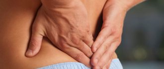
Symptoms of calculous cholecystitis
With calculous cholecystitis, the symptoms of hepatic colic are the same. The main symptom is severe pain with characteristic features:
- sudden onset, mainly in the evening or at night;
- strong intensity and duration – up to 4–5 hours;
- clear localization in the right hypochondrium and pancreas;
- irradiation to the back and under the right shoulder blade.
Symptoms
The presence of stones in the gall bladder may not affect a person’s condition in any way, so at the initial stage the disease is often asymptomatic and latent.
Symptoms directly depend on the stage of development of the disease, therefore acute calculous cholecystitis is characterized by the following signs of biliary colic:
- a sharp decrease in blood pressure;
- the appearance of weakness and cold sweat;
- nausea, vomiting of gastric and bile contents;
- elevated temperature, which is especially characteristic of purulent inflammation;
- acute pain localized in the right side and radiating to the shoulder or right shoulder blade, caused by diet disorders, alcohol, physical activity or stress;
- individual manifestations of jaundice with changes in the color of stool: feces contain a lot of fat and have a discolored appearance, and urine is darker than usual.
The manifestations of chronic calculous cholecystitis are more smoothed out:
- frequently occurring aching pain in the area of the right hypochondrium;
- nausea and belching with a bitter aftertaste;
- violation of nutritional rules can cause isolated attacks of vomiting with bile;
- the appearance of acute pain caused by poor nutrition, which gradually subsides on its own;
- paroxysmal pain that occurs 3 hours after eating fatty, salty or fried foods.
The chronic stage of development of calculous cholecystitis is characterized by the presence of stones in the gall bladder, a mild inflammatory process and periodic manifestations of the disease.
The chronic period can be practically asymptomatic or manifest itself with attacks of hepatic colic. In the latent form of the disease, a person experiences a feeling of heaviness on the right side of the hypochondrium. Suffers from flatulence, diarrhea, heartburn, belching, and feels bitterness in the mouth. After overeating or fatty foods, unpleasant symptoms intensify.
Hepatic colic can occur suddenly, in most cases it is preceded by a violation of diet, heavy physical labor, or psycho-emotional stress. An attack of colic is caused by spastic contractions of the gallbladder and its tract, caused by irritation of the mucous membranes by stones. There is an acute stabbing pain of varying intensity, localized in the right hypochondrium and radiating to the right side of the shoulder, shoulder blade and neck. The duration of the attack can vary from several minutes to a couple of days.
Diagnostics
Laboratory research methods for calculous cholecystitis:
- general clinical blood test;
- biochemical blood test - assessment of liver function, determination of the amount of blood cholesterol.
Instrumental research methods:
- X-ray of the abdominal cavity;
- ultrasonography;
- radionuclide cholescintigraphy.
As a therapeutic and diagnostic procedure, cholangiopancreatography is used, an endoscopic technique during which the mouth of the bile duct is widened, stones are crushed and removed.
Calculous cholecystitis is differentiated from the following diseases:
- myocardial infarction – with the cardialgic form of the disease;
- esophagitis and reflux esophagitis – with the esophageal form of cholecystitis;
- renal colic - always accompanied by dysuric phenomena: acute urinary retention or frequent urination.
Causes of the disease
Official medicine claims: cholelithiasis (GSD) affects about ten percent of the world's population every year.
Moreover, this pathology occurs more often in women. People in the older age group, after forty years, are more often susceptible to the disease. This disease is quite rare in young people and children. The severity of the disease is determined by the size of the stones. The danger of calculous cholecystitis is serious complications. Stones often block the bile ducts, causing severe pain. Concretions, as stones up to 3 millimeters in size are called, can come out on their own. Movement of larger stones will require immediate surgical intervention.
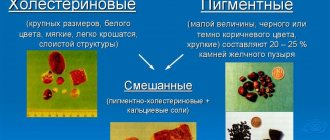
Such “jewels” in medicine are classified according to their composition:
- cholesterol;
- limestone;
- mixed;
- pigmented.
Mixed stones are most common. Treatment is complicated by the fact that in the initial stage of the disease the stones do not cause concern and are not detected during x-ray examination. Only in the case of an acute attack, when characteristic symptoms appear, can this pathology be identified. The shape of concretions can be round, oval, irregular, multifaceted, or branched.
The formation and growth of such stones occurs over a long life period. It is clear that this is provoked by many negative factors. This:
- bile stagnation;
- duct bends;
- bubble bend;
- changes in the composition of the bile itself;
- high cholesterol;
- inflammatory diseases of the gastrointestinal tract, gall bladder;
- cholecystitis.
This pathological condition can be provoked by constant strict diets, fasting, overeating, an abundance of fatty and fried foods, and alcohol. Even regular use of some contraceptives can cause disease and the growth of stones. Modern medicine identifies a number of diseases that contribute to the formation and growth of stones:
- impaired metabolism;
- endocrine pathologies;
- hepatitis;
- cirrhosis;
- oncology;
- pancreatitis;
- duodenitis;
- chronic gastritis;
- Crohn's disease;
- bile duct dyskinesia;
- sedentary lifestyle.
Treatment of calculous cholecystitis
The most striking manifestation of calculous cholecystitis is hepatic colic, which requires emergency medical attention. Treatment should be carried out according to emergency medical care. Pain syndrome is relieved with antispasmodics. Patients are recommended to take a water-tea break for 1–2 days.
In the first stage of the disease, therapy with bile acid preparations is effective, the additional supply of which reduces the relative amount of cholesterol and prevents its accumulation in the gallbladder.
In the absence of pronounced inflammation in the presence of relatively small stones, hardware crushing is performed, followed by removal of small fragments through the intestines under the influence of choleretic agents.
Surgical treatment of calculous cholecystitis involves removal of the gallbladder. The classic method of laparotomy or laparoscopic technique is used. Classic cholecystectomy involves an incision in the anterior abdominal wall, removal of the gallbladder, followed by suturing of the surgical wound. The laparoscopic technique involves removing the gallbladder using an endoscope.
What kind of disease is chronic calculous cholecystitis?
Chronic calculous cholecystitis often occurs due to the formation of stones in the gall bladder.
Chronic cholecystitis is a chronic inflammation of the gallbladder, often combined with a violation of the outflow of bile from the biliary tract (biliary dyskinesia).
Gallstones. Scheme.
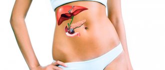
The gallbladder itself is a hollow organ that is located on the lower surface of the liver and acts as a storage site for bile. Bile is extremely important and is responsible for the following processes:
- participation in the breakdown of fats;
- destruction of pathogenic microflora in the intestines;
- stimulation of intestinal perilstatics;
- participation in the removal of excess cholesterol from the body.
The formation of stones in the gallbladder can contribute to its traumatization, hardening and changes in the walls. As a result, the organ cannot accumulate bile in the required volume, and the gastrointestinal tract system is not capable of efficiently processing food. Women suffer from calculous chronic cholecystitis much more often than men.
Features of calculous cholecystitis in pregnant women
During pregnancy, calculous cholecystitis can also develop, the symptoms of which are more pronounced than in other patients. Exacerbations of the disease often occur at the end of the second or beginning of the third trimester, which is due to compression of the gallbladder by the growing uterus. Surgical treatment is carried out taking into account the potential risk to the fetus.
Surgical treatment of calculous cholecystitis involves removal of the gallbladder. The classic method of laparotomy or laparoscopic technique is used.
Complications
Complications of calculous cholecystitis
Chronic manifestation can become a serious problem for the patient if left untreated. With each repeated inflammation in the patient’s body, other diseases may develop, the treatment of which will be more difficult:
- obstructive jaundice;
- hydrocele;
- biliary perforation;
- purulent-destructive changes in the organ;
- pericholecystitis;
- bladder cancer;
- oncological disease of the colon.
Features of calculous cholecystitis in the elderly
Due to age-related anatomical and physiological changes, calculous cholecystitis in most elderly people is asymptomatic. The disease is often complicated by the formation of hydrocele of the gallbladder with the subsequent development of purulent inflammation.
Surgical treatment is carried out using a two-stage method. First, a gallbladder puncture is performed to reduce bile pressure. Then drug treatment is prescribed. After normalization of the functioning of all organs and systems, a planned cholecystectomy is performed.
Diet after cholecystectomy
Patients operated on for calculous cholecystitis are prescribed a diet after surgery. In the absence of the gallbladder, which serves as a physiological reservoir, frequent unloading of the biliary tract is necessary. Therefore, the patient is forced to follow fractional meals throughout his entire life. Otherwise, stones may form again in the bubble stump or in the passages themselves. Meals should be at least 5-6 times a day.
After cholecystectomy, there is a deficiency of bile acids responsible for the digestion of fats. Therefore, it is necessary to exclude lard, lamb and beef fat from the menu. The consumption of fatty dairy products and butter should be limited. To compensate for the lack of lipids, more vegetable oil is included in the diet. It can be used to season porridges, salads, and vegetable purees.
Traditional methods
Traditional methods are aimed at eliminating painful symptoms of colic and increasing the flow of bile. During treatment, it is recommended to use mineral waters for patients with cholelithiasis (Essentuki, Slavyanskaya).
| Ingredients | Method of preparation, application |
| 1 tsp. oregano herbs, 1 cup boiling water | Place the raw materials in boiled water and simmer over low heat for 10 minutes. Leave for 2 hours. Drink ¼ cup of decoction 3 times a day. |
| Corn silk (1 tbsp), boiled water (1 cup) | Brew the raw material in boiling water, leave for 1 hour. Apply 1 tbsp. l. 2 times a day. |
| Plantain herb (1 tbsp), 200 ml boiled water | Place the raw material in boiling water and leave for 10 minutes. Drink in small sips 1 hour before. Take 1 time per day. |
| Herb lemon balm, yarrow, wormwood, celandine, cinquefoil (total weight - 30 g, components are taken in the same ratio), 1 glass of boiled water | Place the raw materials in boiling water and simmer over low heat for 10 minutes. Leave for 10 minutes. Drink 1 glass of medicinal solution in the morning and evening before meals. |
| Sunflower flowers, calamus rhizome, buckthorn bark, mint leaves (total weight - 30 g, ingredients taken in the same ratio), 200 ml boiled water | Place the raw materials in boiling water and leave for 30 minutes. Drink in the morning and evening before meals. |
The shelf life of prepared decoctions is no more than a day. Use after prior consultation with your doctor. Individual intolerance to the components is possible. Uncontrolled self-treatment leads to a worsening of the condition and increased painful symptoms.
For painful symptoms, tubeless tubing according to Demyanov is prescribed, which consists of taking mineral water and warming the gallbladder.
Diet
The diet for cholecystitis should not be high in calories and should not contain fatty, smoked or canned foods. Approximate nutrition is described in diet No. 5 and 5a according to Pevzner.
In addition, the following requirements are imposed on the diet and regimen:
- ban on fried foods (only steamed and stewed), sautéing of vegetables and flour is prohibited;
- you should not eat cold foods, as there is a risk of sphincter spasm;
- Coarse fiber is not recommended to avoid increased motility and stone movement.
Treatment and diet for calculous cholecystitis
In case of a mild form of the disease we are considering, as well as while the patient is in remission, it is necessary to adhere to a specific diet, which is regulated by very important rules. Let's move on to their consideration.
- First of all, you need to create a meal schedule. It is necessary to eat regularly, avoiding long breaks between food intake, since during this time there is a strong thickening of bile, which provokes the formation of stones and increased development of inflammation. The minimum number of meals is five, there should not be less.
- Frequency of meals also means reducing portion sizes as well as reducing their nutritional value.
- It is preferable to consume food at the same time every day, adjusting the so-called biological clock. The processes of bile separation are adjusted to them and adjusted, preventing the mass from thickening and the release of sediment.
It is necessary to create a meal schedule
Following a diet in the event of a chronic form of cholecystitis should be observed by the patient permanently. This is necessary to maintain satisfactory health.
In our special article you will find an approximate menu for a day with cholelithiasis.
There are also dietary basics that you should follow. They are aimed at regulating the contents of the diet.
Table 1. Recommended Products
| Name | Notes |
| Lean meat | It is recommended to eat lean meats, preferably the white parts of turkey and chicken. |
| Fruits and berries | It is preferable to introduce berries and sweet fruits into the diet, as they help lower cholesterol levels in the blood, increase the secretion of bile, and stimulate the secretion of other glands of the digestive system. So, watermelons, strawberries (only ripe and sweet), apples, and dried fruits are perfect for eating. |
| Vegetables | Among vegetables, vitamin-rich items are welcome, such as carrots, tomatoes, white and Chinese cabbage, and zucchini. |
| Oils | It is welcome to add vegetable oils without additives to food, that is, olive, sunflower and others. They need to be seasoned with vegetable salads to stimulate excretory processes in the gallbladder. Oils cope with this task even too well, so it is important to observe moderation. The fact is that an excessive increase in outflow can lead to gallstones moving and blocking the duct, which ultimately prevents bile from leaving the bladder at all. Oils should not be neglected, since some vitamins coming from vegetables and other foods can be absorbed by the stomach only if combined with fats. |
Table 2. Foods that should be consumed in limited quantities
| Name | Notes |
| Full fat dairy products | Full-fat dairy products such as butter and sour cream should not exceed established limits. So, butter is allowed to be consumed in an amount of no more than 20 grams per day, but sour cream cannot be consumed in more than two tablespoons. |
| Fat meat | Under no circumstances should you eat fatty meats such as lamb, pork, beef. Lard is strictly prohibited. |
| Salt | We will also have to limit ourselves in salt, its concentration should be up to 10 grams per day, while products from which we can get salt, for example, bread, meat and other items, are also taken into account. |
| Sugar | The maximum dose of sugar also has clear boundaries. Patients with calculous cholecystitis are allowed no more than 8 teaspoons per day. Again, it is necessary to take into account the intake of sugar from other foods. |
| Baking | Patients burdened with excess weight will also be forced to reduce the amount of carbohydrates in the diet, and sweet pastries and white bread should be completely excluded. |
| Cereals | It is allowed to consume a limited amount of cereals, preferably those containing a significant proportion of protein (buckwheat, quinoa, etc.). |
It is prohibited to eat fried, spicy or baked foods. The only exceptions are those cases when frying is done in a completely dry, non-stick frying pan, without the use of oil.
It is best to eat foods that:
- not thermally processed (vegetables, fruits);
- extinguished;
- steamed;
- cooked.
It is also necessary to pay attention to the temperature conditions of the dishes. Thus, the temperature of cooked food should not be colder than + 16°C or hotter than +55°C.
In remission and the initial stages of calculous cholecystitis, to avoid surgical intervention, the diet is combined with taking medications that contain bile salts. In addition, shock wave therapy is often used to break up stones and remove them naturally.
Treatment and diet for exacerbation of calculous cholecystitis
Acute treatment of chronic calculous cholecystitis involves immediate hospitalization of the patient. In the surgical hospital he receives the following funds:
- antispasmodics;
- antibiotics;
- detoxification drugs;
- antiemetic;
- anticholinergics.
After the condition has stabilized, there comes a time when surgical intervention is necessary. This procedure means cholecystectomy - complete removal of the gallbladder, and with it the stones. It sounds scary, however, medicine does not stand still. Today, instead of serious abdominal surgery, patients undergo a so-called laparoscopic cholecystectomy.
Laparoscopy of the gallbladder
Unlike laparotomy (dissection of the abdominal wall), it is many times less traumatic. Several incisions are made on the abdomen, through which gas is then released into the abdominal cavity:
- carbonic;
- or nitric oxide.
Then trocar tubes are inserted inside, with the help of which the tightness of the abdominal cavity is maintained, as well as surgical instruments. The internal organs are examined, the gallbladder is isolated from the liver bed and removed. A re-examination completes the end of the operation.
Incision after open and closed surgery
Note! Minimally invasive surgery cannot always be performed; in some situations, open cholecystectomy is required, for example, in particularly complicated forms of cholecystitis.
Forms and stages: specifics of the course
The nature of the disease depends on its form:
- Catarrhal
The catarrhal (initial, purulent) phase is accompanied by pain in the right hypochondrium, which increases with physical effort, as well as nausea and repeated reflex vomiting.
- Phlegmous
With the appearance of ulcerative lesions on the mucous membrane of the gallbladder, accompanied by purulent discharge, the disease develops into a phlegmous form. At this stage, the organ is greatly enlarged in size, as it is filled with pus. The patient suffers from severe pain when changing body position, deep breathing or coughing. The temperature rises, the heartbeat quickens, nausea and vomiting appear. The gallbladder is enlarged in size, which is revealed by palpation of the right hypochondrium.
- Gangrenous
If calculous cholecystitis at the phlegmous stage of development is not treated, there is a danger of a gangrenous form. Gangrene is characterized by necrotic damage to the walls of the gallbladder, with the danger of their perforation and leakage of bile into the abdominal cavity. Bile peritonitis develops. The patient experiences pronounced pain that spreads to the entire abdominal cavity. The condition is complicated by disturbances in bowel movements and fainting. May be fatal.
For reference! Gangrene of the gallbladder usually develops in older people whose blood supply is impaired.
Complications and prognosis of the disease
Chronic calculous cholecystitis can lead to the following complications:
- Choledocholithiasis - blockage of the main duct with stones.
- Stenosis of the papilla of Vater.
- Pancreatitis in acute or chronic form.
- Hepatitis, reactive form of cholangitis.
- Abscess in the diaphragm area.
- Bladder perforation and empyema.
- Peritonitis.
It should be remembered that to prevent such complications of the disease, it is necessary to consult a doctor when the first symptoms occur.
It should be remembered that to prevent such complications of the disease, it is necessary to consult a doctor when the first symptoms occur.
The prognosis for this disease is favorable, but if symptoms of cholecystitis appear, especially those manifested by hepatic colic, they will recur in the future. Moreover, within two years, in 40% of cases there is a risk of relapse. Sometimes, with a severe form of the disease, liver cancer or gallbladder can develop.
During the examination, if the diagnosis is unclear, conservative treatment may be prescribed. This plays an important role in the presence of vague signs, a functioning gallbladder and existing contraindications to surgery, which are determined by the patient’s condition.
If the patient is overweight, it is recommended to follow a diet to lose weight. If the gallbladder is not functioning, a diet with limited fat intake is prescribed.
Gallstones. What is the danger?
It is the size of gallstones that determines the symptoms, severity, clinical picture of the disease and possible complications of cholelithiasis (GSD). Chronic calculous cholecystitis is dangerous due to its complications.
In most cases (75%), the stones are located in the body of the gallbladder, but sometimes small stones can get into the bile duct, thereby obstructing or completely blocking it and causing attacks of gallstone colic.
If stones up to 3 mm in size are able to pass through the ducts on their own, then stones from 3 to 7 mm in size when exiting the gallbladder can obstruct (block) the bile duct, and such a complication will require urgent surgical care.
The composition of gallstones also varies. They can be:
- cholesterol - stones of this kind are not visible on radiography;
- limestone;
- pigmented;
- mixed.
Most often, mixed types of gallstones are found; their shape can be very diverse (round, multifaceted, or even branched).
Features of traditional therapy
Self-medication for acute calculous cholecystitis is prohibited and ineffective. Failure to see a doctor in a timely manner leads to serious complications that require extensive surgical intervention. If characteristic symptoms occur, the patient should be urgently hospitalized in a surgical hospital.
During therapy, the following tasks are solved:
- relieving symptoms;
- prevention of complications;
- eliminating the causes of the appearance of new stones.
Treatment is carried out conservatively and surgically. Conservative therapy includes a therapeutic diet, painkillers, antiemetics, and antispasmodics. During the treatment process, the inflammatory process is suppressed with the help of antibiotics, and general detoxification of the body occurs.
It is important to know! The use of folk remedies for the treatment of calculous cholecystitis is strictly prohibited. Herbal choleretic drugs cause the movement of stones and cause unwanted complications.
Often conservative treatment does not eliminate the problem, since it is not able to eliminate the source of the formation of new stones. In this case, such therapy is intended to stabilize the patient and prepare him for surgery.
Treatment
Therapy for this pathology involves an integrated approach to solving the problem. The basis of effective treatment includes the following techniques:
- compliance with dietary recommendations
- the first three days it is recommended to adhere to therapeutic fasting;
- subsequently the patient is transferred to table No. 5;
- relief of a painful attack, as well as biliary colic;
- relief from symptoms of dyspepsia;
- surgical intervention - cholecystectomy
- it can be planned or emergency;
- by type of access (traditional, laparoscopic, mini-laparotomy);
- implementation of litholytic therapy;
- performing lithotripsy.
Treatment of the disease without surgery and surgery
Medication
To block pain syndrome, doctors use the following drugs:
- antispasmodics
- atropine;
- platiphylline;
- no-shpa;
- buscopan;
- papaverine;
- duspatalin;
If the attack cannot be corrected by prescribing one antispasmodic drug, then specialists can use a combination of two drugs, which is supplemented with cerucal and an analgesic to increase its effectiveness.
- analgesics
- analgin;
- baralgin;
- ketorol;
- ketonal
Important! If there is no effect from drug therapy for five hours, such a patient should be taken to a specialized surgical department.
In order to restore the disrupted functioning of the gastrointestinal tract, the following may be prescribed:
- omez;
- cerucal;
- motilium;
- Odeston;
- Essentiale Forte;
- festal
Surgical
Clinical practice shows that to cure cholecystitis in the vast majority of cases, surgical intervention is necessary.
This category of patients who have an acute attack of inflammation or another exacerbation of a chronic process must be hospitalized in a hospital. In the surgical department, the doctor receives all the test results he needs and carries out short preoperative preparation. After this, cholecystectomy is usually performed using a laparoscopic approach.
Important! If the operation is performed on time (and this is the first three days from the onset of pain), then this can significantly reduce the percentage of deaths and serious complications.
A comparison was made with planned manipulations that were performed after the patient had been treated conservatively for 1.5-2 months.