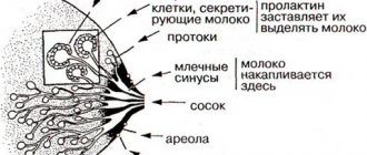
General description of the disease
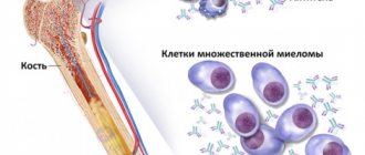
Osteochondropathy is a disease in which a necrotic deformation process occurs in the spongy bones. Basically, the disease begins its course in childhood and adolescence.
Causes of the disease:
- 1 genetic factor;
- 2 pathological functioning of the glands of the endocrine system;
- 3 impaired metabolism of nutrients in the body (in particular, poor metabolism of vitamins and calcium);
- 4 frequent injury;
- 5 various circulatory disorders;
- 6 excessive exercise, due to which the muscles contract intensely;
- 7 being overweight;
- 8 diet abuse;
- 9 exclusion from the diet of consumption of any healthy foods (especially those containing calcium).
Types of osteochondropathy
In traumatology, osteochondropathy is divided into 4 types:
- long tubular bones (the sternal end of the clavicle, hip joint, tibia, phalanges of the fingers, heads of the 2nd and 3rd metatarsal bones are affected);
- short spongy bones (this group includes vertebrae, the lunate bone of the hand, the sesamoid bone of the 1st metatarsophalangeal joint, the navicular bone of the foot);
- epiphyses (the epiphyseal discs of the vertebra, the tuberosity of the tibia and the tubercle of the calcaneus are affected);
- partial osteochondropathy (this group includes damaged articular surfaces of the elbow, knee and other joints).
Course of osteochondropathy
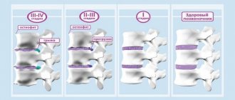
The disease occurs in 4 phases. The full cycle of osteochondropathy can last from 2 to 4 years.
At the first stage, bone tissue begins to die. Duration – several months. During this period, the patient begins to experience pain at the site of the lesion, and disturbances in the functioning of the leg or arm (depending on the location of the lesion) occur. At the same time, the lymph nodes become enlarged. When you touch the sore spot, a strong, sharp pain occurs. An x-ray may not show any changes.
At the second stage, a compression fracture occurs. The bone has sunk and damages the bone beams by simply wedging into them. X-ray shows the affected parts of the bone, the clear contour and structure of the pattern disappears. If the epiphysis is affected, the joint space becomes widened. This period lasts from 3 months to six months.
The third phase is fragmentation. It lasts from six months to three years. At this time, dead areas of the skin are resorbed, replaced by osteoclasts and granulation tissue. There is a decrease in bone height.
Recovery occurs at the fourth stage of the disease. This process can take from a couple of months to several years. First of all, the shape of the bone is restored, and then its structure is restored.
Complications
If the disease is not treated in any way, then after recovery the bone will be deformed. This can subsequently lead to the development of another serious disease – deforming arthrosis.
Basically, the disease affects the growing body during adolescence, less often small children and infants, and even less often young people.
Etiology: reasons for the development of pathology
Osteochondropathy is otherwise called Keller's disease. Among the main causes, doctors identify circulatory disorders in the lower extremities with a predominant localization in the feet. Osteochondropathy is characterized by impaired microcirculation in the bone structures of the scaphoid. As a result, the tissue does not receive enough nutrients and oxygenation is impaired.
The scaphoid bone becomes flattened and fragmented, and may become deformed into a lentil shape. In a child, this leads to the death of bone tissue, and aseptic necrosis develops without the addition of infectious agents. Factors that provoke the development of the disease include:
- circulatory disorders;
- traumatic injuries;
- arthritis;
- osteoarthritis;
- endocrine disorders;
- hormonal dysfunctions;
- heredity.
Diagnostics
Read more…
The doctor makes the diagnosis based on the results of a visual examination and radiographs of the feet. Based on the characteristic changes on x-rays, the orthopedist determines the type and stage of the disease.
- In the first stage of Keller's disease I, osteoporosis of the scaphoid bone is visualized radiographically, resulting from necrotic destruction of its spongy substance.
- Photographs taken later show compaction and flattening of this bone, with visible points of ossification.
- Even later images reveal the breakdown of the bone into separate fragments (defragmentation), due to the progression of necrosis. Signs of a fracture are visible.
In Keller II disease, the radiological signs of necrotic destruction are the same, only they are localized in the heads of the metatarsal bones of the toes.
X-ray changes:
- Stage I may be absent.
- In stage II (impression fracture), the head of the metatarsal bone loses its regular spherical or ovoid shape and becomes flattened, its length decreases by 2-3 times, leading to shortening of the corresponding finger.
- In stage III, there is a pronounced expansion of the joint space, sequestration of the head with the formation of several fragments
- IV (reparations) - resorption of necrotic bone with remodeling of the head, acquiring a saucer-shaped shape.
- Stage V is characterized by the development of secondary osteoarthritis of the affected joint.
Classification: types and forms of the disease
Teenagers more often suffer from type 2 pathology.
Doctors distinguish 2 types of disease when diagnosing osteochondropathy of the foot. The type of pathology depends on the location of the lesion:
- Keller's disease 1. The scaphoid bone located medially is damaged.
- Keller's disease 2. Accompanied by damage to the second or third metatarsal bone connecting to the surface of the digital phalanges. More common in adolescence.
In the clinical picture, depending on the dominant symptoms, 4 forms are distinguished, on which the approach when choosing a treatment method depends:
- Necrotic. Accompanied by aseptic destruction of bone beams.
- Compression fracture. Synthesizing new beam structures is typical. But due to insufficient strength, wedging fractures occur.
- Fragmentation. Osteoclasts finally dissolve the bone beam components.
- Final. The restoration of structure and form begins.
Classification of osteochondropathy
The disease is classified according to the location of the pathological process:
- if the epiphyses of the humerus, the sternal end of the clavicle, the phalanges of the fingers of the upper extremities and carpal bones, the heads of the second and third metatarsals, the head of the femur are affected, category 1 osteochondropathy is diagnosed,
- pathological changes affecting short tubular bones, vertebrae, lunate bone of the arm, scaphoid - foot, sesamoid, are combined into category 2,
- Category 3 is characterized by damage to the apophyses, ossified processes located near the epiphysis, serving for muscle attachment; such pathologies are localized in the heel tubercle, apophyseal rings of the spinal column, tibial tuberosity,
- if the process affects joints - superficial, wedge-shaped (ankle, knee, elbow) - type 4 osteochondropathy is confirmed.
Stages
The clinical picture of the course of the disease corresponds to 5 stages of development:
- Necrotic processes primarily occur in bone tissue. The period of necrosis lasts several months, accompanied by local pain (in the affected area). It is difficult to establish a diagnosis; at first, even with the use of instrumental studies, it is almost impossible to identify changes in tissues.
- The deterioration of the condition of the bones leads to the appearance of spontaneous, impression fractures; bones can overlap one another, wedging, increasing the extent of damage. In other words, under the influence of loads, deformation of the epiphysis occurs. The stage is called impression or moderate.
- At this stage, the tissues that took part in the necrotic process begin to dissolve and are replaced by elements of connective tissue. The name of the stage corresponds to the processes occurring in the patient’s body - the resorption stage.
- The process of self-healing or reparation. Replacement of the damaged epiphysis with healthy bone tissue.
- The final stage is a complete restoration of the bone structure, secondary changes in shape are possible, deforming arthrosis due to impression fractures that accompany the course of the disease at the 3rd stage.
Main manifestations: how not to miss the disease
The leg may hurt if the child runs for a long time.
Preschool boys are predominantly susceptible to osteochondropathy. The peak occurs between the ages of 3 and 7 years. The disease manifests itself on one side and rarely appears on the opposite side. Foot pain is the first and most typical symptom that occurs in patients with chondropathy. Maximum pain appears after sudden exertion and in the late afternoon. The appearance of pain is provoked by long walking or running. Rest does not provide adequate relief.
Chondropathy often becomes an incidental finding during x-ray examination of the foot due to a fracture of the navicular bone.
Upon examination, swelling is clearly visible on the dorsal surface. Palpation of the metatarsal bone of the foot is accompanied by severe pain. Children walk leaning on the outer edge of the limb. Lameness is often associated. The outcome is aseptic destruction and fracture of the scaphoid structure. The disease develops progressively, and as this progresses, the pain syndrome intensifies throughout the year. Then it gradually subsides.
Prevention
To ensure the right approach to prevention and prevent the development of both forms of Keller's disease, parents must, first of all, ensure that children always wear only comfortable and appropriately sized shoes. It is also important to reduce the mechanical load on the feet. To ensure that this condition is met, serious physical activity should not be allowed in preschool children. If you receive any foot injury, you must consult a doctor and undergo the tests prescribed by him. It is also important to pay attention to the presence of foot deformities and be sure to consult a specialist about this.
Effective and safe treatment
A plaster cast does not help stop the destructive process in the bone.
Conservative therapy for osteochondropathy of the scaphoid gives a positive result. It is carried out at all stages. Surgeons must prescribe foot immobilization in children for a period of 1 month. A plaster cast is applied to a height of up to 1/3 of the tibia, even if there is no history of a fracture of the scaphoid element. The main goal is to ensure maximum peace and stop destruction. A scaphoid fracture complicated by opposing fragments is treated surgically.
The lack of effect from conservative measures, as well as unbearable pain, is an indication for surgery. During revascularization osteoperforation, additional channels are created through which new capillaries begin to grow. Trophism is significantly improved and the bone structure is regenerated. Good results for osteochondropathy are achieved by:
- physiotherapy;
- electrophoresis with nicotinic acid and analgesics;
- ultrasound;
- darsonvalization;
- phonophoresis;
- laser therapy;
- thermal procedures.
For rapid regeneration, normalization of blood circulation and metabolic processes, young patients are prescribed medications presented in the table:
Medications
| Group | Action provided | Example |
| NSAIDs | Reduce inflammation, relieve pain | "Nurofen" |
| "Ibuprofen" | ||
| "Nise" | ||
| Chondroprotectors | Restore osteochondral tissue | "Don" |
| "Arthra" | ||
| "Rumalon" | ||
| Antiplatelet agents | Normalize blood flow and clotting | "Dipyridamole" |
| "Pentoxifylline" | ||
| "Heparin" | ||
| Angioprotectors | Strengthens the vascular wall | "Etamzilat" |
| "Dicynon" | ||
| "Solcoseryl" | ||
| Analgesics | Eliminate pain | "Paracetamol" |
| "Calpol" | ||
| Vitamins | Activate metabolic processes | "Folic acid" |
| "Vitamin B2" | ||
| "Vitamin E" |
List of sources
- Traumatology and orthopedics / Guide for doctors: In 3 volumes T.Z / Ed. SOUTH. Shaposhnikova. -M.: Medicine, 1997.
- Movshovich I.A. Operative orthopedics. M.: Medicine. 1994;
- Sorokin, S.A. Diagnosis of the second Köhler disease / S.A. Sorokin // Restorative treatment of children with diseases and injuries of the musculoskeletal system: collection. scientific tr. / ed. B.L. Andrianova. - St. Petersburg, 1991.
- Traumatology and orthopedics. Multi-volume guide for doctors. Ed. N.V. Kornilov. SPb.: Hippocrates. 2006; III;
- Varshavsky G.I. On the differential diagnosis of Keller II disease / G.I. Varshavsky, I.M. Varshavsky // Annals of Traumatology and Orthopedics. 1994. -№2;
Measures to prevent osteochondropathy of the navicular bone of the foot
After removing the plaster cast for osteochondropathy, children are recommended to use special insoles with reduced pressure on the scaphoid bone. This relieves pain and promotes faster recovery. Orthopedic shoes retain their correct shape and are effective during the rehabilitation period. Children are prohibited from running and jumping for 2 months to avoid the appearance of a false joint or fissure. For any injury to the foot and ankle, be sure to contact a traumatologist. Correct adherence to recommendations and high-quality implementation of techniques leads to complete recovery. Lack of treatment causes severe flat feet, osteoarthritis, contracture and deformation of the foot bones.
Treatment methods
Treatment of both types of Keller disease follows the same type of treatment.
| Treatment methods | Details |
| Immobilization of the sore foot | The application of a plaster cast in the form of a boot helps to completely unload the foot, which needs to be worn for 1 month, sometimes longer |
| Drug therapy | Taking non-narcotic analgesics for pain relief |
| Medicines that activate calcium metabolism | |
| Drugs that improve peripheral circulation | |
| One of the vitamin-mineral complexes | |
| Physiotherapy | After removing the plaster cast, foot massage and physiotherapy are prescribed - mud therapy, magnetic therapy, foot baths, reflexology, iontophoresis, electrophoresis |
| Exercise therapy | Exercises specially selected by a physical therapy doctor help develop the foot after long immobilization and restore its functionality. |
| Surgery | If conservative treatment is ineffective, revascularizing osteoperforation is indicated - surgically creating holes in the bone to improve arterial blood flow. Due to them, bone tissue is better supplied with blood, bypassing the affected vessels. |
The recovery period after removing a cast or undergoing surgery should be taken with particular seriousness. It is important to limit the child’s motor activity: avoid jumping, running and any active games. Choose orthopedic shoes for him, when worn, the feet will retain their anatomically correct shape (special insoles will reduce pressure on the bones affected by necrosis).
As a result, nutrition and regeneration of damaged tissues will occur faster.
Physiotherapy
Physiotherapeutic methods for the treatment of osteochondropathy are used for:
- Improved blood supply to the limb;
- Reducing pain (SUV therapy, mid-wave ultraviolet irradiation, in erythemal doses), diadynamic therapy (use of pulsed currents);
- Acceleration of regeneration in the affected area - ultrasound therapy (ultraphonophoresis of drugs), mud therapy;
- Reducing the severity of bone tissue dystrophy (heliotherapy, thalassotherapy, therapeutic massage, air baths;
- Restoration of impaired functions (electrophoresis, radon and sodium chloride baths).
Physiotherapy for heel bone disease
Symptoms
Symptoms of heel bone chondropathy depend on the stage of the disease. The acute period of the disease in some cases may be asymptomatic, and the first manifestations appear several years after the onset of the disease. They are caused by secondary changes in the joints and periarticular tissues.
Osteochondropathy of the heel develops in most cases slowly and is chronic. Main symptoms:
- Pain on the back of the heel when walking, with the pressure of shoes, sometimes swelling in this area;
- Limitation of support on the heel, transfer of the load to the forefoot, as a result of which deformation of the toes and lameness are possible;
- Muscle atrophy is moderate, pain goes away with rest;
- Sometimes body temperature rises.
Forecast
Spinal osteochondropathy is a completely curable disease if it is diagnosed in childhood, when the human body is actively developing. But it is very difficult to make a correct diagnosis in a timely manner, since the symptoms are very similar to those of other pathologies of the spinal bones. Lack of treatment can lead to pathological fractures and disability. In some cases, even with timely treatment, osteochondrosis develops at an older age.
To avoid complications, you must regularly visit an orthopedist. A specialist will be able to detect the disease in time and prescribe appropriate treatment. To prevent complications, the child needs to do gymnastics that strengthens the back muscles, not overload the spine and eat right.
Historical reference
In 1983, pain arising in the posterior part of the heel joint was first described. The disease was described by surgeon Edward Albert, who is considered the founder of the term “Achillodynia.” But in his work he did not put forward theories about the causes and mechanism of development of the disease.
Osteochondropathy of the calcaneus - manifestations
Patrick Haglund in 1928 first outlined his vision that pain in the distal Achilles joint can be caused by a number of reasons. According to his theory, the concept of “Achillodynia” is too general and does not highlight the true root cause of the pain syndrome. He suggested that the pathology is provoked by:
- Achilles tendinitis (inflammatory process in the area of the Achilles tendon);
- Achilles bursitis (inflammation of the synovial bursa surrounding the tendon);
- Pathology of the epiphysis zone responsible for the growth of the calcaneus in children.
P. Haglunda suggested that the factor provoking the development of superficial bursitis was wearing uncomfortable or small shoes, and deep bursitis was one-time or chronic trauma. He suggested treating deep bursitis surgically.
In the works of 1990-1991, terminological confusion remained: in addition to Haglund syndrome, the terms “achillodynia” and “achillobursitis” were used.
In the domestic literature, the following terms were used to describe pathology in the area of the calcaneus: “Haglund’s disease” (an abnormal bone located between the navicular bone of the tarsus and the head of the talus) or “Haglund-Schinz” disease (the apophysis of the calcaneus).
Upper epiphysis
There is a hole in the body of the bone, which is the entrance to the nutrient canal. Many vessels pass through it. The greater and lesser trochanters are located on the proximal epiphysis. The outer surface of the greater trochanter can be easily felt through the skin. Its inner surface has a trochanteric fossa. Between the greater and lesser trochanters, the intertrochanteric line begins and goes down, turning into the pectineal strip.
The posterior part of the superior epiphysis gives rise to the intertrochanteric ridge, which ends at the lesser trochanter. The remaining part of the upper epiphysis forms the head of the femur. On it there is a fossa of the head, which is the place of attachment of the ligaments. The head continues with the neck of the femur, which is most susceptible to fractures, especially in older people. In the case of such an injury, a complex operation is required, followed by a long rehabilitation period.
Diagnostic measures
The patient can be examined using MRI.
When the first symptoms of the disease appear, you should consult a doctor. You cannot try to establish a diagnosis and treatment methods on your own. If the development of osteochondropathy is suspected, the following studies are most often carried out:
- X-ray examination;
- tomography with the results displayed on a computer;
- magnetic resonance imaging;
- skeletal examination using radioisotope method (scintigraphy);
- ultrasound scanning;
- determination of bone tissue density.
Reasons for appearance
Osteochondropathies in children can develop under the influence of various factors. Most doctors consider excessive physical stress on the bone to be the most likely cause. Due to chronic minor injuries, a disruption of the blood supply develops, leading to necrosis of spongy bone tissue. This can occur in athletes, as well as in children who spend a long time in an unnatural position.
But many researchers note that osteochondropathy can also be caused by other reasons:
- hereditary predisposition;
- metabolic disorders - decreased absorption of calcium;
- disturbances in the blood supply to bone tissue;
- hormonal imbalances;
- infectious diseases;
- pathologies of the endocrine glands;
- lack of calcium, magnesium and some vitamins.
At the initial stage, the disease is difficult to detect, since it only manifests itself as pain during physical activity.
What it is?
Osteochondropathy refers to several ailments that occur in the knee area. All diseases differ in location in the knee joint and symptoms. These include:
- Koenig's disease. Characterized by inflammation of cartilage tissue. Damaged pieces of cartilage separate or peel away from the bone. Their erratic movement along the joint capsule disrupts the movement of the joint itself. The inflammatory process occurs in cartilage tissue. In neglected form, its deeper parts die off. The pathology is common between the ages of 10 and 20 years. Moreover, it occurs 3 times more often in boys than in girls.
- Larsen-Johansson disease (osteochondropathy of the patella). With this disease, an improperly ossified patella causes pain, causes swelling, leads to hydroarthrosis, and limits mobility. The affected area is fragmentation of the patella. The risk group includes teenagers involved in sports between the ages of 10 and 15 years.
- Osgood-Schlatter disease. Characterized by pathological changes in the tibia. A growth appears in the tuberosity, which causes pain in the area of the kneecap. The disease causes microtrauma to the bones. It occurs in children and adolescents aged 8 to 16 years. Like Larsen's disease (osteochondropathy of the patella), it most often affects boys.
- Leuven's disease. Causes negative changes in the knee joint. Over time, the disease develops into intractable synovitis. In the early stages it is characterized by intermittent pain of moderate strength. Movements in the joint are preserved in full, but they are painless. Usually the process is one-way. Leuven's disease (osteochondropathy of the patella) develops between the ages of 12 and 14 years.
Confirmed establishment of Osgood Schlatter how to treat
The peculiarity of the problem is the impact of the load on the area of discomfort. Treatment for Osgood Schlatter is based on the same principles as after injuries. This ensures the absence of stress, complete rest, and for both legs at once.
In cases of exacerbation of osgood-schlatter, treatment includes stopping the acute reaction using conservative methods.
How is it carried out when there is Osgooda Schlatter's disease, treatment, what does it include:
- anti-inflammatory therapy;
- painkillers.
The unpleasantness of a long-term symptom of circulatory disorders is corrected using quartz irradiation and thermal effects.
If Osgood Schlatter's disease is diagnosed in adolescents, treatment is supplemented by wearing patellas. After conservative measures, in order to avoid the risk of repeated damage to the lower leg, it is recommended to follow a regimen that limits intense loads and fixes the load zone. These may be removable bandages.
Schlatter's disease in adolescents, treatment, surgery
In case of severe pain and to speed up recovery, surgical dissection in the longitudinal direction of the patellar ligament can be performed. Moreover, if there are displaced ossified areas and an extra zone of the process in the epiphyseal region, they are removed.
Femur fracture
A fracture of the femur is a condition characterized by a violation of its anatomical integrity. Most often, it happens in older people when they fall on their side. Concomitant factors for hip fractures in these cases are decreased muscle tone, as well as osteoporosis.
Signs of a fracture are severe pain, swelling, dysfunction and deformation of the limb. Trochanteric fractures are characterized by more intense pain, which intensifies when attempting to move and feel. The main symptom of a fracture of the upper part (neck) of the femur is the “stuck heel symptom” - a condition in which the patient cannot turn the leg at a right angle.
Fractures of the femur are divided into:
- Extra-articular, which, in turn, are divided into impacted (abduction), non-impacted (adduction), trochanteric (intertrochanteric and pertrochanteric);
- Intra-articular, which include fracture of the femoral head and fracture of the femoral neck.
In addition, in traumatology, the following types of intra-articular hip fractures are distinguished:
- Capital. In this case, the fracture line affects the femoral head;
- Subcapital. The fracture site is located immediately below its head;
- Transcervical (transcervical). The fracture line is located in the femoral neck;
- Basiscervical, in which the fracture site is located at the border of the neck and body of the femur.
If the fractures are impacted, when a fragment of the femur is wedged into another bone, conservative treatment is practiced: the patient is laid on a bed with a wooden board placed under the mattress, while the injured leg rests on a Beller splint. Next, skeletal traction is performed on the condyles of the leg and thigh.
In case of displaced fractures, characterized by deformation and malposition of the limb, surgery is recommended.
Pathogenesis
Normally, the articular surfaces of bones are covered with dense, elastic and smooth hyaline cartilage. This allows the bones to slide smoothly against each other during movement. With osteochondritis dissecans, thrombosis occurs in a small vessel supplying an area of bone. The nutrition of the cartilage located above this area is disrupted, and a zone of aseptic necrosis is formed.
Over time, necrotic cartilage peels off and enters the joint, forming a “joint mouse” - a loose intra-articular body that moves around the joint and, falling between the surfaces of the bones during movements, can cause blockages. The damaged area loses its smoothness, causing overload and further injury to the joint. As a result, osteoarthritis may develop.
Treatment of osteochondropathy of the hip joint
Osteochondropathy of the hip joint can be cured permanently if therapy is started in time. Treatment of the disease is a long process that can take up to 4 years. Therapeutic measures include several methods for improving the condition:
- medications;
- massage;
- physiotherapy;
- alternative medicine;
- surgical intervention.
The main principle of therapy is to stop necrotic changes in bone tissue and prevent the pathological process from spreading to neighboring formations. The patient is also prescribed vitamin therapy to strengthen the body and improve the condition of the skeleton. If conservative methods fail, the patient needs surgical intervention.
Why does Osgood Schlatter disease appear in teenagers?
- Due to the incomplete process of formation of the large medially located tibia bone during its growth, it cannot withstand increased loads. Age 10-14 years.
- This contributes to painful sensations.
- They appear due to inflammation of the ligaments.
Result: disruption of local blood circulation.
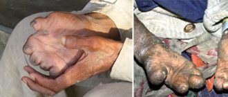
To see what a leg looks like if you have Schlatter's disease, we offer a photo below the text. This is a clear example of changes in the medial bone of the leg.
A noticeable area of protrusion in the medial part of the lower leg, visible to the naked eye
Due to stress loads, the tibia, which is not strong enough in adolescence, cannot cope with them. Therefore, Osgood Schlatter disease practically does not occur in adults. But it remains as a change.
Stages of chondropathy
- At the initial stage of the developing process, tissue necrosis is observed, which is manifested by the first pain sensations in the affected areas. This period lasts for several months. It is quite difficult to determine chondropathy at this stage; even diagnostic research methods do not always reveal the formation of pathology in the muscles.
- At the second stage of the progressive disease, a “fracture” of a compression nature occurs, which involves the involvement of bones in the pathological process, they wedge into each other and characteristic subsidence occurs. The duration of the second stage of the disease is from 2 to 6 months.
- At the third stage, lasting from one to three years, replacement of healthy tissues with granulation cells and “resorption” of bone areas susceptible to necrosis are noted.
- The last stage is the restorative stage, which is characterized by the restoration of the bone’s shape and structure.
Make an appointment with a dentist:
Unfavorable outcome or prognosis without attention:
Impaired blood circulation in the area of a specific type of injury causes the appearance of small microhemorrhages. Complicated variants are described as non-microbial necrosis of hard and cartilaginous areas and separation of the epiphysis (upper). This is the same aseptic option included in the list of the international classification of diseases.
Given the young stage of life, Osgood Schlatter syndrome cannot be ignored. Although many cases ended with the pain going away, it is still recommended to monitor the condition of the bone tissue, with supportive stimulation of its growth. To do this, there are a number of measures to treat Osgood Schlatter, affecting unpleasant symptoms.
Changed state of hard tissue in the image obtained after X-ray diagnostics
Prices
| Disease | Approximate price, $ |
| Prices for diagnosing childhood arthritis | 2 000 — 3 000 |
| Prices for diagnosing childhood epilepsy | 3 100 — 4 900 |
| Prices for pediatric neurosurgery | 30 000 |
| Prices for treatment of childhood epilepsy | 3 750 — 5 450 |
| Prices for treatment of umbilical hernia in children | 9 710 |
| Disease | Approximate price, $ |
| Prices for hip replacement | 23 100 |
| Prices for clubfoot treatment | 25 300 |
| Prices for Hallux Valgus treatment | 7 980 |
| Prices for knee joint restoration | 13 580 — 27 710 |
| Prices for scoliosis treatment | 9 190 — 66 910 |
| Prices for knee replacement | 28 200 |
| Prices for treatment of intervertebral hernia | 35 320 — 47 370 |
Necrosis of the metatarsal heads (Keller's disease 2)
If necrosis affects one or several metatarsal heads at once, these are symptoms of type 2 Köhler disease.
Köhler's disease type 2 is an adolescent necrosis that primarily affects the second or third metatarsal heads (most often the second) in individuals between 12 and 18 years of age. Unlike pathology of the first type, Keller disease II occurs more often in girls.
The reasons are the same as described at the beginning of the article. We can add here that professional sports, circus or ballet activities can become a source of constant teenage stress leading to Köhler’s disease II.
Symptoms of aseptic necrosis of the metatarsal bones
- Enlargement of the heads (one or more): swelling occurs in their area, which can spread to the tarsal bones.
- Painful symptoms: at first it hurts just to walk (walking barefoot or in shoes with too thin or soft soles over stones and unevenness becomes especially painful);
- gradually the leg begins to ache even at rest;
- When pressing on the affected head, the pain intensifies.
Degrees of Köhler's disease type 2
It is best to determine the extent of the disease not by clinical examination, but by analyzing an x-ray. In total, there are five degrees of aseptic necrosis of the metatarsal head:
- First degree. The x-ray shows areas of increased density, but the shape of the head is not changed.
- Second degree: Pronounced dense bone structure of the metatarsal head. The head becomes flatter, its height is smaller, and the articular space of the adjacent metatarsophalangeal joint becomes noticeably wider.
- Third degree. The head is divided into several fragments, different in size and contour.
- Fourth degree. Restoration of structure (reparation): resorption of areas of necrosis;
- merging fragments into one whole;
- the formation of bone beams - rough thickenings that penetrate the entire structure;
- thickening and shortening of the head and a change in its shape: it becomes concave, sharp at the edges;
- narrowing of the joint space.
- a sharp decrease in interarticular space;
Keller's disease II must be distinguished:
- from a fracture of the metatarsal head or its consequences;
- tuberculous or infectious arthritis;
- "marching" foot.
Treatment of type 2 Köhler disease
- Immobilization of the affected leg for a month.
- Restoring foot functions using therapeutic exercises, massage, acupuncture, magnetic and light therapy, shock wave therapy, mud therapy, salt foot baths and other methods.
- orthopedic insoles or shoes for transverse-longitudinal flat feet.
Köhler's disease has a favorable prognosis, that is, complete restoration of bone function is possible, provided proper treatment.
Surgical treatment of childhood and adolescent Keller disease is rarely used.
The cure is based on the combination of natural compensatory restoration of bone (a great miracle of nature) and the creation of favorable conditions for this - a feasible task for Homo sapiens.