Diseases of the blood and hematopoietic organs are a common cause of early death in children and adults. This is explained by the fact that pathological processes in plasma are difficult to treat, which is also very expensive. Timely therapy and preventive measures help reduce the risk of possible disability or death.
Blood diseases are very dangerous for humans
Population Prevalence
In terms of the number of patients, blood diseases are not included in the “15 diseases” defined by the World Health Organization as the most common in the world.
But in Russia, statistics indicate very disappointing figures: compared to 1990, the incidence of blood system diseases in the population has increased 3.6 times.
This has caused the need to develop hematological research, search for means to combat pathology, further expand the number of specialized beds in specialized hospitals, and train doctors of all specialties.
Primary care physicians are required to know what clinical manifestations are associated with pathologies of the blood and hematopoietic organs in order to promptly refer the patient to a hematologist.
The lower blue line shows a graph of the growth of blood diseases by year
Anemia
One type of anemia can be found in every fourth person on Earth, and most often it is caused by a deficiency of vitamins or microelements. Anemia is a disease in which either the number of red blood cells in the plasma decreases or the hemoglobin content inside the red blood cells decreases. The pathology may owe its development to either a poor-quality diet, or damage to the hematopoietic organs, or massive blood loss, in which the level of hemoglobin in the blood cannot be restored to normal after bleeding. There are also other types of anemia, less common, but more dangerous (genetic, infectious).
Clinical manifestations of anemia
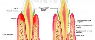
To diagnose anemia, as well as to clarify its type, it is necessary to evaluate the hemoglobin level, the number of red blood cells, hematocrit, red blood cell volume, and the average hemoglobin concentration in a red blood cell.
Table of reference values of blood parameters to exclude anemia
| Index | Reference values | Unit | Note |
| Hemoglobin | 120-140 F 130-160 M | g/l | In pregnant women below 110 g/l |
| Red blood cells | 4.0-5.0 M 3.7-4.8 F | 10^12/l | |
| Hematocrit | 42-50 M 38-47 F | % | |
| Average red blood cell volume | 81-101 | fl | |
| Average hemoglobin concentration in erythrocyte | 32-36 | g/dl | |
| Average hemoglobin content in an erythrocyte | 27-34 | pg | |
| * Eosinophils | 0,5-5 | % | To exclude parasites |
| * Ferritin | 12-150 F 15-200 M | µg/l | To exclude iron deficiency anemia |
| * B12 (cyanocobalamin) | 150-175 | pmol/l | To exclude B12-dependent anemia |
| * B9 (Folic) | 3-25 | ng/ml | To exclude folate-dependent anemia |
Signs and symptoms of anemia
In the presence of helminthic infestations, anemia may develop, since parasites tend to deprive the affected person of a number of nutrients, including those necessary for normal blood composition. If the number of eosinophils exceeds the norm, a specific test is prescribed to find the type of helminth.
Anemia caused by helminthic infestation requires not only anthelmintic treatment, but also the use of a vitamin complex to eliminate vitamin deficiency.
To clarify the nature of anemia, tests are prescribed to assess the level of microelements in the blood - the amount of cyanocobalamin, folic acid and iron in the plasma is examined. If there is a deficiency of one or another component, a medication is prescribed and the diet is adjusted.
Algorithm for separating anemia by erythrocyte indices
Video - Anemia: how to treat
Causes
Many reasons leading to changes in the blood have been studied. But even more mysteries remain. Scientists believe that they have the right to point out risk factors in such cases.
- acute and chronic blood loss lead to disturbances in blood formation and increased cell breakdown in anemia;
- mutations of the human genome under the influence of chemicals (cytostatics, antibiotics, industrial poisons), radioactive radiation;
- hereditary transmission of predisposition to the disease occurs in families where blood diseases occur in children;
- HIV infection and Epstein-Barr virus.
It is impossible to unambiguously associate any of the reasons with the disease of a particular person. Only a combination of disadvantages in life situations can lead to illness.
The role of blood in the human body
Without blood, the life of the human body is impossible. Cells and tissues need oxygen, nutrients and building material every second. The body provides all this with blood. By its origin, it is a type of connective tissue. Its main difference is its liquid state. Blood is heterogeneous in composition. Each of its components is designed to perform its task. Like other types of connective tissue, blood contains cells. Their entire mass is divided into three varieties:
- red blood cells (erythrocytes);
- white blood cells (leukocytes);
- blood platelets (platelets).
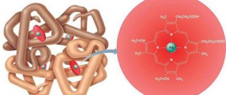
Red blood cells are the most numerous cells. Their content in the blood is one thousand higher than that of other cells. In appearance they resemble a biconcave disk. Thanks to this shape, they are able to pass through even the narrowest vascular tubes - capillaries - without damage. Inside the red blood cell contains a large amount of hemoglobin protein. Iron, located in the center of this structure, is able to attach oxygen and carry it to other tissues and organs. A mature red blood cell does not contain a nucleus as it is unnecessary. Such a cell lives for about one hundred and twenty days, after which it is disassembled into its component parts in the spleen. Many components of hemoglobin are reused.
The red blood cell is the main oxygen carrier in the body
Secrets of Blood - video
White blood cells - leukocytes - do not contain hemoglobin and do not participate in supplying the body with oxygen. They are part of the immune system that protects organs from uninvited guests, pathogens and viruses. Neutrophilic leukocytes attack the enemy directly in close combat. The cell absorbs the microbe and digests it. Under a microscope, a peculiar nucleus of neutrophils in the form of a pearl necklace is clearly visible. Inside they contain multi-colored granules.
Leukocytes are the body's main defenders against foreign agents.
Another type of white blood cell is lymphocytes. These immunity fighters don't like to face the enemy. They usually use more ingenious methods. Lymphocytes produce antibody proteins against microbes. Traveling through the vessels, antibodies find bacteria or viruses and combine with them. The microbe loses its pathogenic properties and is eliminated from the body. The spherical nucleus of a lymphocyte is visible under a microscope. These cells do not contain granules. Platelets are the third type of blood cell. They are thin plates without a core. These blood cells are involved in the process of clotting and stopping bleeding. All blood cells are formed in the red bone marrow and have a common precursor - a stem cell. From it, red blood cells, white blood cells and platelets are subsequently formed.
All blood cells are formed from one precursor - a stem cell
Hematopoietic system - video
In addition to cells, blood contains a liquid part—plasma. It is water mixed with various chemicals - proteins, minerals, hormones. Thanks to these substances, blood clots when necessary, carries chemical signals to target organs, retains water in the vascular bed, and transports nutrients and building material.
Most of the blood plasma consists of water
Classifications
The International Classification of Diseases (ICD-10) identifies more than 90 nosological units for blood diseases.
A simplified clinical classification identifies 4 groups of diseases associated with one leading pathological process:
- anemia - defined as conditions with a reduced level of hemoglobin;
- hemorrhagic diathesis - all diseases with impaired coagulation;
- hemoblastosis - tumor diseases of cell sprouts from the bone marrow and lymph nodes;
- a group of other pathological manifestations.
Each group is further subdivided into many subtypes, depending on the clinical course, damage to specific blast cells, and the leading causes of the disease.
For information about blood diseases, it is important to take into account the primary signs of the disease. Therefore, in practice, a syndromic classification with the identification of leading symptom complexes is more suitable. All symptoms in it can be divided into general and local, which characterize lesions of a specific hematopoietic organ. Many diseases have the same patient complaints and similar clinical manifestations.
Diagnostic methods
The path to making a final diagnosis of a particular blood disease is often long and difficult. Sometimes it is necessary to carry out a lot of analyzes and studies, standard and more complex. Modern medicine has a wide range of studies that help a specialist make a diagnosis:
- standard inspection. During an external examination of the patient, the doctor pays attention to the color and condition of the skin and mucous membranes, palatine tonsils, tongue, the presence of minor hemorrhages and larger hematomas. The liver, spleen, all groups of lymph nodes (inguinal, popliteal, axillary, cervical, submandibular, etc.) must be palpated;
- laboratory blood test according to standard methods. Deviations from the norm in the number of cells and erythrocyte sedimentation rate (ESR) make the doctor suspect a specific diagnosis. Often, along with normal cells, tumor cells (blasts) are found in the blood;
General analysis is a mandatory test for blood diseases - laboratory analysis of blood coagulation. It involves running several tests. It is mandatory if a clotting disorder is suspected;
- bone marrow examination. The most common option for obtaining bone marrow cells is a puncture of the sternum (sternal puncture). The procedure is carried out using a special Kassirsky needle. A small amount of bone marrow is drawn into the syringe. The material is studied in detail under a microscope. Using this method, tumors of the hematopoietic organs (leukemia, erythroblastosis) are effectively diagnosed. For a more detailed examination of the bone marrow, material is extracted with a needle from the ilium (trephine biopsy);
Bone marrow examination - an informative analysis for blood diseases - in some cases it is necessary to examine the cells of the lymph node. The material is taken using a regular syringe without anesthesia, after which it is examined by a specialist under a microscope. In some situations, a section of a lymph node is taken for examination (biopsy) for subsequent histological examination;
Lymph nodes contain white blood cells - examination of the spleen. The material is taken with a needle, after which smears are prepared from it. Studying this material under a microscope is required for some types of anemia and tumors;
- X-ray examination is used to look for changes in the lymph nodes of the chest, as well as large and small bones;
- ultrasound is an informative and safe method for examining the liver, spleen, and lymph nodes;
Ultrasound is a method for examining the liver and spleen - Computer (magnetic resonance) tomography is a valuable method for studying the anatomical structure of internal organs. Using a series of images, the specialist will determine the structure and size of the liver, spleen, lymph nodes, and bones;
- modern technologies make it possible to determine a specific type of white blood cells and blasts using immunophenotyping. The reaction involves the cells themselves and proteins-antibodies to them, artificially created by man;
- Genetic analysis allows us to determine the nature of hereditary blood diseases (for example, hemophilia). The material for research is blood.
Genetic testing is used to diagnose blood diseases
What do blood tests tell you - video
Diseases with anemic syndrome
The group includes all types of anemia associated with a decrease in hemoglobin content, impaired transport and absorption of oxygen and the development of tissue hypoxia (oxygen starvation).
Symptoms:
- persistent headaches;
- dizziness, fainting;
- sensation of “tinnitus”;
- dyspnea;
- tachycardia and stabbing pain in the heart area;
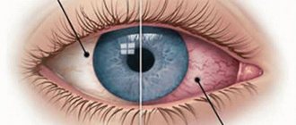
- “darkening or flickering spots” in the eyes;
- increased fatigue;
- decreased memory;
- irritability, insomnia.
In older people, even with a moderate decrease in hemoglobin, heart failure occurs.
Specific symptoms of a particular type of anemia are not included here (perverted taste in iron deficiency anemia, yellowness of the skin and sclera in hemolytic or hemorrhage, in hypoplastic anemia).
Red blood cell diseases
As already mentioned, red blood cells are not completely full-fledged cells that must carry oxygen to organs and tissues, and carbon dioxide from them. Both gases are in a chemical connection with hemoglobin, or more precisely, with its non-protein part - heme. To ensure adequate transport of these gases, the following conditions must be met:
- so that there is a sufficient number of red blood cells, but not exceeding the norm (a large number also has a bad effect on health);
- the membrane must be strong enough so that the red blood cell does not burst when entering a vessel with a changed pH;
- the bond with heme must be strong enough for oxygen to be carried to its destination, and at the same time such that it can detach in the tissues.
Violation of the first two conditions causes blood diseases, which are treated by hematologists. A change in the third situation usually occurs during poisoning; it is not considered a hematological pathology and is treated in toxicology.
Change in red blood cell count
There may be either an increase in the number of red cells (erythrocytosis) or a decrease (anemia). The second type of pathology is much more common.
Anemia is caused by three main reasons:
Bleeding
This anemia is called hemorrhagic and occurs:
- acute, when a fairly large amount of blood was lost at once - more than 1/10 of its total amount;
- chronic: blood is lost often, but little by little (heavy menstruation, frequent nosebleeds, bleeding gastrointestinal ulcer).
Poor quality of the red blood cell membrane, which causes it to quickly disintegrate
These are hemolytic anemias, and there are a great many types of them. Most of them are genetic in nature. This:
- various types of thalassemia;
- sickle cell anemia;
- persistence of fetal (what the fetus has) hemoglobin, the properties of which differ from the same structure in adults;
- hereditary disorders of the shape of red blood cells, in which the quality of their membrane suffers (spherocytosis, elliptocytosis, acanthocytosis);
- membrane weakness that develops as a result of pathology of enzymes that take part in the synthesis of the erythrocyte membrane.
There are also acquired anemias, when red blood cells can disintegrate due to:
- intestinal infection or ARVI (hemolytic-uremic syndrome);
- long walk;
- large volumes of blood transfusions from different donors;
- autoimmune blood pathology;
- diseases of medium and small vessels;
- destruction of red blood cells by a parasite - in malaria;
- poisoning with hemolytic poison;
- enlarged spleen.
Impaired synthesis of red blood cells and/or hemoglobin
There are 5 types of such anemia.
The first are caused by a lack of substances in the blood from which the components of red blood cells or hemoglobin are formed (deficiency anemia):
- iron deficiency;
- caused by a lack of folic acid;
- B-12-deficient;
- occurring with scurvy;
- due to lack of protein in food (kwashiorkor);
- associated with a deficiency of amino acids necessary for the synthesis of globin (as part of hemoglobin);
- caused by a lack of microelements - copper, molybdenum, zinc - which are part of the red blood cell.
The second is a disruption of the bone marrow, which is unable to give rise to a normal number of red blood cells (aplastic anemia). There are several different types: partial, medicinal, Diamond, Fanconi.
Still others arise as a result of chronic pathologies of internal organs (such blood diseases are more typical for adults). Thus, anemia may be the only sign of cancer or chronic renal pathology.
Fourth - when everything is fine with red blood cells, but the process of hemoglobin formation is disrupted. These are diseases called “porphyrias”.
There are also anemias caused not by loss, but by increased consumption of hemoglobin. Here we distinguish anemia of pregnant women, lactation anemia and that which occurs in athletes.
Symptoms of anemia
Anemia of any origin is characterized by the following symptoms:
- pale skin (children have noticeable white ears);
- pale pink lips and gums;
- weakness;
- dizziness;
- noise in ears;
- deterioration of performance and memory;
- increased heart rate;
- there may be a desire to consume inedible things;
- B12 folate deficiency anemia is characterized by a bright pink tongue.
Additional symptoms that occur only with hemolytic anemia are yellowing of the sclera or even the skin. When a large number of red blood cells break down, the urine may darken.
Increased red blood cell count
The erythrocyte lineage is rarely affected by tumors. An increase in the level of red blood cells is usually combined with an increase in the production of other blood cells, and the name for this is polycythemia. A separate increase in the level of red blood cells may occur with erythremia and Vaquez disease, which have a benign course.
Symptoms of this pathology are:
- hot flashes to the face and body;
- the body begins to itch, which intensifies after taking a warm bath or shower;
- "goosebumps;
- facial redness;
- sweating in the absence of fever;
- burning in fingers;
- pain in the feet.
Diseases with manifestation of ulcerative-necrotic changes
Ulcerative-necrotic lesions are possible with a significant decrease or disappearance of the granulocytic lineage of the blood, leukemia. Symptoms:
- sore throat;
- impaired swallowing process due to severe pain;
- salivation;
- attacks of abdominal pain, bloating;
- diarrhea;
- smell from the mouth;
- pain in the anus.
Upon examination, ulcers are found in the oral cavity (stomatitis), on the mucous membrane of the pharynx (angina), during esophagoscopy - in the esophagus, and during colonoscopy - in the intestines.
This is what stomatitis looks like
List of violations from point B
This category assumes a slow progression of the disease. In this case, partial dysfunction or periodic exacerbations are allowed. Similar diseases include:
- hemolytic anemia;
- types of anemia involving congenital structural pathologies of red blood cells and hemoglobin with a stage of compensation;
- hemotylic anemia;
- autoimmune cytopenic syndrome;
- hemostasis disorders;
- pathologies that suggest a long-term effect from the therapy or exclude severe dysfunction.
Here, the decision on suitability is considered on a case-by-case basis.
Diseases with hemorrhagic changes
The syndrome is caused by a decrease in the total number of platelets or their impaired function, increased permeability of the vascular wall, and increased consumption of fibrinogen and platelets during the coagulation process. Characteristic of thrombocytopenic purpura, hemophilia, aplastic anemia, hemorrhagic vasculitis.
Symptoms:
- hemorrhages in the skin, mucous membranes (sclera of the eyes), muscles, joints, injection sites;
- bleeding and bleeding from the nose, gums, intestines, heavy menstruation.
Sources of problems and their treatment
The causes of blood diseases are very different, sometimes they cannot be determined. Often the occurrence of the disease can be caused by a deficiency of certain substances and immune disorders. It is impossible to identify generalized causes of blood pathologies. There are also no universal methods for treating blood diseases. They are selected individually for each type of disease.
Prevention of blood diseases provides a set of specific rules. You should lead a correct lifestyle and limit your exposure to adverse influences. It is necessary to treat any diseases in a timely manner. Timely elimination of helminths and other parasites is of great importance. Nutrition should be balanced and complete.
Treatment of blood diseases requires careful selection of an approach appropriate to the nature of the specific problem and the use of a special, complex system of therapy. Such systems are developed by hematologists, based on the characteristics of the functioning and condition of the body of a particular patient.
Among the innovative methods of treating hematological diseases, it is worth noting positivity emission tomography - a revolutionary method developed by specialists in the field of nuclear medicine. During therapy, positron emitters are introduced into the patient's bloodstream, which spread throughout the body within several hours. Subsequently, based on the data from a specialized scanner, all the necessary data on the state of the blood and the nature of the disease are recorded.
Obtaining the most accurate research results for identifying blood diseases requires the use of computed tomography, which has already become a traditional method for diagnosing the most numerous diseases.
Diseases with enlarged lymph nodes
The syndrome is called lymphadenopathic. Accompanied by the proliferation of lymph node tissue, compression of neighboring vessels and organs. Depending on the location of the enlarged nodes, symptoms appear:
- shortness of breath and dry cough (mediastinal lymph nodes);
- “overflowing” of the intestines, bloating, stool disorders (retroperitoneal and mesenteric nodes).
This is what enlarged lymph nodes in the neck look like
Treatment
The fight against blood diseases is a long process that requires an integrated approach. During treatment, conservative methods or surgical intervention can be used (depending on the type of pathology and the degree of neglect).
Medicines
The main goal of drug therapy is to improve the patient’s condition and restore normal blood functioning. Depending on the type of disease and its severity, the doctor selects individual treatment for a specific pathology.
There is no universal method for all blood diseases, but there are the most commonly used groups of drugs that are used in one case or another:
- Anticoagulants – Aspirin Cardio, Plogrel, Streptase, Aspigrel.
- Hemostatic drugs – Aminocaproic acid, Immunat, Aprotex, Vikasol, Tranexam.
- Antianemic agents – Askofol, Hemofer, Folic acid, Iron gluconate 300, Heferol.
- Plasma replacement solutions – Serum Albumin, Aminocrovin, Peripheral Kabiven, Poliglyukin.
Immunat is a hemostatic drug
The dosage and duration of therapy is determined by the doctor. Treatment usually occurs on an outpatient basis, with the exception of severe tumor pathologies of the blood, when hospitalization is vitally necessary.
Non-drug treatment
Dangerous pathologies of the hematopoietic systems (leukemia, lymphoma) do not respond to conservative therapy.
To combat malignant tumors, effective methods are used such as:
- bone marrow (stem cell) transplantation;
- chemotherapy;
- transfusion of blood components.
The goal of this treatment is to eliminate pathogenic cells of the immune system and prevent the destruction of healthy tissue.
Blood transfusions are used to fight cancer
Possible complications
Blood diseases are dangerous because of their consequences, which do not depend on whether treatment is started on time or not. Pathological processes can greatly affect a person’s ability to work, lead to disability or cause death.
The most common complications include:
- anemia as a result of excessive blood loss;
- sepsis that develops against a background of weakened immunity after chemotherapy or long-term use of medications;
- increased susceptibility to infectious and viral diseases;
- development of concomitant diseases of internal organs (heart, blood vessels, liver, stomach);
- the appearance of hemorrhagic syndrome - a tendency to blood loss (develops against the background of advanced anemia).
Blood diseases negatively affect the cardiovascular system
Long-term untreated blood pathologies can develop into chronic forms of the disease. This condition is more difficult to treat and can greatly affect the patient’s quality of life.
Diseases with severe intoxication
Intoxication manifests itself in different ways in blood pathologies:
- general weakness - in any form is caused by anemia and cell destruction (anemia, leukemia);
- itching of the skin - due to the release of histamine from blood cells, increased levels of basophils (lymphogranulomatosis, myeloid leukemia), impaired microcirculation in the skin (erythremia);
- lack of appetite, weight loss - with malignant lymphomas.
Symptoms of fever and intoxication in some diseases appear during the day, in others - at night
Leukemia
Leukemia is a blood disease, better known as “blood cancer.” The definition of “leukemia” includes diseases such as lymphoma, myeloma, plasmacytoma, and other diseases associated with the appearance of cancer cells in the plasma structure.
Leukemia can be expressed by a whole host of independent symptoms. Therefore, often the disease is determined only by a blood test for diseases in the case of diagnosing other, relatively harmless diseases.
Often, people with cancer of the blood system experience a loss of interest in life, fatigue, enlarged lymph nodes, sudden weight loss, a general decrease in immunity, and frequent lingering colds. A slight regular increase in body temperature can also be a sign of leukemia.
Diseases with pain in joints and bones
Such a symptom as osteoarthropathic syndrome is associated with inflammation of the joints, hemorrhage into the joint capsule, and the growth of tumor tissue into bone tissue.
Symptoms:
- pain in the spine, ribs, iliac bones, skull, less often in the tubular bones of the extremities (myeloma), often independent of anything, intensified by beating;
- radicular radiculitis pain (with lymphogranulomatosis, multiple myeloma) due to germination into nerve trunks;
- joint pain (hemolytic anemia, lymphogranulomatosis, hemophilia);
- swelling and redness of the joints, dysfunction.
Clotting diseases
A blood clotting disorder may have a different name depending on which of the 12 coagulation factor proteins is deficient. So, it could be hemophilia - a hereditary disease linked to the X chromosome that affects only boys, or von Willebrand pathology - a hereditary pathology associated with non-sex chromosomes. Coagulopathy can occur during an autoimmune attack, when one’s own immunity “bombards” coagulation factors. Some types of coagulation diseases appear as a result of poisoning with hemolytic poisons, others - due to an overdose of blood thinning drugs, which are prescribed mainly after cardiac surgery, and others - as a result of liver pathologies.
Coagulopathies are characterized by spontaneous hemorrhages into the joints and into the muscle mass. Bleeding after an injection or surgery, if the deficiency of the factor (taken from the factor prepared in advance at the transfusion station) is not immediately corrected, does not stop and can cause death.
Diseases with impaired plasma protein levels
You can also read: Symptoms of blood cancer
An increase in the content of altered proteins (paraproteins) is observed in myeloma and Waldenström's disease.
Symptoms:
- frequent headaches;
- memory loss;
- numbness and pain in the limbs;
- increased bleeding of the gums, tongue, nose;
- increased blood pressure;
- decreased vision.
Diseases with enlargement of the liver and spleen
The pathology is called hepato-splenomegaly. Develops in infectious mononucleosis, autoimmune hemolytic, sickle cell and B12 deficiency anemia, thrombocytopenia, acute leukemia, chronic lympho- and myeloid leukemia.
Characterized by symptoms:
- heaviness or pain in the upper abdomen;
- abdominal enlargement;
- increasing weakness;
- in later stages, yellowness of the skin.
Other syndromes are less common. Sometimes a blood reaction is caused by the presence of a chronic disease. To identify this at the first manifestations, the patient is asked to undergo a full examination.
Signs that should alert you to a hematological diagnosis
Symptoms that may indicate a blood disorder are:
- long-term elevated body temperature to small numbers, which does not have any special daily fluctuations;
- frequent and sluggish infectious processes: either a scratch does not heal for a long time, or a sore throat appears, the person feels that he is “not getting out” of the disease;
- weakness;
- dyspnea;
- heaviness in the chest;
- fast fatiguability;
- skin itching;
- bone pain;
- change in taste preferences;
- pain under the left rib;
- bleeding gums, nosebleeds.
Only if several of the above signs coincide, you do not need to make a hematological diagnosis. Go to a therapist, tell him about your suspicions, take a general test, as well as a blood coagulogram. If the doctor notices changes here, he will refer you for a consultation with a specialist, who often conducts consultations only in a large (regional or city) hospital.
How to prevent blood diseases
For prevention, you need to try to avoid all harmful factors:
- treat any diseases and conditions accompanied by bleeding;
- take seriously the manifestations and treatment of helminthic infestations in children and adults;
- complete treatment of acute infections;
- Take enough vitamins and minerals with food every day;
- control the radiation dose during x-ray examination, avoid staying near radiation sources;
- reduce contact, use mandatory protective equipment when working with paints, benzene, lead salts, pesticides and other hazardous substances;
- strengthen immunity;
- do not expose yourself to hypothermia or overheating;
- learn to cope with stressful situations with minimal losses.
These measures normalize the process of hematopoiesis and help maintain health and ability to work.
Basic list of malignant blood diseases
Leukemia is a group of serious disorders associated with the transformation of healthy cells into malignant tumors. Hemoblastosis and leukemia spare no one.
Population categories of different ages are at risk. Doctors still cannot give a scientific basis for the reason for the appearance of cancerous bodies in the blood.
However, it has been proven that ionizing treatment, the action of particularly dangerous chemicals, immunodeficiency and a number of other factors provoke the cell to change qualitatively.
All forms of blood cancer are characterized by artificial suppression of hematopoiesis. Functionally active and poorly differentiated cells are replaced by normal bone marrow bodies. Human DNA is disrupted, the genetic code is changed, a certain clone (with minor divisions) multiplies uncontrollably and metastasizes to internal healthy organs.
The disease occurs in extremely severe forms under the close supervision of doctors. If the patient suffers from a chronic form of blood cancer, destruction occurs slowly. Regular chemotherapy is prescribed to prolong life. It should be remembered that blood diseases (which occur in nature) must be treated in a timely manner.
Deviations in the composition of liquid tissue in childhood are similar to neoplasms in adulthood.
Argyrosis
A rare pathology, also called “blue skin syndrome”. It is caused by excess silver in the body. Thus, Californian Paul Karason, who became silver-blue at the age of 57, uncontrollably consumed a homemade balm made from colloidal silver and distilled water. The man died of a heart attack at 62.
In the photo: Paul Karason, “the silver man”
A similar deviation was observed in our compatriot from Kazan. Valery V.'s appearance suddenly changed after harmless treatment of a runny nose with drops containing silver. His skin took on a silver-blue hue and his hair became blond.
Temporary blindness
Melbourne resident Natalie Adler suffers from an unusual condition. Every three days the girl goes blind, that is, she literally cannot open her eyes due to severe muscle spasms. The cycle repeats every three days. This first occurred after a sinus infection complicated by a staph infection.
Natalie Adler periodically goes blind and regains her sight
Since then, Natalie has had to plan her life in such a way as to have time to get everything done during the “sighted” period. “My 18th birthday fell on a blind day, but on my 21st birthday I saw everything and my friends threw me a huge party!”
Autoimmune diseases
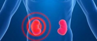
Lupus nephritis
Inflammation of the kidney, primarily the glomeruli (glomerulonephritis). The disease is characterized by the appearance of protein and blood cells in the urine, accompanied by edema and the development of arterial hypertension. Previously, the term "lupus" was used to describe skin erosions that resembled wolf bite marks. Currently used to refer to various diseases, for example, lupus vulgaris, systemic lupus erythematosus, tuberculous lupus, etc.
Predisposition: holders of blood group B (III) are predisposed to this disease to a greater extent than representatives of other groups.
Consequences of blood inflammation
It’s easy to guess why sepsis is dangerous. Firstly, in a short time, inflammation destroys many blood cells, which causes negative changes in the functioning of the immune system, oxygen transport is worse and blood clotting is impaired. Secondly, when bacteria destroy blood cells, they release a lot of toxic waste into the plasma, which in a short time affects the kidneys and liver.
Without treatment, blood inflammation intensifies, the immune system is severely suppressed, and the person has a constantly high temperature. Because of this, the structure of blood proteins is disrupted, the liver and kidneys malfunction, and the person may die.
Analgia
Congenital insensitivity to pain occurs due to a mutation in the SCN9A gene and is quite common in children under 2 years of age.
But there are cases - several hundred for the entire population of the globe - when immunity persists into adulthood. Patients with analgia are significantly more likely to suffer a burn, fracture, or sepsis. Steven Peet from Washington, like his twin brother, knows firsthand about the insidiousness of this syndrome. “I was 6 years old, I was roller skating, I fell and heard my mother scream. I look and a bone is sticking out of my leg. I didn’t feel anything,” he recalled. As a child, he broke his left leg almost every month, until the guardianship authorities removed the children from the family, suspecting violent acts. The parents had to spend a lot of time, effort and eloquence to prove their innocence.
The Pete brothers as children. Right: Steve Peet
Due to constant injuries, Steve developed arthritis by the age of 30. The fate of his brother was much more tragic. Doctors promised that in a couple of years he would be confined to a wheelchair. After this, the young man committed suicide.
Preparing to donate blood for analysis
Depending on the general condition of the body, some infections can be asymptomatic for a long time, disrupting the functioning of some organs and becoming chronic, which is very difficult to cure over time. Experts include such infections as: chlamydia, herpes, syphilis, gonorrhea, mycoplasmosis, ureaplasmosis and trichomoniasis.
In order to protect yourself and people close to you from undesirable consequences, you need to periodically donate blood for hidden infections.
It is important to know that you need to prepare for donating blood for infections; to do this, you must adhere to the following rules:
- exclude alcohol, consumption of fried, fatty and salty foods 2 days before the test;
- do not smoke a couple of hours before the test;
- in the morning, 10-12 hours after the last meal;
- the day before the tests, avoid physical activity;
- for 10-14 days before the tests, antiviral, antimicrobial and other drugs and potent antibiotics are excluded;
- if you plan to conduct any computer diagnostics, x-rays or ultrasound, then you should postpone this until after the test.
These tests are primarily necessary for people who have a large number of sexual partners, who prefer unprotected sexual intercourse, and women planning pregnancy, since many infectious and viral diseases are transmitted from mother to child both in the womb and at birth.
These studies are very important, because if the infection is not cured in time, then any infections that affect developing organs such as the heart, kidneys, liver, mucous membranes of the baby and even the brain can penetrate through the placenta to the fetus. Such proximity to the mother's infected body can lead to birth defects, and sometimes even to the death of the child.
There are cases when you can suspect the presence of an infection yourself, or this is manifested by the following symptoms:
- elevated temperature;
- chills, fever, fever;
- intestinal disorders - diarrhea and diarrhea;
- nausea, vomiting;
- lethargy, loss of strength, drowsiness;
- pale complexion;
- lack of appetite;
- rapid heartbeat (tachycardia).
It is necessary to maintain your immunity by any known means, since it is this that protects us from the negative effects of foreign bodies on the body. A healthy diet, good and healthy sleep, limiting the consumption of alcoholic beverages, moderate physical activity, protected sexual intercourse, lack of stress and periodic testing will increase your chances of protecting yourself from any kind of infection or identifying and treating it in time.
Throughout life, everyone encounters a variety of infections. Some may suffer from acute infectious diseases several times throughout the year, while others suffer from chronic infectious diseases and have to be monitored by an infectious disease specialist for life.
Infectious diseases are considered a fairly large group that can affect not only people, but also various animals.
The development of pathology begins from the moment an infectious agent enters the body, but clinical manifestations develop only after the end of the incubation period.
According to the nature of their course, they can be acute, chronic and hidden; to accurately determine them, tests for infections are carried out.
It is worth noting some points that can lead to the development of an infectious disease:
- Various types of decrease in immune status in immunodeficiency states, in the presence of concomitant diseases, childhood or old age, oncological pathology, pathology of the blood system, the period of time after transplantation surgery.
- The presence of a decrease in the resistance of the entrance gate to infection - mucous membranes, which is noted with frequent colds, after removal of tonsils, surgical treatment, chronic gastrointestinal pathology and others.
- The influence of weather factors - hypothermia, high humidity, strong wind.
- Failure to comply with personal hygiene rules.
- Disregard for the use of barrier personal protective equipment when in contact with a patient with an infectious disease.
The most common symptoms of infectious diseases:
- Fever is the most common clinical manifestation of infectious diseases.
- Changes in the physiological color of the skin and visible mucous membranes, urine and feces are characteristic of infections affecting the liver.
- The appearance of rashes on the surface of the skin, often at the peak of temperature rise.
- Manifestations characteristic of allergies, but when treatment by an allergist is ineffective.
- Dyspeptic symptoms lasting 2 weeks or more.
- Nonspecific symptoms include general weakness, fatigue, headache and others.
Before routine blood sampling for testing, it is important to:
- avoid stressful situations;
- heavy physical loads;
- stop drinking alcohol and smoking. Alcoholic drinks are canceled the day before the blood test. Nicotine – per hour;
- It is important to take a general blood test in the morning, without first eating.
So, what will a clinical blood test show? This test reveals whether the function, integrity and number of blood cells are normal, and also provides insight into other basic parameters:
- Red blood cells are responsible for maintaining optimal levels of oxygen in the bloodstream.
- Platelets give blood the ability to clot and prevent bleeding. If they are less than normal, there is a high risk of bleeding; if more, blood clots are forming on the venous walls.
- Leukocytes form the human immune system, so an increase in their number indicates a decrease in immunity, the presence of inflammation, or a disease of the circulatory system such as leukemia.
- Hematocrit shows the ratio of blood cells to plasma. This is why a clinical blood test is so important.
- ESR is an indicator of the erythrocyte sedimentation rate, which directly shows whether there is an inflammatory process in the body. It is being studied with the addition of anticoagulants - substances that prevent blood clotting.
- Leukocyte formula is a count of all types of leukocytes and the ratio of each of them to the total number, expressed as a percentage.
- Hemoglobin content, which determines the thickness of blood. A low concentration of this substance is typical for anemia of various etiologies, a high concentration is for blood that tends to thicken, or a tumor caused by too rapid proliferation of red blood cells.
- The blood color indicator shows whether there is enough hemoglobin in the red blood cells.
Allergic diseases
Bronchial asthma
An attack of suffocation develops as a result of bronchospasm or heart failure, mainly left ventricular failure.
Predisposition: 0 (I) and A (II).
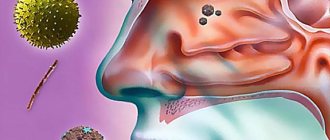
Rhinitis
Allergic rhinitis is an inflammation of the nasal mucosa caused by increased sensitivity to irritating substances.
Predisposition: 0 (I) and A (II).
Arthritis
This is an inflammation of a joint or several of its elements. Rheumatoid arthritis is a systemic inflammatory disease of connective tissue primarily affecting the joints (polyarthritis). Arthritis and osteoarthritis are primary or secondary degenerative changes in articular cartilage that bear a static load.
Predisposition: 0 (I), A (II) type is predisposed to a more acute rheumatoid form of the disease.
Gastroenterocolitis
A combination of acute inflammation of the mucous membranes of the gastrointestinal tract with damage to the small and large intestines. The causative agents of the disease are adenoviruses.
Predisposition: 0 (I), A (II) and AB (IV).
About leukocytes
There are several types of WBC leukocytes in the blood:
- granulocytes are neutrophils, basophils, eosinophils;
- agranulocytes – lymphocytes and monocytes.
Each of these types is responsible for one or another function in the body.
Neutrophils
With a bacterial infection, the number of leukocytes increases due to neutrophils. At the same time, the ratio of young and mature forms changes. Normally, immature neutrophils - band-nuclear - contain only 1-6%, mature - segmented - 45-70%.
With inflammation, this indicator increases in the general analysis and the more young neutrophils, the more severe the consequences and the worse the prognosis for recovery.
If there is an increase in band neutrophils in the blood, then they speak of a shift in the leukocyte formula to the left. This is because on the form the young forms are to the left of the mature ones - this is where this expression comes from. If there is a shift in law, the old forms predominate. This is observed in long-term infections with depletion of cellular reserves.
Table 2. Leukocyte formula and its shift.
An increase in neutrophils occurs during the following processes:
- pneumonia;
- bronchitis;
- sore throat;
- sinusitis;
- purulent focus in any organ, abscess;
- purulent pleurisy, peritonitis;
- osteomyelitis;
- inflammation of the lymph nodes;
- acute abdomen clinic.
When diagnosing, the features of the clinical picture and the results of other studies are compared.
Eosinophils
Eosinophils EOS in the general blood test increase if inflammation is caused by parasitic invasion, as well as when it is accompanied by an allergic component. A blood eosinophil count of more than 5% is a deviation.
Basophils
BAS basophils rarely respond to inflammation, and their concentration in the bloodstream does not change. A significant increase in these formed elements occurs in leukemia.
Lymphocytes
If the increased content of blood leukocytes is associated with the LYM lymphocyte fraction, then this indicates viral inflammation. This occurs in the following situations:
- catarrhal pharyngitis;
- new herpes, herpes stomatitis, generalized forms;
- chicken pox;
- intestinal viruses;
- flu and much more.
Monocytes
MON monocytes do not remain in the blood, but are located inside the tissues. But their significant increase is characteristic of infectious mononucleosis, which is caused by the Epstein-Bar virus.
Diseases included in the lists of point B
This category combines temporary functional disorders, non-systemic disorders in the structure of the blood. This item includes disorders in which a slight limitation in functionality remains after completing the full treatment course.
Consequently, if doctors discover a congenital or acquired pathology of the circulatory system with frequent complications and relapses, the examination automatically takes place under point A. If the disease does not bring much discomfort to the person, diagnostic and medical measures are carried out in advance to objectively assess the state of health. Based on this, a decision is made.
Prevention
Sepsis occurs due to improper wound care or lack of treatment for bacterial infections, so the best way to prevent it is to promptly consult a doctor in case of injuries and inflammatory processes and follow his recommendations. Often, blood inflammation occurs due to the use of home treatment methods for various infectious diseases, so you should also avoid them.
Inflammation of the blood develops rapidly, disrupts the functioning of the body and, without treatment, can be fatal. It occurs due to improper wound care or lack of treatment for infections. You need to see a doctor immediately if you discover symptoms of sepsis, because home treatment is powerless.
Infectious diseases
Cholera
Acute intestinal infection. It is characterized by a fecal-oral mechanism of infection, damage to the small intestine, watery diarrhea, vomiting, rapid loss of fluid and electrolytes by the body with the development of varying degrees of dehydration up to hypovolemic shock and death.
Predisposition: owners of blood group 0 (I) are especially sensitive to cholera vibrios, although representatives of other groups remain quite susceptible to this dangerous infection.
Chicken pox
Acute viral disease with airborne transmission. Usually characterized by a febrile state, papulovesicular rash with a benign course.
Predisposition: A (II).
Salmonellosis
In case of salmonellosis, the sources of the infectious agent are domestic animals - cattle, pigs, poultry, etc. Salmonella can be found in poultry eggs. Infection occurs when food processing technology is violated and stored incorrectly. The disease develops 6 hours (latent period up to 3 days) after consuming contaminated products.
Predisposition: people with blood groups A (II) and AB (IV) suffer especially often from salmonellosis.
Dysentery
Acute intestinal infection. The first symptom of the disease is a fever accompanied by stool disorder. Since dysentery pathogens primarily affect the large intestine, signs of the onset of the disease are regularly recurring pain in the lower abdomen, most often in the left iliac region.
Predisposition: those with blood group B (III) suffer from dysentery more often than representatives of other groups.
Symptoms of blood inflammation
Symptoms of sepsis appear 3-4 days after its onset. During this time, the first infected cells are destroyed, toxic bacterial waste is released, and inflammation begins. A person experiences the following symptoms at this time:
- a sharp deterioration in health;
- dizziness;
- elevated temperature 38-39 degrees;
- nausea, yellowing of the skin;
- loss of appetite.
After some time, the patient's condition worsens significantly. Due to the large amount of free bilirubin in the blood, which is released from infected red blood cells, the functioning of the liver and kidneys is disrupted, the sclera of the eyes turns yellow, stool and urine color change.
It is advisable to go to the hospital immediately if the temperature of 38-39 degrees lasts longer than 2 days, but there are no symptoms of a cold or flu. Timely therapy will help to quickly cope with sepsis and avoid complications on the kidneys and liver.
Platelets
During the inflammatory response, PLT platelets are also consumed. They form microthrombi in the affected capillaries, causing blood stagnation, swelling, and redness of the inflamed area of tissue.
Normal platelet counts are 180-320*109. Symptoms of thrombocytopenia occur when levels are below 50*109. But this value occurs during complications - sepsis, peritonitis, systemic inflammatory response syndrome, when disseminated intravascular coagulation develops.
Diseases of the cardiovascular system
Aneurysm
Pathological protrusions of thinned areas of the heart or blood vessels. The formation of cerebral aneurysms is facilitated by congenital defects in the vascular walls of arteries and veins.
Predisposition: holders of blood groups A (II) and AB (IV) more often than others suffer from cardiac aneurysms, which are usually located in the wall of the left ventricle.
Congenital heart defect
Anomaly in the development of the heart and major great vessels, which can occur in the 28th week of pregnancy.
Predisposition: It has been clinically and statistically proven that heart defects develop in children of all four blood groups.
Rheumatism
Rheumatism, rheumatoid arthritis are systemic inflammatory diseases of connective tissue with a predominant lesion of the cardiovascular system.
A predisposition to systemic lupus erythematosus, systemic scleroderma, and periarthritis nodosa is more pronounced in those with blood groups B (III) and AB (IV).
People with type A (II) blood suffer from heart disease of non-rheumatic origin.
Skin diseases
Pyoderma
Painful skin conditions, most of them hereditary. This name refers to all dermatoses that are caused by streptococci, staphylococci and other pathogens and are characterized by purulent inflammation of the skin.
Predisposition: A (II) and AB (IV).
Psoriasis
Chronic recurrent dermatosis of unknown etiology, manifested by the appearance of plaque-papules on the patient’s body.
Predisposition: primarily affects those with blood group 0 (I).
Kaposi syndrome
Dermatosis in the form of multiple grouped blisters, accompanied by general intoxication of the body.
Predisposition: more often registered in those with blood group A (II).
Lyell's syndrome
It is a dermatosis of a medicinal nature. It is characterized by rapid necrosis of superficial areas of the skin and mucous membranes. Usually accompanied by the formation of large, rapidly opening blisters and severe intoxication of the body.
Predisposition: an allergic skin reaction to certain medications is typical primarily for those with blood groups A (II) and AB (IV).
Atopic dermatitis
Allergic inflammation of the skin, accompanied by redness, swelling, exudation, crusting and peeling.
Predisposition: A (II).
Eczema
One of the most common skin diseases. It is expressed by redness, itching and rashes in the form of spots, blisters and pustules. Eczema can be a manifestation of phenylketonuria, a hereditary disease characterized by a deficiency of the enzyme responsible for the transformation of amino acids.
Predisposition: A (II) and AB (IV).
Hives
The disease occurs when taking certain medications or foods and is characterized by a sudden rash of itchy blisters on the skin surrounded by an area of redness.
Predisposition: A (II) and AB (IV).
Scheme of autohemotherapy
With the classic treatment option, blood is taken from a vein (volume from 5 to 25 ml) and immediately injected into the gluteal muscle. If you miss the moment, clots will appear that can no longer be used. 1-2 days – break between procedures. As a rule, the result is achieved after 8-12 injections. It is unacceptable to administer more blood than the specified volumes; this can cause inflammatory reactions, chills, and muscle pain. In addition to the classic version, there are others - stepwise, with ozone, the use of blood subjected to various chemical influences, laser treatment.
With ozone
This method is more modern, superior in efficiency to the classical one. On average, treatment requires no more than 5-7 procedures. Course – 1-2 times a week. Before use, the blood is mixed with ozone in a certain concentration. Specialists use:
- Minor autohemotherapy . About 10 ml of blood is drawn from a vein into a syringe containing an ozone-oxygen mixture and injected into the patient.
- Major autohemotherapy . In a sterile container, stir 100 to 300 ml of the mixture and approximately 100-150 ml of blood. After mixing, use as directed.
- How to treat bumps on the buttocks after injections
- How to get rid of boils at home
- Injections for weight loss
Stepped
Stepped autohemotherapy involves the introduction of a small amount of blood - about 0.1-0.2 ml. It is pre-mixed with several homeopathic medicines. As a rule, the procedure takes 4 stages. For injections, you can use one syringe, the main thing is that after each injection a small amount of blood remains in it. From stages 2 to 4, the contents are intensively shaken and administered to the patient.
Drugs for stepwise autohemotherapy are selected individually for each person. Sometimes it is enough to use complex remedies containing nosodes; a little less often, ampoule homeopathic monopreparations and symptomatic medications are prescribed. Stepped autohemotherapy has proven itself as a proven way to get rid of viral infections, arthrosis, chronic eczema, migraines, and toxic liver damage.