Features and sizes of ovarian fibroma
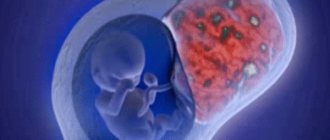
A benign neoplasm of the internal sex glands in women is called ovarian fibroma.
The tumor does not produce sex hormones and develops from the connective tissue membrane of the ovary.
The disease is characterized by slow progression (over several months and years). Sizes range from microscopic to the size of a child's head.
Fibroids account for up to 10% of all benign ovarian tumors. The clinical feature of the disease is its more frequent development during pre- and postmenopausal periods (after 40 years). The average age of patients is 47-55 years. Fibrosis does not occur in girls or young women before puberty.
Fibroma occurs in both the left and right ovary.
Types of ovarian fibroids
There are two main types of ovarian fibromas:
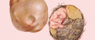
- Limited - the tumor has a connective tissue membrane that separates it from healthy ovarian tissue.
- Diffuse. In this case, the neoplasm completely affects the gonad. Unfortunately, this type of fibroma is much more common - in about 90% of cases.
Fibroids also differ in their consistency; they can be:
- Hard, feels like stone. The fibroma “turns to stone” if it is completely calcified.
- Soft. Softening occurs due to tissue swelling.
- Elastic, tight. Most common. Their cavity usually contains multiple cysts. Cystic cavities can be filled with liquid contents - serous or hemorrhagic.
Why does fibrosis occur?
No single cause for the development of the disease has been established. Local changes in the structure of the ovaries associated with gynecological diseases predispose to the formation of fibromas. Pathology of the genital organs in women of any age is important, the long-term consequence of which can be fibroma.
In 90% of patients, changes in the ovarian lining begin in the premenopausal period. Risk factors for developing the disease:
- Inflammatory diseases of the ovaries (oophoritis);
- Menstrual irregularities;
- Uterine bleeding;
- Salpingo-oophoritis;
- Polycystic ovary syndrome;
- Pelvic injuries;
- Pathology of the ovarian artery;
- Primary and secondary infertility;
- Habitual miscarriage;
- Ovarian operations;
- Suffered inguinal hernia.
REFERENCE. Fibroids can develop secondary to uterine fibroids, endometriosis, and ovarian cysts. The connection of the tumor with other gynecological disorders is explained by an imbalance of sex hormones in these diseases.
Symptoms and clinical picture
Symptoms of ovarian fibroma depend on the size and stage of the pathological process. They are very characteristic. If the tumor does not reach 2-4 cm in size, symptoms are completely absent or are represented by isolated scanty manifestations. The reasons for the further development of a specific symptom complex are compression of surrounding tissues by fibroids.
A typical picture includes the following features:
- Pain syndrome. The intensity of discomfort varies from patient to patient. Usually we are talking about weak, aching pain in the lower abdomen. Ovarian fibroma rarely makes itself felt with strong discomfort, especially in the early stages of development.
- Accumulation of fluid in the abdominal cavity. Also known as ascites. An increase in abdominal volume is visually noted. At advanced stages, if the formation has reached a significant size, the patient can be confused with a pregnant woman.
- Appetite and digestive disorders. Caused by compression of the stomach.
- Constipation. The cause of constipation is compression of the structures of the large intestine by neoplasm.
- Problems with urination.
- Bloating not associated with flatulence.
- Weakness, lethargy and weakness.
- Heart rhythm disturbances (manifested by tachycardia).
- Respiratory system disorders (shortness of breath).
When the formation ruptures or torsions, symptoms of acute peritonitis with abdominal pain, nausea, vomiting, and a drop in blood pressure occur. The menstrual cycle and fertility remain normal. However, ovarian fibroma rarely acts as an isolated pathology. Much more often it “neighbors” with other neoplastic processes. In this case, the clinical picture is more diverse and includes the entire set of symptoms.
How is fibroma diagnosed?
Most fibroids are detected by chance, during a preventive gynecological or ultrasound examination.
A neoplasm can be suspected based on the above complaints.
The complex of diagnostic examinations includes a survey, examination, gynecological examination, ultrasound of the pelvic organs and biopsy.
Survey
The patient has a history of gynecological diseases (often from childhood), pathologies of pregnancy and childbirth, pelvic injuries, and acute infections.
Inspection
A general examination can reveal a unilateral tumor formation in the pelvic area and ascites. When listening to the lungs, dullness of the pulmonary sound on the affected side (hydrothorax) is determined.
Gynecological examination
When examined in the projection of the fibrous ovary (slightly above the middle of the inguinal ligament), a neoplasm with the following characteristics is revealed:
- Smooth or knotty surface;
- Round sizes;
- Rocky density;
- Mobility (the tumor “rolls” under the fingers);
- Moderate pain.
Symptoms
The clinical picture is influenced by the clinical form of the disease. Most often, feminizing formation with increased estrogen production is diagnosed during menopause. Virilizing defects that increase the amount of androgen are extremely rare.
In childhood, the first type of pathology leads to early puberty, enlargement of the genital organs, the appearance of pubic hair, in the armpits, and growth of the mammary glands. Moreover, blood is often discharged from the vagina.
As you grow older, your menstrual cycle becomes disrupted and your periods become longer and heavier. During the postmenopausal period, the patient rejuvenates, the vagina ceases to be dry, libido reappears, and vaginal discharge is noted, reminiscent of menstruation.
With virilizing thecoma, menstruation stops, and hair grows on the face and body, like a man’s. Patients also suffer from shrinkage of the mammary glands, infertility, and deepening of the voice.
Treatment methods
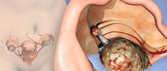
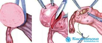
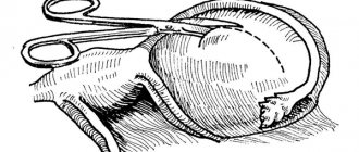
Surgical removal of the entire tumor (together with the capsule) is the method of choice in the treatment of this disease.
Types of operations performed:
- Laparoscopy is a low-traumatic intervention without an incision in the pelvic cavity. It is carried out when the tumor is small, when the structure of the ovary is preserved;
- Laparotomy removal is an open operation with an incision into the pelvic cavity. It is performed when the ovary is extensively involved in the process. The operation is often supplemented by excision of the tissue of the ovary itself, and, if indicated, the uterus.
Torsion of the tumor stalk with infection is an indication for emergency abdominal surgery. In such cases, not only the fibroid is removed, but also the ovary, adjacent ligament, fallopian tube and, as a rule, the uterus.
Treatment after surgery is aimed at accelerating recovery and preventing adhesions. Set of measures:
- Early activation of the patient (2-3 days after surgery);
- Electrophoresis with longidase (an enzyme preparation that prevents the development of adhesions);
- Taking vascular drugs (nicotinic acid, Actovegin, pentoxifylline);
- Enriched nutrition (calorie content - up to 2500 kcal).
The full course of rehabilitation lasts up to 1-3 months.
Prognosis and complications
As a rule, in uncomplicated cases a favorable outcome is possible. If the tumor is not large in size, there is a high probability of maintaining reproductive function.
The risk of recurrence of ovarian fibroids is minimal, but this does not eliminate the need for regular examination after surgery.
Complications of the disease arise, as a rule, only when fibroids are detected late and medical recommendations are not followed. Possible adverse effects:
- formation of adhesions with abdominal organs;
- torsion of the fibroma stalk with subsequent necrosis;
- the addition of purulent inflammation leading to melting of the tissue of the affected ovary;
- peritonitis (inflammation of the peritoneum);
- internal bleeding (when the leg is torn off, the integrity of the capsule is violated).
Ovarian fibroma is an insidious disease. A seemingly “harmless” tumor is fraught with dangerous, sometimes life-threatening consequences. A simple way to avoid them is to regularly visit a gynecologist, follow all his recommendations, and remove the tumor at the earliest possible stages.
Does ovarian fibroid prevent you from getting pregnant?
When planning pregnancy, patients with fibroids are recommended to undergo surgical treatment. Conception is planned after surgery and completion of a full course of rehabilitation.
If a tumor is discovered during pregnancy, the woman is monitored dynamically. Fibroma does not affect the development of the fetus, since it does not produce hormones. The course of pregnancy may be disrupted:
- If the tumor is large and begins to compress the fertilized egg;
- With the development of ascites.
In this case, the tumor is removed without waiting for the moment of birth. In all other cases, delivery is carried out naturally.
Diagnostics
First of all, you should visit a doctor and undergo a gynecological examination. Bimanual examination can reveal a unilateral round tumor with a smooth surface. It is imperative to check the state of hormonal levels, since during illness the amount of testosterone and estradiol increases.
It is also important to donate blood for tumor markers, undergo ultrasound examination, and Doppler testing. Magnetic resonance imaging is prescribed as an auxiliary method, which makes it possible to clarify the diagnosis and determine malignant degeneration. After resection of the tumor, a biopsy is necessarily performed, thanks to which the type, nature and calcification of the defect is revealed.
When diagnosing, it is important to distinguish the disease from stromal thecomatosis. A rare benign growth of tissue structures of the appendage appears in older women, accompanied by acne, excessive hair growth on the face and body, baldness typical of men, and obesity.
Symptoms of Frenkel syndrome resemble the virilizing type of formation.
Causes
Xanthomatous theca cell fibroma can be bilateral or unilateral. The shape of the tumor resembles the ovary itself. On average, such neoplasms are 7–8 cm in diameter. They are yellow or orange in color and sometimes contain liquid cytoplasm. Approximately 90% of tumors are unilateral, which means they form on only one side of one ovary.
The causes of the disease have not yet been clarified. But doctors put forward several factors that can trigger the development of a pathological condition. A benign tumor can develop due to genetic predisposition and hormonal imbalance.
There is also a theory that ovarian fibrothecoma can develop as a result of an unhealthy lifestyle. For example, if a woman smokes or eats poorly.
The development of benign formations in the appendages is also influenced by radiation and systematic exposure to stress factors. But most often, thecofibroma is formed against the background of hormonal disorders that develop due to other tumors in the pelvis and injuries.
Prevention
Due to the lack of specific causes, there are no special methods for preventing tumor development. The most important thing here is to detect the tumor in time and prevent its possible negative consequences. Therefore, it is very important not to forget for all women to undergo a preventive examination with a gynecologist at least once a year. This will help to identify risk factors in a timely manner. If necessary, the doctor may prescribe additional methods to clarify the diagnosis.
You also need to monitor your general condition, treat all other gynecological diseases in a timely manner, and be attentive to the choice of sexual partners and contraception. This will prevent unwanted pregnancy and sexually transmitted diseases.
General recommendations include:
- Fight bad habits.
- Maintaining proper body weight.
- Moderate physical activity.
- Strengthening the immune system.