What's happened
Teratoma is a lipid-cellular formation arising from the primary cellular structures of the gonads. Germ cell tumors have a heterogeneous structure. As a result, hair, teeth or eyeballs sometimes grow from this defect, making the disease look terrifying.
Embryoma is a benign neoplasm, but there is a risk of malignant degeneration. Because of this possibility, you should not hesitate to visit a specialist if severe symptoms appear.
Kinds
A distinction is made between teratoma of the left and right ovary. In rare cases, the pathogenic process affects both sides.
Most often, pathology is diagnosed on the right, since the right ovary has an active blood supply. On the left, ovulation occurs rarely, the load is not so great, therefore tumor-like or cystic formations develop less frequently than on the right side.
Depending on the histological structure, mature and immature ovarian teratoma are distinguished. The first type is small single, solid and cystic. Solid formations reach large sizes, are heterogeneous, have a lumpy surface, and high density.
Cystic defects look like accumulated multiple bubbles filled with liquid contents, cellular structures of sweat and sebaceous glands. Such formations are extremely dangerous, as they often grow rapidly, causing twisting of the base of the teratoma and tissue necrosis.
An immature tumor is an extremely rare phenomenon, prone to transformation into cancerous teratoblastoma. The risk group includes young girls.
The defect is characterized by a smooth surface, absence of cartilage and epithelial cells. Often, immature formation accompanies gliomatosis and provokes metastasis.
What is a dermoid cyst and ovarian teratoma?
A non-malignant formation that develops in utero and is formed from fragments of embryonic tissues.
In gynecological oncology, it is customary to divide cystic formations into cystadenomas and dermoid tumors. When revealing the question of what an ovarian teratoma is, the structure, size, growth characteristics and localization of the formation are considered. Another name for the disease is ovarian dermoid cyst or dermoid, as well as mature teratoma.
Cystic teratoma of the ovary is classified as a benign tumor that can actively grow and increase in size. It is considered a remnant of underdeveloped germ layers. Increased activity has been recorded during hormonal changes, especially during puberty, pregnancy and the menopausal cycle.
Germ cell cystic teratoma in women is benign and there is no malignancy. Usually the tumor is medium in size and does not exceed 40–50 mm in diameter.
In cross-section, the tumor is single-chambered; in rare cases, multi-chambered is observed.
It is worth noting that the dermoid cyst in most cases is localized in the right ovary, which is due to the structural features of the internal reproductive system and the blood supply to the appendages. It is visualized not only in adult patients, but also in prepubertal girls.
What does a dermoid look like?
Visually, the dermoid cyst of the left ovary is round or oval, the surface is smooth, slightly bumpy with a gray tint. The tumor is densely elastic, contains two or more chambers, and has additional capsules inside. The teratoma is attached to the left or right ovary using a stalk that directly feeds and innervates the formation.
Benign teratoma tends to grow due to relentless cell proliferation. Contains a translucent liquid, in places a dense jelly-like substance. A complex tumor includes several different types of germ layers, so underdeveloped structures of the body are found: cartilage and bone formations, hair, teeth, sebum, limb buds, etc.
The dimensions of the dermoid vary from a few millimeters to 10–40 cm; the larger the lesion, the greater the likelihood of the process becoming malignant.
Causes
The true causes of ovarian teratoma have not yet been identified. However, the following provoking factors should be highlighted:
- Various chromosomal abnormalities.
- The presence of abnormal processes during the development of identical twins, when one embryo is absorbed by the other.
- Hormonal disorders.
- Taking contraceptives .
- Surgical interventions in the female genital organs.
The appearance of the disease can also be influenced by the menstrual cycle. Pathology sometimes develops at the beginning or end of menstruation.
Causes of ovarian teratoma formation
The causes of teratoma have not been fully studied, but scientists have put forward several hypotheses. At the moment, none of these hypotheses have been clinically proven and have not become basic.
A probable theory is abnormal embryogenesis, when a malfunction occurs in the chromosomes. As a result of the failure, the epithelium becomes the raw material for germ cell tumors.
According to another, more exotic hypothesis, a teratoma is an embryo within an embryo. The reason for putting forward such a theory was medical practice, when doctors discovered embryonic body parts in a tumor.
This rare type of teratoma is called a parasitic tumor, which forms due to abnormal coordination of stem cells and surrounding tissues. According to the hypothesis, at some stage of embryogenesis there is a pathological “niche” when a failure in the induction of two embryos occurs.
One of them (the weaker one) is absorbed by the tissues of the stronger and more active embryo. The probable cause of teratoma is chromosomal abnormalities in the early stages of pregnancy (4-5 weeks).
Symptoms
Mature formations often occur even in newborn girls. Most often, the pathological process affects the right ovary. Minor formations are asymptomatic, as a result of which in most cases they are diagnosed accidentally during an ultrasound examination for another reason.
If the tumor begins to suppurate or the leg becomes twisted, the patient experiences acute pain in the lower abdomen. Large defects with a diameter of seven centimeters significantly worsen the quality of life.
Large tumors in the patient cause displacement of the pelvic organs, a feeling of heaviness, pain in the affected side, urination problems, constipation, abdominal enlargement, and severe anemia. Immature atheroma leads to drowsiness, weakness, and fatigue. The appearance of fever indicates the onset of suppuration or necrosis.
Signs of teratoma
In the initial stage, the tumor practically does not detect itself at all, similar to other oncological diseases. Late diagnosis is the main danger of the disease. If signs appear in the early stages, this indicates a large size of the tumor (7-10 cm), as a result of which the tumor presses and displaces neighboring organs, or it is a signal of a malignant nature and metastasis.
Teratogenic tumors do not depend on the hormonal system and do not affect it in any way, although according to statistics, in most cases their growth occurs during puberty, pregnancy or menopause.
Teratoma is characterized by the following symptoms:
- urinary disturbance;
- feeling of heaviness in the lower abdomen;
- problems with bowel movements (usually constipation, rarely diarrhea);
- abdominal enlargement in thin women;
- anemia with large mature teratoma.
Vivid symptoms are produced by a dermoid cyst, which is prone to suppuration, inflammation, and complications. When inflamed, the dermoid is accompanied by chills, weakness, and abdominal pain. The pain can radiate to the rectum or leg. The general symptoms of teratoma are similar to those of other benign tumors.
Diagnostics
If you have any alarming symptoms, you should immediately visit a gynecologist. The specialist will examine the complaints and conduct a visual examination of the cervix using a special mirror.
After this, an ultrasound examination of the pelvic area is required. During pregnancy, this diagnostic method can also detect disturbances in the intrauterine development of the embryo.
To clarify the diagnosis, fluoroscopy, Dopplerography is performed to examine the blood supply to the defect, irrigoscopy, sigmoidoscopy, computed tomography or magnetic resonance imaging.
To determine the type of tumor formation, a puncture of the abdominal cavity with a biopsy is performed for subsequent histological examination of the samples taken. It is also important to take a blood test for tumor markers.
Can it develop into cancer?
Despite its benign nature, teratoma can transform into a malignant tumor. Malignancy occurs extremely rarely, but it is better not to advance the disease to such a stage. Cystic mature and immature tumors are most prone to degeneration.
As a result, during diagnosis it is necessary to perform a biopsy and send the collected biological material for histological examination. If the transformation process does occur, then microscopic examination of the biopsy will reveal melanoma and adenocarcinoma cells.
In addition, affected ovaries sometimes become filled with cellular structures characteristic of thyroid cancer.
Treatment
Conservative therapy is not effective, so they resort to surgical intervention. If surgery is performed in a timely manner, the risk of degeneration into a malignant neoplasm is significantly reduced.
On this topic
- Female reproductive system
Differences between cytology and colposcopy
- Natalya Gennadievna Butsyk
- December 6, 2020
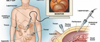
During laparoscopic enucleation, only pathogenic cells are eliminated. To preserve reproductive function, partial resection of the affected organ is performed along with formation. In rare cases, complete removal of the ovary and uterus is performed.
Before the surgical procedure, the patient must undergo a complete examination. To do this, it is necessary to undergo general and biochemical blood tests, as well as for the presence of infectious and viral pathologies. In addition, it is important to do a coagulogram, electrocardiogram, and take a vaginal smear to check for STIs or inflammatory processes.
Large tumors are removed using a classic incision in the abdomen. During the manipulation, the surgeon is obliged to check the condition of the pelvic organs and exclude other neoplasms or adhesions. After resection, the abdominal cavity is sanitized.
Most often, laparoscopic surgery is performed, which leaves minor scars. In case of extensive damage, in addition to the teratoma itself, the ovaries, uterus, and fallopian tubes are excised through punctures.
This method often allows one to preserve reproductive function, minimally damages the skin, and extremely rarely causes complications, blood loss, or adhesions in the intestines.
If a histological examination has diagnosed a malignant pathology, then chemotherapy, radiation treatment are used, and special antitumor drugs are prescribed. Hormonal therapy is also used if the defective receptors are sensitive to hormones.
On this topic
- Female reproductive system
Echosigns of adenomyosis
- Olga Vladimirovna Khazova
- December 4, 2020
A few days after surgical removal, the patient is allowed to stand up and walk. After discharge and removal of the stitches, it is important to take care of your body for another week, get plenty of rest, and take a short walk in the fresh air. It is necessary to avoid sexual intercourse for several months to prevent ruptures and internal bleeding.
Pregnant women should be especially attentive to this pathology, since self-medication can lead to miscarriage and even death. Therefore, the patient should strictly follow the doctor’s instructions, undergo a gynecological examination and ultrasound examination every month, do not make sudden movements, do not turn or bend over. In the absence of complications, laparoscopic surgery is performed after the 17th week of pregnancy.
Treatment methods
To eliminate immature ovarian teratoma, surgery is most often required. Its implementation has the following advantages:
- complete elimination of pathology;
- prevention of tumor malignancy;
- eliminating the risk of metastases in nearby organs;
- reducing the likelihood of relapse of immature ovarian teratoma.
During the intervention, partial resection of the ovary or its complete removal may be performed. The latter type of operation is recommended for mature women who do not plan to have children. This prevents the occurrence of other neoplasms on the cured appendage.
Complications
If ovarian teratoma occurs and is not treated in a timely manner, the disease will lead to dangerous consequences. Necrosis of tissue structures often develops, the stalk of the neoplasm becomes twisted, and extensive internal bleeding occurs, leading to anemia.
If a cystic defect ruptures due to mechanical damage, the liquid contents leak into the abdominal cavity. This situation threatens with fatal peritonitis.
Large formations compress neighboring pelvic organs, disrupting their functionality. In advanced cases, a benign teratoma transforms into a malignant disease and metastases develop.
Dermoid during pregnancy
In the case of a teratoma of the right or left ovary during pregnancy, gynecologists decide on further tactics for managing the woman. Dynamic observation is preferable for volumes less than 50 mm.
If education is stable, then during pregnancy there are no problems with the health of the baby and mother.
Sizes over 50 mm are subject to careful monitoring and planned removal of the ovarian dermoid cyst. A low-traumatic and minimally invasive laparoscopy technique is preferred. Elimination of the pathological focus is simply necessary due to the fact that the pregnant uterus will continue to grow and create pressure on nearby organs, including a cystic tumor. In this case, there is a high risk of torsion of the teratoma leg or rupture of its capsule.