Uterine fibroids are among the most frequently diagnosed gynecological pathologies in women of reproductive age. The disease is not dangerous from an oncological point of view, however, in some cases it can seriously impair the quality of life.
The disease is otherwise defined as leiomyoma, fibromyoma or fibroma. Uterine fibroids are characterized by the formation and growth of a tumor-like nodule in the myometrial tissue.
Uterine fibroid nodes have different sizes and are defined as large, medium or small. They can be located both in the body of the uterus and in its cervix. In addition, myomatous nodes are attached to muscle tissue through a wide base or stalk.
Gynecologists classify uterine fibroids as:
- retroperitoneal, growing from the neck;
- intramural or interstitial, progressing inside the myometrium;
- subserous, appearing under the serous membrane;
- intraligamentous, located between the so-called leaves of the uterine ligament;
- submucosal or submucosal, developing under the mucous membrane.
Like other types of pathology, submucous fibroids are considered a benign tumor, which is detected in approximately 30% of cases. Submucosal neoplasms grow towards the uterine cavity.
Due to their location, such tumors progress quickly, and even small nodes are accompanied by pathological signs such as infertility, pain and bleeding.
Submucous fibroids often present in multiple forms. Experts distinguish several types of submucosal or submucosal fibroids.
- Such a pedunculated tumor is located directly under the mucous layer. There are no signs of growth into the myometrial fibers.
- There is insignificant growth of part of the node into the myometrium.
- Ingrowth of a significant part of the tumor into muscle tissue is diagnosed.
- Experts note the absence of muscle tissue between the fibroma and the mucous layer.
Interstitial-submucous uterine fibroids are quite common. This type is otherwise called intramural-submucous uterine fibroid. With the interstitial or intramural-submucous variety, the fibroid is located in the uterine cavity, and the node is located in the thickness of the myometrium. Submucosal leiomyoma in such cases also has signs of an interstitial or intramural tumor. Some time ago, submucous fibroids required surgery, which involved removing the nodes simultaneously with the uterus. Modern gynecology uses a sufficient number of effective techniques that make it possible to preserve the organ and restore lost reproductive function.
Causes
The pathology is hormone-dependent. Doctors believe that the growth of the tumor is based on a disruption in the production and balance of certain sex hormones, in particular estrogen, as well as an increase in the sensitivity of estrogen receptors in the uterus.
However, the interstitial or intramural-submucous form is poorly studied. Possible reasons for its appearance include some congenital structural features of the myometrium, as well as the effect of frequent operations on muscle tissue.
There are also provoking factors, the combination of which can trigger the development of the disease. Among the factors that provoke submucous fibroids of the interstitial or intramural form are:
- endocrine disorders;
- metabolic disorders;
- early menstruation;
- hypertension;
- lack of activity and obesity;
- venous congestion due to irregular intimate life;
- first birth after 30 years;
- prolonged insolation;
- stress;
- uncontrolled use of COCs;
- influence of heredity;
- untreated inflammatory processes in the pelvis.
Before starting treatment for intramural or interstitial submucous fibroids, unfavorable factors should be eliminated if possible.
https://youtu.be/m0Vxqi0yWgk
Symptoms
In general, myomatous neoplasms are characterized by the absence of symptoms until the fibroma reaches a large size. However, a feature of the submucosal type of tumor, located intramurally or interstitially, is the early appearance of the clinical picture. The severity of symptoms of submucous fibroids also depends on the size of the formations and on the individual characteristics of the woman.
Symptoms of submucous fibroids localized intramurally or interstitially include:
- a noticeable increase in the amount of bleeding during menstruation;
- bleeding that is acyclic in nature and not associated with menstruation;
- anemia as a result of constant blood loss;
- pain in the pelvis like contractions, aggravated by physical activity, sexual intercourse, menstruation;
- feeling of pressure in the lower abdomen;
- frequent urination and constipation, which develop due to compression of neighboring organs;
- urinary tract infections;
- increase in abdominal volume;
- impossibility of conception;
- miscarriages or premature births;
- complications during childbirth.
This phenomenon is accompanied by severe pain and signs of intoxication. If emergency surgery is not performed, necrosis develops. If symptoms of an “acute abdomen” appear, you should immediately seek medical help.
Symptoms of submucous fibroids
The symptom depends on the time of formation, size and growth rate of the node. The first stage is characterized by the absence of symptoms. The tumor is found involuntarily during an examination by a gynecologist and an ultrasound scan. Menorrhagia appears first. This is heavy menstrual flow with blood clots. In time they exceed the duration of standard menstruation. Blood discharge may also appear during the period between menstruation. Regular blood loss in large quantities provokes the development of anemia and general malaise. The skin becomes pale, pain and dizziness occur.
With a submucosal position of the node, according to doctors in the field of gynecology, pain is observed in 40% of patients. They appear during menstruation and resemble the process of contractions. They can be located in the lower abdomen and extend to the lumbar region. A submucosal tumor differs from subserous fibroids in that it does not affect nearby organs. Reproductive function fails in 30% of cases. Girls cannot get pregnant or have a miscarriage.
Types of uterine leiomyomas
At the initial stage, there are no specific signs of fibroids. Only during a gynecological examination is it possible to detect nodes in the uterine cavity. Symptoms depend on the location and size of the growths. Submucous myoma (ICD-10 code D25.0) is interpreted:
- hyperpolymenorrhea (long and strong bleeding);
- disruption of the menstrual cycle;
- bleeding from the uterine region;
- infertility and miscarriages.
Interstitial subserous fibroids exhibit symptoms:
- Menorrhagia is prolonged bleeding during menstruation.
- Metrorrhagia – bleeding between menstrual periods and smears.
- Regular pain like contractions during and before menstruation.
- Increased body temperature.
Due to severe blood loss, women develop anemia with iron deficiency. Anemia causes constant fatigue, headaches and dizziness. The mucous membrane takes on a pale tint. Hair loss and fragility of the nail plates have been noticed. Pulse readings increase and blood pressure decreases. A blood test reveals a decrease in red blood cell volume and hemoglobin.
Signs of the intraligamentary form of the tumor are menorrhagia and metrorrhagia, lumbar pain and infertility. The subserous form is expressed by disruption of the menstrual process, causeless miscarriages and the inability to conceive. The tumor puts pressure on the bladder and rectum. As a result, their direct functions are disrupted. Even in a calm position, women feel severe pain in the lower perimeter of the abdomen.
Diagnostics and methods
Detection of fibroids is often a diagnostic finding due to its latent course. With the submucosal form, the symptoms are usually quite pronounced, which forces the woman to visit a gynecologist and undergo an examination.
Large submucosal tumors can be identified during a gynecological examination. The doctor may notice a characteristic increase in abdominal circumference, and by palpation palpate the submucosal nodes and determine the uneven, deformed contour of the uterus.
To prescribe treatment, it is necessary to obtain additional information, which is possible using the following diagnostic methods:
- Ultrasound transvaginal or transabdominal;
- MRI;
- CT;
- hysteroscopy;
- laparoscopy;
- Dopplerography.
Submucous (submucosal) uterine fibroids
Such nodes are located under the mucous membrane and grow into the uterine cavity. Submucosal fibroids are the most unpleasant in terms of symptoms, since they lead to heavy periods, during which a woman loses a lot of blood, uterine bleeding, infertility, miscarriages, and premature birth.
A small nodule—only 2–3 cm in diameter—can lead to severe symptoms. Sometimes the bleeding is so severe that the woman has to be hospitalized and receive a blood transfusion.
Chronic bleeding from submucosal fibroids leads to anemia. Characteristic manifestations of this condition: pallor, constant feeling of fatigue and weakness, headaches and dizziness, shortness of breath, tinnitus, fainting, rapid heartbeat.
Subserous fibroid
Unlike submucosal nodes, subserous nodes are usually “quiet”. They can grow large without causing symptoms. When people talk about giant fibroids, the size of a melon or a watermelon, they are usually talking about subserous nodes. They grow outward into the abdominal cavity - there is much more space there than in the uterine cavity.
When a subserous myomatous node, which can be located on the anterior or posterior wall of the uterus, reaches a large size, it begins to compress neighboring organs: the bladder, rectum. A woman is worried about frequent urination and constipation.
Complications
A benign tumor can cause complications such as painful menstruation, accompanied by significant blood loss; menstruation turns into uterine bleeding, which, if intense, is life-threatening.
- Another complication is compression of neighboring organs located in the abdominal cavity. The consequence is constipation, problems with urination, cystitis, pyelonephritis.
- It is extremely rare for a tumor to transform into a malignant one.
- For a woman planning to become a mother, fibroids can become an obstacle to the happiness of motherhood.
The disease should be treated - you can’t let everything take its course.
Intramural fibroids
Intramural, or interstitial, uterine fibroids are located in the thickness of the organ wall. By and large, all nodes initially grow as intramural, closer to the cavity or outer surface of the uterus, but then they begin to predominantly protrude into the uterine cavity or towards the abdominal cavity, depending on the nature of growth.
Until interstitial fibroids reach a critical size, they usually do not cause symptoms. In the future, it can begin to protrude into the uterine cavity and lead to uterine bleeding, like submucosal nodes.
Classification of the International Federation of Obstetrics and Gynecology
The division of myomatous nodes into three types - submucosal, intramural and subserous - is a somewhat simplified classification. In fact, fibroids can occupy different positions and protrude to varying degrees into the uterine cavity or abdominal cavity. In order to cover all options, the International Federation of Obstetrics and Gynecology has developed a special classification. In accordance with it, ten types of fibroids are distinguished:
Submucosal nodes:
- Type 0 is a pedunculated submucosal fibroid located in the uterine cavity.
- Type 1 - the node protrudes into the uterine cavity by more than half.
- Type 2 - the node protrudes into the uterine cavity by less than half.
Other types:
- Type 3 is an intramural fibroid that does not protrude into the uterine cavity, but is adjacent to its mucous membrane.
- Type 4 is an intramural fibroid, which is located deep in the muscles.
- Type 5 is an intramural subserosal node that protrudes less than half of the way out on the surface of the uterus.
- Type 6 is an intramural subserosal node that protrudes more than half of the surface of the uterus.
- Type 7 is a pedunculated subserosal node that is entirely above the surface of the uterus.
- Type 8 - fibroids that have a specific localization (for example, in the cervix).
- Hybrid nodes that affect both the mucous membrane of the uterus and its outer surface. They are designated by two numbers.
Classification depending on the number of fibroids
Depending on the number of nodes, there are three types of fibroids:
- Single - 1-2 nodes, which can have different sizes.
- Multiple. More than 3 nodes are detected. Treatment usually helps reduce symptoms, but it is often not possible to remove all fibroids, and some may grow large over time.
- Mixed. There are many fibroids, but at least one of them is large and dominant. For example, multiple fibroids with a large subserous node may be diagnosed. It can be removed, but another node may grow strongly in the future. Multiple and mixed fibroids pose a particularly big problem in women of reproductive age before menopause, since sex hormones promote the growth of nodes.
Treatment and prevention
Since submucous uterine fibroids develop quite actively and rapidly, treatment with a conservative method does not completely overcome this disease.
It is advisable to take medications when the size of the uterus with a submucosal node does not exceed 12 weeks, and the tumor is 2 cm in diameter. The action of the drugs is aimed at alleviating the symptoms of the disease and slowing its progression. If the formation is characterized by dynamic growth, then we are talking about the size of the fibroid for surgery, and usually reaches 12 cm in diameter and above in weeks.
In this case, hormonal or non-hormonal therapy may be prescribed. One way or another, most drugs are aimed at regulating hormones that promote the growth of estrogen and progesterone.
Considering that the submucous form is quite active, when diagnosing it is important to understand how quickly it develops. If relatively slow, women who are about to reach menopause can do without surgery. If the node regresses, there is a chance that it will not bother you until menopause. And during menopause, the formation usually dries out.
In addition, drug therapy may be prescribed if the nodes are too large, since in this case surgery is dangerous.
Previously, submucosal leiomyoma (fibroids) was completely treated only by removal along with the uterus. Modern medicine has developed methods that allow you to get rid of the disease, preserve all important female organs, and conceive and give birth to a child. Depending on the diagnosis, a radical method may be recommended - without organ preservation.
Which fibroids need to be operated on?
Women are concerned about the question of whether it is necessary to have surgery, whether it will be possible to save the uterus, what is the optimal size of submucous uterine fibroids for surgery.
Size isn't really as important as some other factors. Main indications for treatment:
- Myoma causes problems: bleeding, miscarriages, infertility, urination problems and constipation, and abdominal enlargement.
- A woman wants to get pregnant, but the node, due to its size and location, can interfere with pregnancy and create certain problems for the expectant mother and fetus.
- The node is growing according to 2-3 ultrasounds performed with an interval of 4-6 months.
Previously (some gynecologists still adhere to this tactic) all such women were candidates for surgery. If it worked, the surgeon removed only the fibroid; otherwise, he removed the entire uterus.
Currently, there is a less invasive method of treating uterine fibroids - uterine artery embolization (UAE).
During the procedure, the doctor inserts a special catheter through a small puncture in the skin into the vessel feeding the node, through which an embolic drug is delivered. The latter blocks the lumen of the vessels, the fibroid stops receiving oxygen, dies and turns into connective tissue.
Embolization of the uterine arteries is indicated for most women with myomatous nodes; it allows you to save the uterus, conceive a child and carry out pregnancy in the future. Contact us to learn more about this treatment method.
Last update: 09/18/2019
Most modern women are faced with benign formations in the muscular layer of the uterus, what is it? These neoplasms are fibroids (or otherwise fibroids, leiomyomas) of the uterus and consist of intertwined muscle fibers. The tumor can invade all layers of the uterus and form nodes of various sizes on the surface. In most cases, fibroids develop in the body of the uterus, much less in the cervix.
Treatment with folk remedies
Traditional methods of treating fibroids when they are small in size can be very effective. The herbs used in the recipes have an anti-inflammatory effect and help normalize hormonal levels:
- Nettle infusion works well. The plant (a tablespoon of crushed leaves) is poured with 2 cups of boiling water and cooked in a water bath for 10 minutes. Then filter and drink throughout the day.
- You can pour a glass of boiling water over several large calendula flowers. Drink the infusion on an empty stomach in the morning.
- The infusion of boron uterus, which is sold in pharmacies, has proven itself well. Take 30 drops before meals three times a day.
Any treatment lasts at least a month. Before starting it, you should consult a doctor. And we must remember that traditional methods are auxiliary. The main treatment is prescribed by a gynecologist .
Let's get acquainted with general information about the intramural node
So, what are intramural uterine fibroids? This is a benign tumor that forms in the muscular part of the uterus. Because this is the largest area of the uterus, it is most vulnerable to tumor development.
Intramural myomatous node is the most common diagnosis among these pathologies and has its own characteristics of the development of the disease. And treatment tactics are selected individually, based on a preliminary examination.
There are several types of nodes in the body of the uterus:
Submucosal form is a nodular form of fibroids, which is located in the mucous layer of the uterus. The growth occurs predominantly in the abdominal side. Submucosal nodes cause severe pain with intense frequency. The uterus quickly increases in size.
Interstitial form - the formation of nodes is located in the interligamentous apparatus. As a rule, there is a multiple accumulation of nodes in the muscles, which causes heavy menstruation.
Intramural uterine fibroids - formations are found in the muscle layer.
Intramural-subserous fibroids - form on the outside of the uterus, affecting the pelvic area. They are characterized by mild symptoms, which is the reason for late detection. In this case, a diagnosis of “subserosal fibroids of the uterus” is made.
Depending on the degree of disease, single and multiple appearances of tumor nodes can differ.
Kinds
There are many types of classifications. Nodes are divided into groups according to size, number, location, and of course, tissue composition. The most important indicators are the size and histology of the node (its tissue composition). Based on this factor, treatment is prescribed. The approach to therapy itself, as well as the drugs or type of intervention, depend on it. Based on histology, there are two most common varieties.
Intramural-subserous
Actually, intramural uterine fibroids are located in the muscle layer - the myometrium. However, there is another variety of it. Namely, intramural subserous uterine fibroids. This is located partly in the serous layer, partly in the myometrium. The serous or subserous layer is the outer lining of the uterus. The one that contacts directly with other internal organs.
With this arrangement, tumor growth is usually observed in the direction from the uterus to the peritoneum. It is precisely such formations, when progressing to large sizes, that most often compress neighboring internal organs. Sometimes the intestines, sometimes the bladder.
Intramural-submucosal
As mentioned above, the intramural node is completely located in the muscle layer. But it can also be located in two layers, as in the previous case. When intramural submucous uterine fibroids are diagnosed, it means that a neoplasm has formed in two layers - intramural and submucous.
What is this, submucosal layer? This is the inner lining of the uterus, its submucosal layer. If the node is partially located in it, it may appear more pronounced. It contains blood vessels, which is why it sometimes bleeds. It can also cause more severe pain. This form is a little more difficult to treat.
Reasons for appearance
The following reasons can lead to the development of this pathology:
- Hormonal imbalance.
- Any nervous system disorder.
- Factor traumatic to the uterus: abortion, curettage, surgery.
- Hormone-dependent diseases – endometriosis and adenomatosis.
- A chronic inflammatory or infectious process in the uterus.
- The presence of diseases such as diabetes, excess body weight.
- Having irregular sex life.
- Endocrine diseases.
- Wrong lifestyle and nutrition, bad habits.
- Menopause period.
- Hereditary predisposition.
Causes of the disease
The main reason for the formation of nodes is hormonal imbalance . Doctors note a change in the functioning of the hypothalamic-pituitary system: an imbalance in the production of hormones, especially FSH and LH. Factors predisposing to the development of the disease are:
- obesity;
- diabetes;
- polycystic ovary syndrome (when many follicles grow, but they stop growing, do not release an egg and eventually turn into small cysts);
- incorrect use of COCs;
- sedentary lifestyle;
- chronic inflammation of the uterine appendages, the uterus itself;
- absence of pregnancies.
In addition, stress and chronic lack of sleep are of great importance. Poor heredity increases the likelihood of fibroids . In other words, if the mother had a hormone-dependent tumor, then the daughter will most likely have to deal with it too.
But the exact causes of fibroids are unknown. Why do some women, despite the presence of several provoking factors, never develop nodes, while others, even without suffering from concomitant diseases and having more than one child, know firsthand what fibroids are? There is a lot here that has not been fully studied. But fibroids are easily diagnosed and successfully treated.
Symptoms
The most unpleasant and dangerous thing is that intramural fibroids can be asymptomatic, not making themselves felt until the fibroid nodes increase in size. In such cases, there will be pressure on neighboring organs and disruption of their functions, resulting in deformation of the uterus. In this case, the symptoms of the disease begin to clearly appear.
The most common symptoms of intramural uterine fibroids are:
- Changes in the menstrual period of various types: an increase in the duration or amount of discharge, the presence of blood clots.
- The presence of painful discomfort during menstruation: there is severe pain and a feeling of heaviness in the area of the uterus.
- Heavy bleeding, which can subsequently cause increased fatigue, weakness, headache and dizziness, and pale skin. This usually indicates anemia.
- Constant presence of pain in the lower abdomen.
- There may be problems with the functioning of the bladder.
- Presence of bleeding during the intermenstrual period.
The manifestation of signs of the disease depends on:
- Node size;
- Its location;
- The degree of its growth.
5 myths about uterine fibroids video
https://youtu.be/OMlEMMQDrLI
Types of surgery to remove a tumor
The choice of method for removing fibroids is determined by your doctor. Which operation is preferable: abdominal or laparoscopy depends on a number of factors:
- dimensions in millimeters;
- location of nodes;
- how much does the procedure cost?
- age and wishes of the patient.
Price
The clinic will tell you exactly how much the surgical intervention will cost and will calculate it for free, but it ranges from twenty to one hundred thousand rubles and depends on a number of factors:
- degree of neglect of the disease;
- node sizes;
- place of residence of the patient;
- individual characteristics of the human body;
- type of leiomyoma removal;
- location of education.
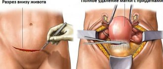
Hysterectomy
The operation is characterized by eliminating the formation through an incision in the abdominal cavity. In this case, the organ is amputated completely or with preservation of the neck. Typically, hysterectomy is performed on patients after forty years of age.
The following types of surgical interventions to remove the uterus are distinguished:
- subtotal amputation - in this case, the neck and appendages are preserved;
- extirpation (total removal of the uterus along with the cervix) - is carried out with a high risk of malignant formations, as well as with hypertrophy;
- hysterosalpingo-oophorectomy - in which the uterus is removed along with the appendages. After such an operation, early menopause is possible;
- radical amputation - in this case, the reproductive organ is removed along with the cervix, ovaries and fallopian tubes.
The reproductive organ is removed for the following reasons:
- Heavy and prolonged bleeding, leading to low hemoglobin levels in the blood.
- Prolapse of the uterine organ.
- Degeneration into a cancerous tumor.
- Tissue necrosis.
In other cases, organ-saving techniques are used.
Surgical intervention to amputate the uterus is fraught with a number of negative consequences:
- Termination of childbearing function.
- Cystitis.
- Severe vaginal bleeding is possible after amputation.
- Lack of menstruation.
- Decreased bone density, early aging of the body.
- Malignant formations in the mammary glands.
Myomectomy
This type of surgery belongs to the organ-preserving method and is carried out if the size of the cyst has reached six centimeters. Myoectomy is performed on patients under forty years of age. There are three types of myomectomy:
- laparotomy - an incision is made in the lower abdomen;
- laparoscopic myomectomy (endoscopic technique) - three small holes are made in the peritoneum area. To conveniently operate surgical devices and avoid injury, carbon dioxide is injected into the reproductive organ before the operation. The optimal parameters for laparoscopy are 8–9 weeks (from 30 to 40 mm). Using this method, intramural and interstitial subserous tumors are removed;
- hysteroscopy - removal of the cyst through the vagina using a resectoscope. This method is prohibited if the patient has inflammation and cervical abnormalities.
Larapotomy is an operation to remove large fibroids if the fibroids have reached more than 60 mm and have a negative impact on nearby organs. An incision is made in the abdominal area through which the formation is removed. The surgeon's work time to remove the nodes is about one and a half hours.
Indications for abdominal surgery:
- Severe anemia due to heavy bleeding.
- Pressure on nearby organs.
- Cervical twisting.
- Degeneration into a cancerous tumor.
- Inability to get pregnant.
Which operation is better: abdominal or laparoscopy?
Each type of surgery has its own advantages. The leading role in choosing a method is played by the patient’s reluctance, but by the degree of neglect of the disease.
The positive aspects of abdominal surgery to remove fibroids are:
- convenience for the doctor during the operation;
- visual examination of organs;
- if any pathologies are detected, the duration of the operation may be changed;
- removal of the tumor and the area around it, which reduces the risk of its spread.
The advantages of laparoscopy include:
- the likelihood of complications is much less compared to cutting the abdominal cavity;
- minimal recovery period;
- absence of postoperative scar.
X-ray surgery
Minimally invasive manipulation occurs by blocking the blood vessel of the malignant tumor, as a result of which its growth is reduced. The positive characteristics of this technique are:
- anesthesia is administered while maintaining consciousness;
- rapid recovery of the body;
- effectiveness of treatment.
There are a number of contraindications to using this method:
- period of bearing a child;
- malignant process in the reproductive organ;
- individual intolerance to administered drugs;
- heart disease;
- serious pathologies in a woman’s body.
Before undergoing minimally invasive manipulation, a woman must undergo an X-ray examination.
MRgHIFU method
This type of surgery is a highly effective way to destroy pathologies through remote exposure to an ultrasonic laser. The action of the laser beam is monitored using a tomograph.
Indications for:
- detection of signs of nodular formations;
- if a woman has chosen this method to remove fibroids;
- no contraindications to magnetic resonance imaging.
MRgHIFU technology (other names: MRgFUS or otherwise MRI-guided FUS ablation) is prohibited in a number of cases:
- if a woman is carrying a child;
- if there are inflammatory processes in the female reproductive system;
- in the presence of serious pathological processes in the body and in case of intensification of long-term diseases.
Treatment tactics
The course of treatment depends on the woman’s age, location and size of the tumor, as well as the presence of concomitant diseases and the causes of this pathology.
If the size of the intramural node is small - up to 2 cm - wait-and-see tactics with regular monitoring are acceptable.
A woman’s age is important in determining the preservation of reproductive function and the need for the patient to preserve internal organs to bear a child. Treatment can be either medication or surgery.
During a medical course of treatment, patients are prescribed hormonal drugs to normalize the menstrual cycle and reduce the size and growth of nodes. As a rule, the hormonal course is 8-9 months. For large intramural nodes, hormonal therapy may also be prescribed before surgery to reduce their size.
Treatment methods
Embolization is a drug blocking blood access to a tumor for its subsequent death. Within two months, the damaged layers are restored.
Myomectomy – removal of a node by surgical intervention. The affected area is cleaned and taken for histology. Restoration of the organ and pregnancy planning are possible a year after the operation.
Vaporization is the effect of sudden changes in temperature on a node, which leads to its death.
Hysterectomy – complete removal of the uterus. The operation is performed if a woman does not need to preserve her reproductive function. Also, large bleeding fibroids or suspected malignant tumors undergo this operation.
Modern methods of treating intramural nodes help protect a woman from having her uterus removed. After such operations, a long recovery period is not required, and the possibility of infection of the body from the external environment is reduced.
After any operation, a restorative course of treatment is required in compliance with all recommendations of the attending physician.
A woman who has been diagnosed with an intramural node must register with a dispensary and undergo regular medical examinations.
Consequences
If treatment is not started in a timely manner or if it is not treated independently, the following complications may develop:
- Anemia;
- Large blood loss;
- Dizziness, attacks of nausea and vomiting;
- Pressure of the tumor on nearby organs;
- Development of bladder problems;
- Renal dysfunction;
- Development of frequent constipation:
- Inability to bear a child;
- Infertility.
Treatment
The method of treatment may differ depending on the stage of the pathology, its size and location. If the tumor is less than 5 centimeters and is located in the intracavitary area, surgery is not required. A large formation is treated in three stages: removal of the accessible part, taking hormonal drugs, removal of the remaining part. Sometimes hormonal therapy is prescribed first, followed by elimination.
Drug therapy
Conservative treatment is prescribed for small fibroids (less than 2 cm). In this case, the uterus should not be more than 12 weeks pregnant. The most commonly used groups of drugs are:
- Androgens. Used to reduce the amount of female hormones by increasing male hormones. The group includes Omnadren 250;
- Antigonadotropins. Necessary for inhibiting the production of gonadotropic hormones. Drugs – Gestrinone, Danazol;
- GnRH agonists. They block the production and activity of luteinizing and follicle-stimulating hormones. The drugs stimulate the occurrence of false menopause. The group includes Buselerin, Triptorelin, etc.
Hormonal treatment may cause some side effects, please take this into account.
Hysteroresectoscopy
Hysteroresectoscopy of uterine fibroids is a fairly gentle method that involves inserting a special instrument with a small camera into the vagina. The new direction makes it possible to remove fibroids, normalize menstruation, and preserve a woman’s reproductive function.
Hysterectomy
Hysterectomy (removal of the uterus) is a radical method that requires amputation of the entire organ, and sometimes the ovaries and fallopian tubes. Naturally, after such an operation the woman will no longer be able to give birth. Hysteroscopic removal is performed with a special instrument and is prescribed in severe cases when it is impossible to save the reproductive organs.
Fuse ablation
The method was introduced recently, but is already actively used for the treatment of gynecological pathologies. Ultrasound is applied to the affected area, and the entire process is observed under a tomograph. When heated, ultrasound causes necrosis of fibroid tissue, causing its death.
Laparotomy
Band surgery is performed when multiple and large fibroids are detected. The process uses a scalpel and cuts the peritoneum and uterus. After treatment, characteristic scars remain, and recovery lasts 2-3 weeks.
Myomectomy
Submucous fibroids are removed using the unscrewing method. The device fixes the tumor through the cervical canal and removes it. The operation takes 1 hour; surgical treatment is prescribed for large tumor sizes. The method preserves the uterus and other organs, which allows the woman to give birth in the future.
Pregnancy and intramural node
Pregnancy may well occur and develop fully without any special features in the presence of intramural fibroids. But sometimes, miscarriage and other complications can occur.
During pregnancy, the location of the fibroid and the node is very important. Myomatous nodes can be located in such a way that they can provoke the development of complications during this important period.
In addition, the size of the node is important. Small nodes will not prevent conception; if its growth is not observed, then the pregnancy period will pass without complications.
If the formation is located near the placenta, it can provoke miscarriage caused by intrauterine infection of the fetus or insufficient supply of nutrients from mother to child.
If there are large nodes, pregnant women may face the following complications:
- Untimely onset of labor;
- Spontaneous miscarriage;
- Rejection of the placenta;
- Heavy bleeding.
For these reasons, ultrasound is performed quite often during pregnancy to monitor growth and prevent the development of complications.
When indicated, surgical removal of the node is prescribed, namely for:
- Rapid increase in the size of the tumor node;
- Impossibility of independent childbirth due to the location of the tumor;
- Antepartum hemorrhage;
- Acute fetal hypoxia.
As a rule, neoplasms during pregnancy, if they do not cause complications and do not cause inconvenience or pain to the expectant mother, are removed after childbirth.
If an intramural node has developed due to a hormonal imbalance, during pregnancy the disease can heal itself as the hormone levels are restored. However, this does not indicate that the fibroid has resolved without a trace. Because muscles stretch during pregnancy, nodes may not be noticed during examination. Also, ultrasound examination when the nodes are located in the muscle layer may not be reliable. After childbirth, fibroids may reappear.
It should be taken into account that this pathology significantly complicates the course of childbirth. As a rule, delivery in such cases is performed by caesarean section.
Dimensions for surgery
The operation is prescribed in cases where the node is large (more than 11 mm) and continues to grow. If the tumor is small, conservative therapy is usually used. The size for the operation is determined by the doctor.
If the uterus reaches 12-15 weeks of pregnancy, surgical intervention cannot be avoided. In addition, indications also include dysfunction of organs, concomitant pathologies, bleeding, pain, and torsion of the legs.
Submucosal fibroids and pregnancy are a huge danger to the fetus. Bearing is possible only with a small size of the formation. A benign tumor is rarely found in expectant mothers, as it interferes with conception.