The ovaries are the most important components of the reproductive system of the female body. They are located on the sides of the uterine organ, at the same symmetry relative to each other. In the cavity of these organs, the processes of maturation of eggs occur, their release from the follicular membranes and subsequent movement along the fallopian tube, where the moment of its meeting with the sperm and fertilization occurs. Due to the fact that pathological changes in the functionality of the ovaries can lead to serious changes in the body’s fertility and general health, the normal size of the ovaries during menopause plays an important role, especially with ultrasound of the pelvic organs.
Normal ovarian sizes during the fertile period
The size of the ovaries in a young and healthy female body in the fertile period can change under the influence of hormonal levels and general health. Also, the sizes of both ovaries can differ up to several millimeters in normal conditions. A sharp and disproportionate growth of the ovaries is evidence of the development of any neoplasm of various etiologies or an inflammatory process.
Indications of the size of these organs depend on a certain number of reasons that tend to influence the reproductive glands of women at various stages in the menstrual cycle.
For the most accurate examination of the condition of the ovaries and the correct determination of their size, ultrasound research methods are carried out on days 5-7 of menstruation. The main indicator that you should pay attention to is not the width and length of the ovaries, but the volume of their cavity. Judging by them, the development of a tumor-like neoplasm, cystic lesion, inflammation, or whether this is a normal condition is established.
Normal indicators of ovarian volume are considered:
- volume readings from 4 and not more than 10 cm3;
- lengths – 21-36 mm;
- width – 17-31mm;
- thickness – 16-23 mm.
The range in ovarian normal indicators is quite large, so the data obtained from an ultrasound examination of the reproductive system cannot be the only basis for making an accurate diagnosis. This requires other diagnostic methods.
Normal sizes and structure of the organ
The ovaries are almond-shaped and whitish in color. The size of the ovaries is normal: length about 4-4.5 cm, width 2-2.5 cm, thickness 1.5-2 cm. Normal weight of the organ is 6-8 grams, and volume is within 12 mm3.
Important! Normally, the volume of this organ does not exceed the specified parameters. If there is a discrepancy with the standards, the patient is advised to visit a doctor after the ultrasound.
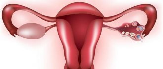
The organ itself is covered with a layer of rudimentary epithelium, under which is the tunica albuginea. Even deeper is the layer of the cortex, in which follicles at different stages, corpus luteum and other structures are located. The medulla of the ovaries contains blood vessels and nerve fibers.
The diagram shows the internal structure of the organ.
Causes of changes occurring in the ovaries
Throughout the life of the female body, the ovaries tend to change slightly in size, which depends on:
- age indicators;
- number of births and abortions;
- day of menstruation;
- use of contraceptives containing hormonal substances;
- taking hormonal medications.
With the onset of puberty, the ovaries begin to become involved in the functioning of the woman’s reproductive system and subsequently, within normal limits, can change in size. During pregnancy, these organs, under the influence of increased blood flow necessary to ensure adequate nutrition of the fetus, increase in size. Moreover, with the increasing period of pregnancy, the ovaries can change their location, since the growing uterine organ with its dimensions displaces all nearby organs and tissues to a certain level. The size of the woman’s gonads increases by a couple of millimeters, and the previously occurring ovulation processes cease during pregnancy. Instead, the ovaries begin to produce progesterones, which play a critical role in normal gestation and an easy delivery process.
With delivery, the size of the ovaries in an involutionary mode begins to decrease, along with the uterus.
The circulatory processes in the placenta stop, the speed of general blood flow decreases, which leads to a gradual return of the ovaries to their original form. This, in turn, leads to the resumption of estrogen production and the subsequent preparation of the female body for the full functioning of the entire reproductive organ system, if the woman does not feed her baby with breast milk. In the event that breastfeeding is still used, the restoration of the reproductive functionality of the reproductive system will occur only after the end of lactation processes in the mammary glands.
As women age, reproductive functionality begins to gradually fade away. This also affects the size of the ovaries, which begin to decrease at a leisurely pace. And by the period of premenopause, both glands become the same in all sizes.
The norm in the premenopausal stage of menopause is the following values of ovarian size:
- In volume from 1.5 to 4 cm3;
- Length – from 20-25 mm;
- Width – 12-15 mm;
- Thickness: 9-12mm.
The first two to three years of the postmenopausal period may be accompanied by the production of single follicles, despite the fact that there is no menstrual cycle. This explains the slight changes in size indicators in the ovaries.
What size ovaries are considered normal?
Ultrasound of the ovaries is one of the informative studies, as it allows you to identify various forms of pathological and other changes in the early stages. For example, if the procedure is carried out in the first phase of the menstrual cycle, then the size of the dominant follicle can be determined. This information will help to identify a follicular cyst in the early stages and suggest possible problems with infertility.
If we consider the normal size of the ovaries, we focus on the following indicators:
| Width | Length | Thickness | |
| In a woman of reproductive age | 18-28 | 20-37 | 15 |
| During ovulation | 15-30 | 25-40 | 20-25 |
| During the postmenopausal period | 12-15 | 18-25 | 10-12 |
Minor deviations from the norm are not an absolute indication of the development of pathology. The doctor analyzes the woman’s general condition, age, pregnancy and other indicators that may affect the volume and size of the ovary.
Enlarged ovaries
A significant increase in the size of the ovaries is a possible symptom of the development of a serious disease. These may be the following pathologies:
- The development of infectious, viral diseases that provoke inflammation and swelling of tissues.
- Ovarian cyst. A common pathology in women of all ages, both single and multiple neoplasms can be observed. It is important to detect the cyst in the early stages so that ovarian tissue is not damaged when it is removed.
- Oncological formations. Dangerous conditions when malignant tumors develop on the surface of the ovaries. They can be primary or formed due to metastasis.
Some enlargement of the ovaries may occur during pregnancy. Although the gonads begin to work in a different mode, ovulation does not occur; due to increased blood supply in the pelvic organs, and in particular in the ovaries, a slight increase in width and length is possible.
Pathological causes of changes in the gonads
When determining the possible development of a pathological process, it is necessary to take into account the indications of the norm of the ovaries in the fertile period. Evidence of the onset of development of a pathological change is the size of the ovaries doubling two or more times.
When determining the volume of the ovaries, pathology includes their increase by 1.5-2 mm3.
When determining such indications during an ultrasound examination of the reproductive system of organs in the female body, this may be evidence of the development of the following pathological processes:
- Cystic lesions of the ovarian cavity with various etiologies and localizations.
- The development of polycystic disease, that is, multiple formation of tiny cysts.
- The appearance of benign neoplasms.
- The appearance of neoplasms with a malignant course.
- Development of metastases.
- Hereditary factor or congenital pathological development of the reproductive organs.
The reason for urgent surgical intervention may be pathologies such as purulent inflammation of the ovaries in menopause or their torsion. With such a course of dysfunction of the genital organs, if timely surgery is not performed, then everything can become complicated to the point of irreversible damage or death.
The most dangerous pathological change for a woman’s life is oncological processes.
- Cancer , localized in the organs of the reproductive system of the female body, ranks second among all causes leading to death, after cancer of the mammary glands. If an ultrasound specialist is able to discern the development of a cancerous tumor in the first stages of its development, then the woman has every chance to continue living, actively fighting against cancer. And sometimes even a full recovery is possible.
- The clinical picture will be much worse if the malignant neoplasm reaches an impressive size and causes symptoms of metastases. Therefore, a timely ultrasound examination will help to promptly identify the pathology and take the necessary measures to eliminate it.
A sharp decrease in the size of the ovaries during the fertile period is also dangerous. Such changes in the ovaries are generally called premature menopause, since a woman’s gonads simply fade away and cease to perform their functionality in the reproductive performance of the female body. Such a pathological change can occur from 36 to 40 years. Moreover, the uterine organ begins to shrink, and the uterine walls become thinner; not a single follicle is observed in the ovaries themselves. Under the influence of these atrophic processes, natural menstruation stops. After which, after a short period of time, menopausal symptoms may begin to develop in the female body:
- Increased sweating.
- Psycho-emotional state disorder.
- The appearance of insomnia.
- A sharp decrease or gain of extra pounds.
- Attacks of hot flashes and heat.
If these manifestations are diagnosed in a timely manner, then by taking hormone replacement therapy it will still be possible to restore reproductive functionality and safely conceive and give birth to a child.
Ovarian changes during menopause
Atrophic changes characteristic of the female body during the menopausal period also apply to these organs of the reproductive system.
The size of the ovaries decreases during menopause. Their structure also undergoes changes, during which hormone-secreting tissues begin to be replaced by connective tissues. The number of follicles is reduced, until they disappear completely.
The development of a functional cyst in menopause should not occur. All neoplasms that arise at this age are already referred to as tumors.
Taking into account the fact that after the age of 55, the likelihood of developing cancer in women increases several times, medical specialists should pay special attention to the state of a woman’s health during diagnostic testing during the menopausal period, especially her mammary glands and reproductive system.
Every woman, in turn, should not forget that the absence of a menstrual cycle does not mean that there cannot be problems with gynecological health.
Regular visits to the gynecological office (at least once every six months) will help eliminate the likelihood of developing many serious pathologies, even preventing the development of oncology to a stage that is no longer amenable to any treatment method.
Any cystic ovarian lesion in menopause should be treated with surgery to prevent complications.
Normal sizes of ovaries and uterus in pregnant women
During pregnancy, a woman's internal genital organs undergo major changes. Namely the uterus and ovaries.
The ovaries stop producing eggs during this period of time. But its size increases, literally by a few millimeters. This is due to increased blood circulation in the pelvis. The uterus changes significantly. Its size increases as the fetus grows.
It should be noted that there are norms for the size of the uterus during pregnancy. If they are abnormal, a pathology of pregnancy or fetus can be suspected, as well as a multiple pregnancy can be detected.
For example, if the gestational age is 12–13 weeks, then the height of the uterine fundus is 12–13 centimeters. At 37 - 38 weeks, the fundus of the uterus is under the ribs and presses the diaphragm (fundal height: 36 - 37 centimeters). After this (from 38 to 40 weeks), the bottom of the organ begins to gradually descend. Thus, the body prepares for the upcoming birth.
Did you like the article? Share it with your friends on social networks:
The ovaries (gonads) are female reproductive glands located in the pelvic area. These organs of the reproductive system ensure ovulation and the ability to become pregnant.
Every month, a follicle with an egg emerges from these gonads, which is intended for fertilization with male seminal fluid.
The size of the ovaries in women is precisely the indicator by which gynecologists and medical workers are guided regarding the state of health of the reproductive system. If they increase, this indicates the presence of an inflammatory process caused by one of the diseases.
Symptoms of pathological changes in the ovaries
The most insidious thing about the development of tumors in menopausal women is that they do not cause any clinical manifestations. And only sometimes (no more than 30%) can they make themselves felt with blurred manifestations, relating equally to neoplasms of both a malignant and benign nature.
In most cases, among those representatives of the fairer sex who ignore the need for regular examination by specialists, such diseases are detected only in the event of complications characterized by torsion or rupture of the ovary, or acute pain symptoms in the lower abdomen. Also, the increasing manifestation of ascites and symptoms of compression near located organs indicates the development of metastases against the background of the main pathological process.
Diagnostic methods
Ultrasound diagnostics with an additional Doppler method of vascular condition will help you find out what is happening to the ovaries. The following may also be carried out:
- CT scan;
- Magnetic resonance imaging.
But these methods are expensive and are not particularly effective, which is why they are used much less frequently than conventional ultrasound.
Malignant neoplasms are distinguished by a number of characteristic manifestations that contribute to the detection of a cancerous tumor during ultrasound, these are:
- increase in blood flow speed;
- bilateral localization of the lesion;
- polyp growth.
If an ultrasound examination reveals the presence of a neoplasm, a blood test is prescribed to determine the content of cancer markers. The obtained blood test results, in combination with ultrasound results, provide a more complete clinical picture, on the basis of which the subsequent treatment regimen is developed.
After surgical removal of a tumor in the ovaries, a histological examination of the extracted tissue is performed, on the basis of which a final diagnosis and further treatment are made.
Useful video on this topic: