Uterine leiomyoma is a benign tumor that is formed from the muscle tissue of the organ and in certain situations can cause oncology. Actually, the tumor is benign in nature, but with a protracted course and neglected therapy it can become irreversible. The disease most often occurs in every fourth woman aged 30-40 years.
What is it, types
To understand the specific concept of leiomyoma, one should understand the anatomical structure of the uterus. This organ can carry and push the baby out of the body during labor. A similar complex mechanism of activity occurs thanks to the myometrium - the inner layer of the uterus. This durable frame is created from various types of muscle tissue in combination with connective matter.
Externally, the myometrium is covered with a serous layer, which resembles the abdominal cavity in its composition. The inner layer of the uterus is called the endometrium, which is made up of several layers of epithelium. At a certain phase of the cycle, this layer begins to renew itself, and menstruation begins. All actions of the endometrium occur under the control of female hormones produced by the ovaries.
What is nodular uterine leiomyoma?
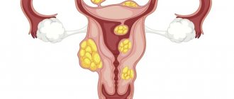
This disease is characterized by the appearance of a myomatous node. If there are several of these nodes at the same time, then the fibroid is called a multiple tumor. Such growth may vary in volume, structure or typology. The nodular form is often not expressed in any way, and a woman may not know about this formation for several years if she does not visit a specialist on an ongoing basis.
Intramural form of the disorder
This type of leiomyoma can occur frequently. With it, the neoplasm appears only in the thickness of the uterine muscles. When it receives volumetric parameters, the tumor may be located with other types of disease.
Initially, a woman may not even be aware of the formation of intramural uterine leiomyoma, since symptoms do not appear at all. The first thing a woman notices is disruptions in the menstrual cycle. Critical days last longer than usual and are more abundant than usual. This phenomenon is due to the fact that the myometrium loses its own ability to contract, as a tumor formation develops there.
As the disease progresses, women note the appearance of pain and heaviness in the abdominal area. Such signs can vary depending on where the tumor is oriented.
Subserous form of the disease
This type of neoplasm in women is detected more often than others. The tumor in this case forms under the serous layer of the uterus, which lines the organ from the outside. The tumor increases towards the peritoneum or pelvis.
Subserous leiomyoma always has a stalk. This is the name given to the extensive or narrow base, which is formed from the serous layer. Vessels and nerve endings pass through there.
If the tumor is small, women will not feel any symptoms. When the tumor begins to grow and reach a certain size, pressure is exerted on nearby organs, which provokes unpleasant signs of the disease.
The most common symptoms with this type of uterine tumor are:
- Pain of varying nature and intensity, which can be noted in the lower abdomen and lower back. This occurs because the tumor begins to put pressure on the nerve endings and organs located in the peritoneum.
- Difficulties in defecation and emptying the bladder, if the disorder has formed in the area where the organs that are responsible for these processes are located.
- It is not possible to conceive a child because the tumor compresses the fallopian tubes.
The most negative consequence of subserous leiomyma is twisting of the node's stalk. Because of this, a malnutrition of the tumor occurs, which leads to its necrosis.
In this case, the patient feels excessively strong pain in the abdominal area, the temperature rises, and general intoxication of the body appears, as the matter inside begins to break down. As a result, peritonitis may occur. Twisting of the node is considered a complete indication for immediate surgical treatment.
Submucosal form of the disease
Submucosal leiomyoma is the growth of uterine tissue under the mucous membrane. It is also called the submucosal form. Such a neoplasm is the most harmful and can provoke quite pronounced symptoms if its size is large. The submucosal type of leiomyoma requires only surgical treatment.
With this disorder, the tumor is partially or entirely covered by the mucous membrane of the uterine cervix. Based on this, experts distinguish 3 types of the disease:
- “Zero”, when the neoplasm is entirely covered with a membrane and is not associated with the muscular tissue of the uterus.
- The first is that the knot has penetrated halfway into the muscles.
- The second is that the tumor penetrates more than half of the myometrium.
What signs does submucous leiomyoma cause:
- Irregularities in the menstrual cycle, menstrual periods become longer and excessively abundant.
- Uterine bleeding appears.
- Formation of anemia due to heavy bleeding. Hemoglobin drops.
- Excruciating pain in the area above the pubis.
- Difficulties in conceiving a child arise.
- Pregnancy is difficult, miscarriages and placental pathologies occur.
A risky consequence of this type of leiomyoma can be uterine inversion. This tumor is more likely to become malignant than all other types.
Transformation of leiomyoma into cancerous tumor
If there is aggravated heredity, a predisposition to cancer, or other negative factors influencing the development of the disease, there is a possibility that the tumor will transform into cancer, for example, cervical cancer. Therefore, after detecting a tumor, doctors categorically recommend seeing a doctor once every 3 months. Monitor tumor growth and adhere to the treatment plan.
In the early stages, tumor growth can be stopped with medication. The course of medication is aimed at reducing estrogen in the patient’s blood. In addition, hormonal medications (oral contraceptives) are prescribed to restore balance.
It is possible to use medical procedures within a hospital, these are baths, douching, injections and intravenous drips.
Surgery to remove fibroids will not be necessary. But if doctors have doubts, then to determine the quality of tumor cells, tissue is plucked and sent for a biopsy. This is the most reliable method for detecting cancer cells. Under such circumstances, uterine fibroleiomyoma, an oncological disease, may develop.
Leiomyoma will have to be treated throughout its existence. Since fibroids do not have progressive growth, they do not cause inconvenience to the body and do not require removal.
If there are no additional factors that provoke the growth of leiomyoma, it can grow by 1 mm over several years. This is why it is important to constantly monitor the size and growth trend of the tumor. It can have the same size for years, but grow within a short period of time. This may be triggered by stress, deteriorating health, menopause, decreased immunity, or genetic diseases.
Leiomyoma, located in a cavity close to the cervix of the uterus itself, can interfere with fertilization and provoke inflammatory processes.
We must not forget that the female reproductive system is a barometer of the whole organism. Considering that the uterus performs a vital function, is responsible for childbirth and hormonal levels, special attention should be paid to its health.
Often, women who have already given birth neglect their health and rarely visit a gynecologist. This may bring negative results in the future. You may not notice the stages of fibroid growth and find it has already transformed into fibroma, which is highly undesirable.
Even if a woman does not plan to give birth again, she must remember that the work of the pelvic organs is the basis of a woman’s health.
Where does leiomyoma come from?
The possibility of the disease occurring against the background of prolonged hormonal imbalance and undergoing regression during menopause certainly points to hormonal causes of the disease. But not every patient with a hormonal imbalance has leiomyoma, so they talk about predisposing conditions.
There is an opinion that the increase in nodes is carried out in three main ways:
- Central
Sex hormones affect all movements in the uterus. The ovaries are controlled by central structures - the pituitary gland and hypothalamus. Any manifestations that lead to pathologies in the brain will also lead to pathologies in the ovaries. These may include severe psycho-emotional and vascular disorders and injuries.
- Ovarian
The functioning of the ovaries will be abnormal with prolonged inflammation (salpingitis), cystic degeneration and similar conditions that change the usual function of the ovaries. Not only the secretion of estrogen and progesterone changes, but also their ratio. This option may be more common than others.
- Uterine
The disease can also occur against the background of normal ovarian activity. The production of hormones occurs normally, but the uterus does not perceive them, since it has damaged receptors due to the tumor.
Etiology of the disease
The reliable reasons for the development of uterine leiomyoma have not been established. According to scientists, the following risk factors play a significant role in the occurrence of benign neoplasms of the uterine wall:
- Hormonal imbalance. Today, the question of the primacy or secondary nature of hormonal imbalance is open.
- Late onset of menstruation.
- A history of two or more abortions.
- Chronic inflammatory diseases of the female genital organs.
- Obesity and a sedentary lifestyle.
Important to know: Uterine cancer - radiation and its consequences
Signs and symptoms
To understand what leiomyoma is, you need to know and understand at least a little. Leiomyoma is formed from smooth materials, often found in the uterus, and quite rarely on the walls of the stomach and rectum.
The disease develops under the influence of internal and external causes, the symptoms are mild. What symptoms may appear:
- Excessive bleeding during menstrual periods;
- Increase in body weight;
- Spotting blood after sexual intercourse;
- Discharge between critical days;
- Pain in the lower abdomen;
- Urinary incontinence.
When large in size, uterine fibroids can put pressure on the ureters, provoke kidney problems, the formation of kidney stones, and cause the formation of infectious processes in the urinary tract. During the examination, the specialist is able to determine the position of the uterus by its density, hardness, and volume. When carrying a child, the uterus takes on a soft appearance. When a tumor forms, it is hard.
To confirm suspicions or refute them, the specialist prescribes the following types of examination to the patient: ultrasound, laparoscopy, histology, colposcopy.
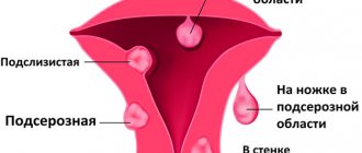
Submucosal uterine fibroids
Intracavitary growth. Submucosal uterine fibroids are one of the most common variants of the disease: the myomatous node begins to enlarge towards the uterine cavity, which is manifested by symptoms that cannot be ignored. There are 3 types of benign muscle tumor growth:
- The node on a thin stalk is completely located in the uterine cavity;
- The tumor is partially (less than 50%) located in the muscular wall of the uterus;
- Most of the fibroids (more than 50%) are detected intramural, and a smaller part is found in the cavity.
The first option is classic submucosal uterine fibroids. The other two are different types of interstitial benign neoplasms with centripetal growth of the node. All main manifestations of pathology are characteristic of a submucosal node on a thin stalk.
Differences from fibroids
A correctly diagnosed disease is of great importance for selecting a treatment regimen. A benign tumor of muscle tissue is called fibroid, but there are several types of muscles in the human body. For this reason, the correct term for a tumor that grows from the body of the uterus is leiomyoma. The differences between these diseases lie in the indication of the histological structure of the materials, which helps the specialist assist in treating the disease.
Fibroids are tumors of muscle tissue that can develop anywhere in the human body where there are muscles.
Leiomyoma is a benign neoplasm of smooth muscle formed by internal organs.
Classification
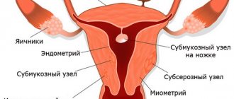
The classification is based on the number of tumors - a single node or multiple lesions - and the location of the fibroids.
Based on location, the following groups of fibroids are distinguished:
- Submucosal neoplasms or submucosal leiomyoma are located closer to the internal cavity of the uterus. This node has a long stalk and is capable of falling out of the uterus into the cervix or vagina.
- Intramural leiomyoma of the uterus - seals are located in the middle muscular layer of the organ. The most common type of disease.
- Subserous leiomyoma of the uterus - this neoplasm is located on the outside of the organ closer to the peritoneum.
- Intraligamentary myoma - the location of the neoplasm is located on the ligamentous apparatus of the organ.
- Cervical fibroids.
The influence of tumor location on the dynamics of progression
Enlargement and placement are considered the main conditions that determine the staging of the disease. In addition, the location of the tumor affects symptoms and pain threshold, especially during sexual intercourse and when bearing a child.
Leiomyoma found in a woman during pregnancy can become a prerequisite for miscarriage, intracavitary copious bleeding, premature labor, and changes in fetal presentation.
A small tumor is difficult to diagnose because it does not have any visible signs. Almost always the neoplasm is benign; quite rarely (1 case per 1000 women) it is oncological. Such neoplasms are called leiomyosarcoma. Doctors believe that the oncological nature of the tumor, as well as the increased risk of its development, is not affected in any way by the existing fibroids. The accompanying conditions are to blame.
Important ! The nodular form of the disease does not affect the degeneration of the neoplasm into other forms, for example, cancer.
Leiomyoma is a typical cause of infertility; this diagnosis accounts for approximately 3% of cases with the inability to conceive a child. Usually, often in case of infertility, leiomyoma can be diagnosed, which is located under the mucous membrane of the uterus. This location prevents the mucous membrane from fulfilling its key role - attaching the fertilized egg to the placenta.
Causes
The exact causes of this disease have not yet been identified. Doctors identify a number of factors that provoke disturbances in the fibers that make up the muscles of the uterus:
- various hormonal disorders in the reproductive system, adrenal cortex, thyroid gland and related diseases. Outwardly, this manifests itself in menstrual irregularities, including amenorrhea and hyperandrogenism;
- lack of regular sex life during reproductive age;
- anorgasmia in women;
- spontaneous or medical abortions;
- diagnostic curettage procedure;
- childbirth with complications;
- hereditary factor;
- excess weight;
- hypertonic disease;
- diabetes;
- chronic stress condition;
- adynamia.
Diagnostics
Taking into account the complaints of women, the presence of leiomyoma can only be assumed. Options when a violation is detected by chance are quite common.
With a two-handed examination, it is possible to determine an increase in the volume of the uterus against the background of a change in its texture (it becomes more dense). In some situations, you can feel the uneven contour of the uterus, which is deformed by the node. Usually, the uterus begins to “grow” along with the leiomyoma, for this reason the dynamics of changes in its size is an important diagnostic criterion.
The volume of the uterus with leiomyoma is assessed similarly to pregnancy - in weeks. The day of the cycle is selected and palpation is performed regularly in a certain month. If over the past period of time the uterus has not increased in size by more than 4 weeks, the growth of the neoplasm is regarded as slow.
When examined with a speculum, submucosal nodes may be visualized. To clarify the detected sign, colposcopy is recommended.
The most accurate examination is the conclusion of an ultrasound scan. It makes it possible:
- “Consider” the nodes, determine their number and topography;
- Establish the structure and age period of leiomyoma;
- Assess the typology of tumor growth;
- Find accompanying changes in the endometrium;
- Diagnose the condition of the ovaries.
Laboratory diagnosis helps to establish the causes of the disease. Smears and bacterial cultures for flora and oncocytology, blood tests and hormonal examination will be required.
The list of modern diagnostic capabilities is quite large; for this reason, for each specific option, the choice of diagnostics is carried out individually.
Diagnostic measures
Large tumor sizes can be seen during a routine examination by a gynecologist. But when there are many tumors and they are small in size, a simple examination in the mirrors will not help. In this case, doctors prescribe additional examination methods.
As a rule, for any gynecological diseases, the patient is prescribed an ultrasound examination. This diagnostic method allows you to fully examine the uterine cavity and other pelvic organs.
If the diagnosis has already been made, but it needs to be confirmed, then Doppler ultrasound is additionally used (helps to assess blood flow).
If submucosal leiomyoma is suspected, it is mandatory to undergo hysteroscopy.
Additional events:
- pathohistological examination of the material, carried out after diagnostic curettage;
- MRI is prescribed;
- laparoscopy, which helps to identify formations in the pelvis, as well as in the abdominal cavity.
Standard diagnostics include: general blood and urine analysis, hormone analysis.
Treatment of uterine leiomyoma
The treatment plan for leiomyoma depends on the signs that worry the woman, the size of the tumor, location and the patient’s question of childbearing. As a rule, treatment is carried out conservatively, but in certain situations it is necessary to use a surgical method.
Conservative treatment
Conservative therapy for uterine leiomyoma is carried out in the following ways:
- A woman is planning to give birth to a child.
- The disease goes on latently.
- The tumor grows slowly.
- The volume of the tumor does not exceed 12 weeks of pregnancy.
- The tumor is located in the wall of the uterus.
- There are violations when it is not recommended to use general anesthesia and perform surgical interventions.
- Surgical treatment has been performed and a period of rehabilitation is required.
A conservative treatment method consists of using hormonal drugs to suppress tumor formation. In addition, other means are used together with them, which make it possible to simplify the symptoms of the disease, that is, relieve pain and inflammation. After completing conservative treatment, the woman will need to visit a specialist for 6 months.
Surgical treatment
If the disease has already been detected with a large volume exceeding the size of the uterus at 12 weeks of pregnancy or has begun to grow very quickly, then specialists prescribe surgical intervention. In addition, a similar solution is used in cases of pathology of internal organs due to compression of the tumor.
Modern medicine involves several methods of getting rid of a tumor. These include:
- Myomectomy . With its help, only the affected areas of the uterus are eliminated; healthy tissue is not affected. This method is used when the patient wants to have another baby, the medications she is taking are not working, and a tumor is discovered on the leg. After this operation, scars will remain on the uterine surface, which require careful monitoring during pregnancy.
- Embolization of the uterine arteries . This method of surgical treatment is also carried out if a woman wants to preserve the reproductive role of the uterus. However, this method is not recommended for use in cases of pedunculated fibroids and malignant tumor degeneration.
- Hysterectomy . This operation consists of complete removal of the uterus. One comes to such a radical method when other methods have failed to help.
A favorable prognosis for leiomyoma will be when the disease is detected early and effective treatment is carried out.
Folk remedies
Traditional medicine methods are used to treat and prevent a wide variety of diseases, including in the gynecological field. But you should not use such products yourself, since even their natural origin cannot protect against allergies or various other consequences.
Aloe
To prepare the medicinal solution you will need:
- 700 g honey;
- 0.5 l aloe juice;
- 20 mg mummy;
- 0.02 ml celandine juice.
Everything must be mixed well and allowed to brew for 3 days. The course of treatment lasts for a month. The first 10 days take 1 tsp 3 times a day, all subsequent days 1 tbsp.
In addition, mumiyo can be used in the manufacture of tampon mixtures. To do this, mix everything with honey and sea buckthorn oil. Leiomyoma therapy is effectively carried out using tampons that contain aloe, propolis and honey.
Burdock
To treat leiomyoma with burdock you will need:
- Fresh juice from burdock leaves – 1 l;
You should pick fresh burdock leaves, wash them well and dry them with a towel. Then transfer through a meat grinder or use a blender. The resulting mixture must be squeezed out. As a result, you will get approximately a liter of juice. It should be consumed three times a day before meals, 30 minutes, 1 tablespoon. The course of therapy is a month.
Chaga
To get rid of leiomyoma and avoid surgical treatment, you should follow the following recipe using chaga:
- Dry chaga – half a packet (sold at the pharmacy);
- Water – 1 l.
Pour water into a saucepan and add half a packet of chaga. Bring the mixture to a boil and boil for 2-3 minutes. Then let it brew for 10 minutes. You can drink it instead of tea, diluting half a glass of chaga decoction with half a glass of water. 1 package is enough for 3 weeks. After 3 weeks, you can brew a new portion.
Herbal infusions
The following have a reliable beneficial effect on leiomyoma:
- Borovaya uterus.
- Red brush.
- Flax-seed.
The herbs need to be brewed. To do this, take 100 g of all ingredients, add 1 liter of water, and put on high heat. Bring to a boil, leave for another 3 minutes over medium heat. Cool, strain and drink 30 minutes before meals.
Potato
Potato juice is quite useful and not only for gynecological problems. How to drink it to alleviate the condition of uterine leiomyoma?
On an empty stomach in the morning you should drink 100 ml of freshly squeezed potato juice. You must drink this juice every day for 3 months. Sometimes the time frame can reach up to 6 months. If the condition of the leiomyoma has not changed, then you should pause for 4 months and carry out this procedure again.
Celandine
Celandine has many healing properties, including antiseptic, anti-inflammatory and regenerating effects. How to use celandine for uterine leomyoma. The most common option is to take an alcohol tincture orally. The preparation is very simple. To do this, dig up the celandine with its roots. Wash it well and cut it completely into small pieces, along with the root. Next 2 tbsp. l. Place the crushed plant in a dark bowl and fill with 100 ml of vodka. The product must be infused in a dark and cool place for 30 days. Take 2 drops per 100 ml of water. The next day, the dosage is increased by 2 drops. This should be done every day until you reach 20 drops in a glass. After this, reduce the dosage by 2 drops until you reach the original volume.
Motherwort
Motherwort tincture is very popular. Preparation:
- Combine crushed leaves and inflorescences of the herb with vodka in a ratio of 1 to 5.
- Close the lid tightly.
- Leave for 30 days in a dark place.
- Shake the container once every day.
- After a month, strain the tincture.
Use the mixture 30-50 drops every day. Take 3-4 doses of tincture per day after meals; the infusion can be diluted with water.
How long do people live with this disease?
After conservative treatment of fibroid lesions, patients must be examined by a gynecologist annually. Scheduled preventive examinations allow timely detection of disease recurrence and adequate treatment. Uterine leiomyoma, the survival rate of which is about 100%, when operated on surgically, does not require special oncological monitoring.
Uterine leiomyoma is mainly diagnosed during a routine gynecological examination and after a final diagnosis is made, the patient will be offered appropriate treatment. It must be remembered that a benign neoplasm has a high potential for cancerous degeneration.
Treatment during menopause
During menopause, leiomyoma may begin to regress. In most situations this is what happens. However, if examination reveals that the tumor is growing, treatment may be required.
General treatment recommendations are the same as for the normal condition. Which targeted symptomatic treatment would be appropriate for menopause:
- Prescribing vitamins and minerals to maintain normal processes in the body and general well-being.
- Take hormonal medications only as prescribed by a gynecologist.
- Taking sedatives to alleviate the symptoms of menopause.
As for the cure, the prognosis is negative - leiomyoma cannot completely go away during menopause if it has increased to a large size. It is not life-threatening. Regular diagnostic examinations make it possible to detect and monitor the course of the disease.
https://youtu.be/2FpPzJD9fI0
What is regression and its causes
Regression is a gradual decrease in the tumor due to the onset of menopause or appropriate therapy. At the regression stage, hormonal changes occur in the body, which create certain conditions for the growth to disappear or decrease.
The process can occur for several reasons:
- Climax.
- Pregnancy and lactation.
- Appropriate treatment.
- Impaired blood flow in the uterus.
In any case, the tumor does not disappear, it subsides for some time and can always be activated again. To exclude this, a woman must visit the clinic on time and follow the doctor’s recommendations.
Symptoms of leiomyoma
Uterine leiomyoma can develop asymptomatically until it reaches a size of 2-3 cm. The growth of neoplasms leads to painful sensations due to tissue stretching and impaired contractility of the uterus.
Menstrual irregularities appear. Menstruation becomes long and heavy. Without knowing the reason, the woman begins to take hemostatic and painkillers. This helps relieve unpleasant symptoms, and a visit to the doctor is postponed. The regularity of the cycle is gradually disrupted, and blood loss increases.
Signs of bladder dysfunction (cramps, frequent urination) and digestive disorders appear. This occurs due to the compressive effect of the tumor on other pelvic organs.
Due to the increase in the size of the nodes, the woman’s belly begins to grow, like a pregnant woman. It is customary to estimate the size of leiomyoma both in centimeters and in “obstetric weeks”. The size of the abdomen as the tumor enlarges corresponds to its size at a certain week of pregnancy.
Note: An enlarged abdomen due to fibroid growth is sometimes mistaken for a sign of pregnancy. Even the presence of menstruation does not bother you, since spotting also occurs during this period.
What are the possible complications?
The severity of complications depends on the size and location of the leiomyoma. Tumors are most often detected after the size of the nodes increases to 5-6 cm or more.
Depending on the type of tumor
Submucous uterine leiomyoma is the most common and noticeable cause of complications, such as:
- Menstrual disorders, manifested by prolonged heavy bleeding not only during menstruation, but also between them.
- Infertility. The tumor interferes with the normal formation of the endometrium, which makes it impossible to retain the embryo in the uterus, as well as the formation of a normal placenta. It may block the cervix or fallopian tubes.
- Birth of leiomyoma. Under the influence of contractions of the uterine muscles, large fibroids, located near the cervix and having a thin base, can prolapse into the vaginal area.
- Damage to tumor vessels, leading to dangerous uterine bleeding, the occurrence of an “acute abdomen” and anemia.
Subserous. Disorders of menstrual function do not appear. But complications can be no less severe. Twisting of the thin stalk of such a tumor leads to necrosis of its tissue. Decomposition of a dead tumor in the abdominal cavity causes peritonitis.
The pressure of the nodes on the adjacent pelvic organs leads to disruption of their functioning, compression of blood vessels, inflammation, severe abdominal pain, nausea, and vomiting. Such tumors are more difficult to detect and are often discovered only when complications occur.
Intramural. As the tumor enlarges, the state of the vascular network and the structure of the muscle layer are disrupted. This leads to a decrease in uterine contractility, as a result of which the excretion of menstrual blood takes longer than usual. Blood stagnation in the organ cavity, endometritis and endometriosis may occur.
Dull, constant pain in the abdomen is a characteristic sign of the presence of such a tumor.
Leiomyoma during pregnancy
When large submucosal nodes form in a woman, pregnancy becomes difficult due to disruption of the structure of the uterine mucosa. Excess estrogen, which provokes the formation of a tumor, leads to the appearance of anovulatory cycles, in which the egg does not mature and conception is impossible. The implantation of the embryo in the uterine wall is hampered not only by the immaturity of the endometrium, but also by the increased contractility of the uterus during the formation of submucous fibroids.
If the nodes are small, then pregnancy is possible, but the enlarging tumor interferes with the growth of the fetus and impairs its blood supply and nutrition. Labor may begin several weeks before your due date, and heavy bleeding may occur.
After a neoplasm such as uterine leiomyoma is detected in a pregnant woman, its development is carefully monitored. If it increases so much that it interferes with the growth of the fetus, it is removed (most often after the 16th week of pregnancy). Childbirth is carried out prematurely using cesarean section.
Leiomyoma during menopause
In women over 50 years of age, such a tumor cannot normally form. And even vice versa, a pre-existing node often resolves on its own. However, when endocrine diseases occur, uterine tumors still appear, and the risk of their degeneration into cancer increases significantly, especially in the presence of a diffuse type tumor.
Warning: A woman should immediately contact a gynecologist if she experiences bleeding from the genitals during postmenopause. Sometimes this is vital.
Symptoms of the submucosal location of the node
Benign muscle tumor, like a foreign body. Regardless of size, submucosal uterine fibroids are manifested by the following symptoms:
- Heavy uterine bleeding occurring outside of menstruation and/or associated with the cycle;
- Pain syndrome (cramping sensations, painful menstruation);
- Lack of pregnancy (uterine factor infertility);
- Inability to maintain the resulting pregnancy (miscarriage);
- Progressive anemia with weakness and dizziness.
The myomatous node acts as an intrauterine contraceptive, preventing the conception of a new life. The uterus tries to push out this “foreign body,” and the woman perceives these unsuccessful attempts as painful contractions. Severe pain will occur during the “birth” of the node, when the pedunculated tumor moves along the cervical canal.