Skin cancer is one of the most common types of cancer today. All types of skin cancer are effectively treatable if diagnosed early, but you need to know what to look for. There are many types of skin cancer, each of which can be visually and symptomatically different. These photographs contain examples of skin cancers as well as other benign tumors. Based on individual characteristics, skin cancer may differ from the photos we provided as an example. This is why it is important to see a doctor if you have any bumps, swellings, spots, ulcers, or other marks on the skin that are new or changed.
Skin cancer: causes
Currently, there is no clear answer to the question of what causes skin cancer. Like many other oncological diseases, skin tumors are considered a multi-etiological pathology. There are several predisposing factors, the presence of which increases the risk of developing a tumor focus. These include:
- Excessive exposure to ultraviolet rays on the skin. A similar situation occurs with prolonged and frequent exposure to sunlight, visiting a solarium, or working outside. Residents of the southern regions are at risk of developing skin cancer.
- Having light skin. Lack of melanin production increases the likelihood of skin tumors.
- Skin burn. A high degree of burn is accompanied by scarring of the skin. This process contributes to the emergence of latent carcinogenesis.
- Irradiation. Exposure to radioactive, ionizing rays has a detrimental effect on the skin. The risk of radiation dermatitis increases.
- Presence of skin contact with toxic substances. This group of carcinogens includes arsenic, aluminum, titanium, nickel and other heavy metals.
- Immunodeficiency. Conditions in which the body's protective functions decrease predispose to the formation of a tumor focus.
- Age. Most often, skin tumors affect people over 50 years of age.
- Concomitant systemic diseases. Doctors identify a group of pathologies in which the risk of developing skin cancer increases significantly. These include systemic lupus erythematosus, leukemia, and chronic skin diseases.
- Heredity. The presence of a skin tumor in previous generations of relatives is not a major risk factor. However, family history in combination with other predisposing conditions increases the possibility of developing skin cancer.
- Tattooing. In this case, there are two risk factors. This is a violation of the integrity of the skin and the introduction of paint with carcinogenic substances. Cheap tattoo ink may contain impurities of aluminum, titanium, and arsenic.
- A large number of nevi. Doctors urge you to monitor the condition of moles and contact a specialist if there is the slightest change. Traumatization of nevi increases the possibility of developing skin cancer.
- Excessive alcohol consumption, smoking. Chronic intoxication has a detrimental effect on the body as a whole. Against this background, the risk of tumor formation increases several times.
- Eating foods high in nitrates.
Oncologists identify several precancerous conditions, the presence of which significantly increases the risk of developing skin cancer. These include:
- Xeroderma pigmentosum.
- Bowen's disease.
- Paget's disease.
- Hyperkeratosis.
- Cutaneous horn.
- Late stage radiation sickness.
- Dermatitis and dermatoses.
Expert opinion
Author:
Alexey Andreevich Moiseev
Oncologist, chemotherapist, Ph.D.
Every year, 9,000 cases of newly diagnosed melanoma are registered in Russia. This aggressive malignant tumor is responsible for the death of 40% of patients. Such statistics indicate that the country's population is not sufficiently aware of the symptoms of melanoma. As a result, a visit to a doctor occurs in the later stages, when treatment is considered ineffective.
Doctors at the Yusupov Hospital determine the location of melanoma and prescribe treatment appropriate to the stage of tumor development. An individual approach to the problem of each patient allows us to reduce the number of deaths. A tumor detected in time is characterized by a favorable prognosis for recovery. The later melanoma is detected, the longer the treatment process will be. Its success depends on many factors. The main treatments for melanoma are surgery, radiation and chemotherapy. Depending on the condition, symptomatic therapy is carried out.
You can diagnose the presence of skin cancer yourself at home. Doctors recommend regularly examining the skin, especially nevi, for the appearance of pathological formations.
More details about the types of disease
The skin is characterized by three groups of oncological diseases: benign, locally destructive and malignant.
Skin cancer is a malignant oncological disease that is characterized by changes in the structure and function of stratified squamous epithelial cells. The tumor can occur against the background of other diseases, which are called precancerous. These include: Bowen's disease, xeroderma pigmentosum and Paget's disease. There are several types of the disease, so let’s look at the photo to see what skin cancer looks like .
Doctors distinguish two main forms of the disease (which determines what skin cancer will look like):
- The exophytic form is papillary.
- The endophytic form is ulcerative-infiltrative.
Statistics provide the following figures: among cancer diseases in men, skin cancer ranks third, after lung and stomach cancer; among women - second place (breast cancer is in first place).
Skin cancer: symptoms and signs
The clinical picture of skin cancer development depends on its type. Often the first signs of a tumor are mistaken for other skin diseases. This results in untimely consultation with a doctor and the spread of the malignant process. Common signs for all types of skin cancer are:
- The appearance of a small spot or lump on the skin. The color of the formation can be pinkish, gray-yellow. It is noteworthy that the color of the neoplasm differs from moles and freckles.
- Uneven shape and asymmetric contours of the pathological focus.
- The appearance of itching and slight discomfort in the area of the tumor process. This symptom appears some time after the formation of cancer.
- Progressive growth of the tumor.
- Fatigue, weakness, sudden loss of strength.
- Decreased appetite, resulting in sudden weight loss.
- As cancer develops, regional lymph nodes are affected.
If any of the above symptoms appear, it is recommended to seek medical help. Depending on the type of skin cancer, the following symptoms are distinguished:
1. Basalioma. It is the most common form of skin tumor. It is characterized by a favorable prognosis when detected at the initial stage. Appears in the form of a nodule, painless on palpation. It has a grayish-pink color. The surface of the nodule is usually smooth. Scale formation is possible. As the tumor process spreads, the affected area increases. The neoplasm is covered with a bloody film. The predominant localization of basal cell carcinoma is the face, neck, and arm area. Basalioma does not cause a significant change in condition. This is the reason for late seeking medical help.
2. Squamous cell carcinoma. At an early stage, it looks like a dense red or brown nodule. A tumor of this type is prone to rapid decay, and therefore, as the process progresses, an ulcer forms. Its edges are heterogeneous, the ulcer often bleeds. Squamous cell carcinoma quickly grows into nearby tissues. If detected at a late stage, metastasis to regional lymph nodes or organs is possible.
3. Melanoma. Refers to an aggressive type of tumor. Most often, melanoma occurs at the site of an injured nevus, which increases in size, changes color and acquires uneven contours. A characteristic feature of this type of cancer is the asymmetric form of formation, as well as a tendency to bleed. The malignant neoplasm itches and increases in size. As the process progresses, the tumor turns into an ulcer. Melanoma progresses rapidly and often metastasizes.
4. Adenocarcinoma. At the initial stage of development, it looks like a dense nodule. It is most often localized in the armpit area, under the breast. With the development of the tumor process, adenocarcinoma grows into nearby tissues. This type of tumor is detected extremely rarely and is characterized by slow growth.
Melanoma
Melanoma is a type of skin cancer. Despite the fact that melanoma is a rarer disease than other forms of tumor, it is highly dangerous for the patient. The process is extremely malignant, characterized by progressive tumor growth and a high ability to metastasize. Since the disease is life-threatening, it is important to know, including. Here you can see the main symptoms of melanoma:
- changes in moles - if you notice any changes in the shape, color, or texture of a mole, you should urgently consult a specialist;
- asymmetry - one part of the mole differs in color, texture, shape, height from its other part;
- the edges of the mole have an uneven shape;
- large sizes - if the mole has grown in size and reaches six millimeters, then it is necessary to be examined by a doctor;
You can see how it can be distinguished from other forms.
Melanoma is diagnosed based on objective and subjective history, and the main method is laboratory diagnosis, which helps to make a residual diagnosis and determine a treatment regimen. The main method for diagnosing cancer is a biopsy.
A biopsy is the removal of a piece of tissue for histological examination. During the analysis, the doctor examines a piece of tissue under a microscope and determines the structure of the cells. If the cells are atypical (cancerous), then a diagnosis of cancer is made, but if the cells have a normal structure, then the neoplasm is called benign.
Types of skin cancer
Depending on the type of cells from which the tumor is formed, there are several main types of skin cancer. Determining the structure of the tumor is necessary to clarify the diagnosis and carry out appropriate treatment. In addition, the speed of its spread and further prognosis depend on the type of cancer. Oncologists distinguish the following types of skin cancer:
1. Basal cell (basalioma). Basal cell carcinoma is the most common form of skin cancer. It originates from the basal cells of the epithelium. Basalioma is characterized by slow, sudden development and a relatively favorable course. Unlike other types of skin cancer, it rarely metastasizes. Basal cell carcinoma is usually localized in the area of the nose, bridge of the nose, eyebrow, upper lip and nasolabial fold. A rare location for this type of cancer is the neck and ears. There are several forms of basal cell carcinoma:
- Nodal. The most common location of nodular basal cell carcinoma is the head and neck area. At an early stage it looks like nodules up to 5 mm in size. As the tumor process develops, the nodules merge with each other. At a late stage, nodular basalioma is a deep ulcer with necrotic areas.
- Superficial. This type of basal cell carcinoma is located on the trunk and limbs. The tumor looks like a round pink spot with ulcerations. Superficial basal cell carcinoma is considered the most favorable and non-aggressive form of skin cancer.
- Scleroderma-like. Refers to an aggressive type of cancer. Located in the deep layers of the skin. The most common location for this type of tumor is the head and neck. Basalioma has the appearance of a plaque, which in the later stages of the process has the appearance of a depressed scar.
- Cystic. A rarely detected form of basal cell carcinoma, externally similar to a cyst.
- Fibroepithelial. Located in the lumbar region. Basal cell carcinoma has the appearance of a pedunculated polyp. The fibroepithelial form is extremely rare and has a favorable course and prognosis.
2. Melanoma. Refers to an aggressive type of skin cancer that originates from melanocytes. Melanoma is characterized by rapid growth and invasion into neighboring tissues. This type of skin cancer metastasizes not only to regional lymph nodes, but also to organs. A favorable prognosis for melanoma is only possible if the tumor is diagnosed at an early stage. Women with fair skin are at risk for developing skin cancer. Often injured nevi, warts, and pigment spots degenerate into melanoma. Distinctive features of this type of tumor are its uneven shape, asymmetrical edges, pain and bleeding in the early stages of the process. Melanoma is often located on the skin of the face, chest, arms and legs.
3. Squamous. Squamous cell carcinoma is the 3rd most common type of skin cancer. It develops from the outer layers of the skin. It is characterized by slow development, which increases the possibility of detecting a tumor at an early stage. At the initial stage, squamous cell skin cancer looks like a red lump up to 2 cm in diameter. Because of this, squamous cell carcinoma is often mistaken for inflammatory diseases of the skin. It is localized in the eyes, mouth, lips, genitals and mucous membranes. Squamous cell skin cancer has a tendency to metastasize.
The above forms of skin cancer are diagnosed most often. They account for about 90% of all detected oncological processes of the skin. In addition, there is skin appendage cancer, which is extremely rare. This type of tumor includes:
- Adenocarcinoma. Comes from the sweat and sebaceous glands. Characterized by slow growth. Outwardly it looks like a dark blue node, localized in the groin and chest area. Adenocarcinoma does not tend to grow into adjacent tissues, and metastasis rarely occurs. The tumor is removed surgically.
- Verrucous carcinoma. Refers to a very rare form of squamous cell carcinoma. Outwardly it resembles a wart, so detection at an early stage is difficult. A distinctive feature of verrucous carcinoma is its bleeding.
Bowen's disease
- It is the earliest form of squamous cell skin cancer. Bowen's disease usually appears as rough patches on the skin, sometimes on the surface of the anus, or on the genitals. Malignant spots are usually larger and redder than age keratosis. Bowen's disease is difficult to distinguish from squamous cell carcinoma and can sometimes become invasive, so doctors recommend prompt treatment.
Skin cancer - what the initial stage looks like
Symptoms of a skin tumor depend on its type. Growth rate, color, and tendency to metastasize are characteristic of each form of cancer. However, at the initial stage, several symptoms are common to all types of tumor. These include:
- The presence of a formation on the surface of the skin with unclear shapes and asymmetrical contours.
- The appearance of itching and changes in the color of the lesion as the process progresses.
- A sharp loss of strength, increased fatigue.
- Loss of appetite, sudden weight loss.
- The presence of pain in the area of the tumor.
Skin cancer develops through 4 stages. Each reflects the size of the lesion, its spread to surrounding tissues, and the presence or absence of metastases. Depending on this, the following symptoms of skin tumors are distinguished:
- First stage. At this stage of skin cancer formation, there are no specific signs. The size of the tumor focus is up to 2 mm. In this case, the general condition does not change. Detecting cancer at the first stage is possible only with a thorough self-examination of the skin.
- Second stage. The growth of the tumor leads to the appearance of the first subjective signs. These include itching and pain in the area of the pathological focus. The cancer increases in size up to 4 mm.
- Third stage. The tumor focus has a diameter of more than 4 mm. At this stage of skin cancer, the process spreads to regional lymph nodes.
- Fourth stage. Late stage skin cancer is characterized by the appearance of metastases. The tumor grows into nearby tissues. The clinical picture is accompanied by the appearance of symptoms of tumor intoxication (cachexia, weakness, lack of appetite, headache).
Lack of medical care in the early stages of skin cancer leads to rapid progression of the process. As the tumor develops, nearby organs and lymph nodes are affected. The appearance of the lesion in the form of an ulcer contributes to the addition of a secondary bacterial infection. There are often deaths due to the development of sepsis.
Skin structure
Skin is the largest organ of the human body, which performs protective, respiratory, thermoregulatory, immune, and receptor functions. Human skin consists of three layers, each of which performs its own tasks:
- The epidermis is the outer covering that is regularly regenerated (renewed) and contains melanin (the hormone that gives our skin color and protects it from solar radiation). Melanomas develop from cells containing melanin;
- dermis - the second layer of skin, which contains hair follicles, consists of connective tissue. At stage 2, the tumor deepens into this layer of skin;
- subcutaneous fat is the third, lower layer of skin, consisting of adipose tissue, and performing thermoregulatory and storage functions. If a skin tumor penetrates this layer, they speak of stage 3 skin cancer.
How to identify skin cancer: diagnosis
An integrated approach to diagnosing skin cancer is the key to timely detection of the tumor process. In order to determine the presence of skin cancer, it is necessary to carry out the following
- Inspection. The first signs of skin cancer can be detected on your own. For this purpose, the ACORD principle is used.
- A – asymmetry. A lesion that has an uneven and asymmetrical shape requires close attention from a doctor.
- K – edge. A malignant neoplasm of the skin is characterized by jagged edges. The appearance of such a sign requires immediate contact with a dermatologist.
- O – coloring. Skin cancer is characterized by the appearance of a red, dark blue, or black lesion.
- R – size. The tumor process often has a diameter greater than 6 mm.
- D – dynamics. The growth of education in size requires a visit to a doctor.
The appearance of any of the above symptoms requires immediate medical attention. At the appointment, the specialist examines the formation using an epiluminescent microscope, which allows him to differentiate various skin diseases.
- Collecting anamnesis of illness and life. At the doctor's appointment, complaints are collected, the time of occurrence of the pathological formation, and the presence of predisposing factors. Precancerous diseases are determined, and the rate of development of the lesion is determined.
- General analysis of urine and blood. Laboratory diagnostics allows us to determine the presence of an inflammatory process in the body.
- Blood test for tumor markers. Specific markers for the presence of skin cancer are TA 90 and SU 100. Their determination is diagnosed already in the early stages of the tumor process.
- Tissue biopsy of pathological formation. It is one of the most important methods for diagnosing skin cancer. Histological examination makes it possible to differentiate the neoplasm, determine its stage and type. In cases where there is a metastatic lesion, a biopsy of the affected areas is performed.
- MRI and CT diagnostics. Instrumental research methods are carried out to identify metastases.
- PET-CT. This diagnostic method allows you to determine the localization of metastases. The study is carried out using a contrast agent injected into the body before tomography.
- Ultrasound. Ultrasound diagnostics helps to identify damage to the lymph nodes, as well as changes in other organs if metastases are suspected.
Detection of skin cancer at the initial stage of development allows for timely treatment and provides a favorable prognosis. Therefore, if you have the first signs of the disease, you should seek medical help. The Moscow Yusupov Hospital has modern equipment that allows you to accurately determine the type and stage of cancer, as well as the presence of metastases. Highly qualified doctors, based on the obtained diagnostic data, select individual correct treatment.
Diagnostics
This is, first of all, a clinical examination that allows the doctor to determine whether the existing tumor may be malignant.
This uses:
- Dermoscopy. This is an examination of the skin with a dermatoscope. It allows you to see the structure of skin lesions and clarify the diagnosis.
- Biopsy. If the doctor suspects cancer, he takes a skin sample from the site of the suspicious growth and sends it for histology, which helps determine the presence of atypical cells.
- Other tests. If a biopsy shows cancer, your doctor may suggest other tests to help assess the stage of the disease's progression and how far it has spread.
In the presence of melanomas, the following is carried out:
- Search for enlarged lymph nodes.
- Chest X-ray.
- CT.
- Liver scintigraphy.
The scope of diagnostic procedures by doctors in Belgian clinics is specified individually. Here you will not be prescribed unnecessary examinations.
Skin cancer: treatment
The answer to the question whether skin cancer is curable or not depends on several factors. When selecting treatment, the type and stage of the tumor process are taken into account. The earlier cancer is detected, the better the prognosis. The main types of treatment for skin tumors are:
- Surgical intervention.
- Radiation therapy.
- Chemotherapy
- Cryosurgery.
- Laser therapy.
- Drug treatment.
Surgery
Surgical removal of the tumor is the most affordable method of treating skin cancer. Surgery is most effective in the early stages of cancer development due to the lack of tumor growth into nearby tissues. The surgeon removes the tumor and all affected areas. In the presence of metastatic lesions of the lymph nodes, their removal is indicated. If an aggressive form of the tumor develops, surgical intervention alone is not enough to completely get rid of the pathological focus.
A modern method of surgery to remove skin cancer is micrographic surgery using the MONS method. In this case, tumor cells are removed layer by layer under microscopic control. Each removed layer is sent for histological examination. The operation is stopped only after histological confirmation of the absence of malignant cells. Minimal cosmetic defect of the skin is an advantage of this method of surgical treatment.
Chemotherapy
It is carried out both as an independent method of treatment and in combination with surgical intervention. Prescribing chemotherapy before surgery is intended to reduce the pathological focus. The purpose of this method of treating skin cancer after surgery is designed to completely destroy cancer cells. The disadvantage of this method is the inability to exclude the effects of drugs on healthy cells. The question of the need for chemotherapy is decided by the attending physician based on the individual characteristics of the development of the tumor process.
Radiation therapy
This treatment method is used in cases of ineffectiveness of a surgical operation, in combination with it, and also when surgical intervention is impossible. The positive effect of radiation therapy occurs at the initial stages of cancer formation. The method is based on the effect of ionizing radiation on cancer cells. The disadvantage of this treatment is the detrimental effect of ionization on the entire body.
Cryosurgery
This treatment method is most often used when cancer is localized in the facial area. The tumor is cauterized using liquid nitrogen. After this, it is removed without severe trauma to the skin. Cryodestruction is effective in the early stages of the tumor process. In the case of a late stage, as well as in the presence of aggressive forms of cancer, this method of treatment is not used.
Laser therapy
This method was developed relatively recently. The feasibility of using radiation therapy without prior surgery is still being debated. The treatment method is based on the impact on the pathological focus using a laser beam. Before this, photosensitization is performed, thereby increasing the sensitivity of cancer cells to light.
Drug treatment
As the tumor process progresses, corresponding clinical symptoms appear. The main ones include pain and itching. To symptomatically combat these symptoms, non-steroidal anti-inflammatory drugs are prescribed to relieve pain due to skin cancer.
| Stage | TNM | Treatment method |
| 1 | T1-4N0M0 | At the initial stage of the tumor process, treatment consists of surgery with excision of the pathological focus. |
| 2 | T1-4N0M0 | At this stage, the tumor does not metastasize or grow into nearby tissues. Therefore, surgical intervention is considered sufficient treatment. |
| 3 | рTN1-3M0 | The stage is characterized by growth into nearby tissues and damage to regional lymph nodes. Therefore, the scope of treatment is:
|
| 4 | pTрNM1 | The spread of the tumor process affects the lymph nodes and organs. The fourth stage is characterized by multiple metastases. The volume of therapy depends on the general condition and is selected strictly individually. Treatment is as follows:
|
Localization
Skin cancer can occur on any part of the skin and mucous membranes. Those areas of the skin that are actively exposed to sunlight are often affected: face, arms, legs.
- What does skin cancer look like on the face ? The face is constantly exposed to solar radiation, so malignant neoplasms can occur in this area of the body. On the face, skin cancer appears as a small formation with jagged edges of different colors (from red to brown) or as an ulcer with edges that bleeds. If the tumor is not treated in time, the process spreads to neighboring tissues. For example, if the tumor is localized on the nose, then the process can spread to the nasal cartilage and destroy it.
- What does skin cancer on the leg look like ? At the first and second stages, spots or small “nodules” appear. Then a focus of necrosis appears in. As in the case of tumor localization on the face, without adequate medical care the process will spread and occupy an increasingly larger area of the skin.
- What does skin cancer look like on the arm ? There are no significant differences depending on localization. But depending on which joints are nearby, the danger of the process spreading to the joint is determined.
Forecast
The average life expectancy of patients diagnosed with skin cancer depends on several factors. These include the type of cancer, its stage, and the presence or absence of metastases. Detection of a tumor focus at the initial stage contributes to timely treatment and minimal risk of complications. In addition, each type of cancer has its own prognosis. Squamous cell and basal cell carcinomas are characterized by a favorable course and prognosis. Melanoma is an aggressive type of tumor that tends to grow rapidly and metastasize. The average five-year survival rate after skin cancer is diagnosed is:
- 80-100% when skin surface cancer is detected in the first stages.
- Up to 50% when skin cancer is detected at a late stage.
- Up to 25% in the presence of metastases in regional lymph nodes and other organs.
In order to minimize the risk of developing a tumor, it is necessary to prevent skin cancer. It is recommended to limit the time spent under ultraviolet rays and avoid excessive consumption of alcohol and nicotine. If there is an occupational hazard, it is required to undergo an annual medical examination to detect pathology at an early stage. In addition, it is necessary to wear protective suits that limit skin contact with carcinogenic substances. One of the cancer prevention measures is self-examination of the skin. Any changes require immediate medical attention to diagnose the disease.
First symptoms
In the case of cancer, the earlier the tumor is detected and treated, the better the survival prognosis for the patient. At the present stage of medicine, tumors of the first and second stages are highly treatable. In order to be able to recognize a malignant process in yourself or someone you know, you need to familiarize yourself with the symptoms and signs of skin cancer in the photo.
Various rashes and neoplasms may appear on the skin, and this may not necessarily be a tumor. But if any abnormalities occur, you should immediately consult a doctor in order to exclude the progression of a malignant disease.
So, the symptoms that you might notice about skin cancer are:
- the appearance of a neoplasm (its rapid growth rate);
- skin ulcers (bleed easily and may have a whitish film);
- weight loss;
- fast fatiguability;
- loss of appetite.
In the photo you can see what early stage skin cancer looks like. External manifestations depend entirely on the type of cancer:
- red spots that are covered with a crust;
- an ulcer that has edges around the edges;
- formation similar to a wart (characteristic rapid growth).
If you notice any of these symptoms, be sure to consult your doctor. It is better to disturb a specialist once again than to start the disease.
Skin cancer treatment in Moscow
The Moscow Yusupov Hospital has been diagnosing and treating skin tumors for many years. Thanks to modern equipment, skin cancer can be detected in the shortest possible time, and its type and stage can be accurately determined. This is necessary to determine further treatment tactics. Highly qualified doctors of the medical institution select individual therapy, taking into account the obtained diagnostic data. In their work, specialists at the Yusupov Hospital use current recommendations for the treatment of skin cancer. To make an appointment with an oncodermatologist, and to learn more about prices, please call
Author
Natalya Aleksandrovna Vyaznikova
Oncologist
conclusions
Let's summarize all the information.
- Skin cancer is a process that is difficult to diagnose in the early stages, but since the symptoms appear immediately on the skin, by being careful, you can detect the disease in time;
- If there are any changes in moles or the appearance of new unclear spots, you should consult a doctor;
- do not forget that even benign neoplasms have the ability to malignize (turn into malignant);
- The use of sunscreens and foundations with SPF protection reduces the risk of skin cancer. We remember that sun exposure promotes mutation of skin cells;
- if you notice that you have “minor signs of cancer syndrome” - fatigue, loss of weight and appetite, strong cravings for sweets, persistent low-grade body temperature (37-38 ° C), then consult a doctor, this may indicate the onset of a cancer process;
- Regular preventive examinations are mandatory. Thanks to these examinations, it is possible to identify the pathological process in the early stages, when there is no obvious clinical picture of the disease;
- Don't panic if you are diagnosed with skin cancer. At the present stage, medicine copes with the treatment of malignant skin tumors much better than before.
Bibliography
- ICD-10 (International Classification of Diseases)
- Yusupov Hospital
- Cherenkov V. G. Clinical oncology. — 3rd ed. - M.: Medical book, 2010. - 434 p. — ISBN 978-5-91894-002-0.
- Shirokorad V.I., Makhson A.N., Yadykov O.A. The state of oncourological care in Moscow // Oncourology. - 2013. - No. 4. - P. 10-13.
- Volosyanko M.I. Traditional and natural methods of preventing and treating cancer, Aquarium, 1994
- John Niederhuber, James Armitage, James Doroshow, Michael Kastan, Joel Tepper Abeloff's Clinical Oncology - 5th Edition, eMEDICAL BOOKS, 2013
Oncology treatment
The question of whether skin cancer is completely curable or not is relevant. In the early stages, when there are no metastases yet, an absolute cure is quite possible. The following treatments are used to treat this cancer:
- surgical intervention to remove the tumor and metastasis with capture during excision of adjacent healthy tissue (as an alternative, diathermocoagulation is used in the initial stages);
- radiation therapy (radiotherapy) is used for targeted irradiation of hard-to-reach tumors;
- chemotherapy. Used for anti-relapse purposes;
- laser destruction of tumors;
- cryosurgery;
- For small basal cell carcinomas, application therapy is used. This method involves applying colhamine or prospidin ointment to the tumor.
In the final stages, they are treated through combination treatment: combining surgery with chemotherapy and radiation therapy.
Remember! Skin cancer cannot be treated with traditional methods.
Considering that most tumors are benign neoplasms, they are treated in a clinic by simple mechanical removal and subsequent sending for histology.
If melanoma is diagnosed, the operation is performed under general anesthesia in an oncology hospital, and the excised tumor is also sent for histology.
If the tumor is on the phalanges of the limbs (arms, legs), then amputation of the fingers is performed. If the tumor is located in the upper thirds of the ears, they are removed entirely.
After operations, plastic surgery is used to eliminate skin defects.
Life expectancy with cancer
Life expectancy is determined for an individual patient or group of patients; it is either the exact or average period that the patient managed to live from the starting point of diagnosis or completion of some special treatment. In addition to the oncological process itself, the life expectancy indicator is influenced by age, gender, physical condition and concomitant diseases, as well as genetics and previous lifestyle, anti-cancer therapy, that is, individual variables. It is possible to transpose the standard life expectancy indicator to a specific person with very large reservations.
Is it possible to rely on the life spans indicated in the results of clinical trials of methods and drugs? It is imperative to take into account that at the end of clinical trials, the process of monitoring patients often ends, funding has ended and the participants “go about their business”, and to evaluate the result of the experiment they resort to a mathematical calculation of expected survival. It is clear that the estimated life expectancy of patients derived by statisticians will not always reflect reality.
Treatment
The main method of treatment for skin cancer at stages 1 and 2 is surgical removal of the tumor with the capture of healthy tissue. In cases where the cancer is localized in areas of the body such as the wings of the nose and eye areas, surgery is contraindicated, so alternative methods are used. Some small tumors are removed using laser coagulation and cryodestruction. Radiation therapy is sometimes used.
In clinics in Europe and Israel, surgical removal of tumors has been taken to a fundamentally different level.
This operation involves excision of the tumor with a thin layer of surrounding tissue. Then, layer-by-layer samples are taken for histological analysis, on the basis of which doctors further remove areas of skin with cancer cells.
If necessary, surgeons perform plastic surgery to eliminate the defect created by the intervention. Surgery using the Moss method ensures patient survival of 97% of cases.
Some types of melanomas or squamous cell carcinomas are successfully removed using cryodestruction, a method in which the tumor is exposed to liquid nitrogen. Using ultra-low temperatures, tumors on the torso are eliminated; this operation is contraindicated on the face.
Radiation therapy (close-focus X-rays) is also used to treat tumors in the early stages. If the tumor is small in diameter and has not spread beyond the primary lesion, complete cure with radiation therapy can reach 97%. This type of exposure is used when surgical treatment is contraindicated or impossible for medical reasons.
In recent years, in specialized hospitals dealing with skin cancer, a method such as photodynamic therapy has been widely used. It is based on the ability of tumor cells to absorb photosensitizing substances.
After these compounds are introduced into the blood and accumulate in the tumor, the tumor is exposed to light. Photosensitizing elements break down into compounds that cause the death of cancer cells.
Photodynamic therapy has the following benefits:
- acts exclusively on the tumor without affecting healthy tissue;
- ensures rapid healing;
- safe in cosmetic terms;
- can be reused.
At the recovery stage after radical intervention, local and general chemotherapy and immunotherapy are used to activate the body's defenses and prevent relapses of the disease.
Prevention
Prevention consists of limiting the impact of carcinogenic factors that cause skin cancer on the human body. The use of protective creams is recommended even for people with dark skin. For those working in hazardous industries, it is recommended to visit a dermatologist more often for preventive examinations, follow safety precautions and use personal protective equipment.
If moles or nevi begin to change - their size, color - immediately contact a specialist to identify the onset of the disease and accurately determine its type. It is recommended to be very careful with self-medication methods for all skin diseases, injuries and injuries. Some folk remedies can provoke the formation of the disease.
Metastasis and life expectancy
Most often, melanoma metastasizes with skin cancer, spreading throughout the body through the blood and lymphatic pathways. After surgical excision of melanoma, 9 out of a dozen patients develop metastases over a 5-year period, which are usually localized in the subcutaneous tissue and on the surface of the skin.
With such metastasis, the patient’s life expectancy ranges from six months to one and a half years. If metastasis occurs in internal organs or the brain, then the life expectancy of patients with skin cancer is reduced to 3-6 months.
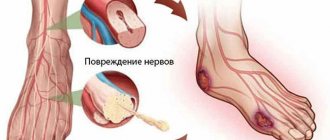
What are the reasons for the development of skin cancer?
Several exo- and endogenous factors of this disease have been proven:
- Exposure to ultraviolet rays, natural and artificial - such as solariums.
- X-ray and gamma radiation, which sooner or later leads to skin radiation dermatitis.
- Infrared rays are most often associated with work (occupational disease).
- Skin viral diseases - human papillomavirus (HPV)
- Exposure to carcinogenic factors, such as: petroleum products, herbicides, coal, mineral oils.
- Frequent use of hair dyes.
- Arsenic intoxication. There are acute and chronic forms; this oncology is most often caused by the chronic form.
- Mechanical damage to the integument.
- Burns, if this is a repeated burn, then the likelihood of illness increases.
- Chronic inflammatory processes on the skin. The etiology can be different: skin tuberculosis, trophic ulcers, fistulas, eczema, etc.
The most popular reason is Ultraviolet Rays (UV) received from the sun; in people who move from a cold country or city closer to the sea or warmth, this pathological neoplasm is detected.
What it is?
Skin cancer is one of the most aggressive forms of malignant tumors, which has a tendency to rapidly progress with the formation of metastases. Every year there is an increase in the number of cases of this pathology, which is also called melanoma, so it is very important to know what the disease looks like, its symptoms and signs at the initial stage. Our article will help with this, which presents photos of skin cancer of different stages, as well as photos of healthy and borderline moles.
As the name suggests, this cancer occurs on the skin. If we talk about the number of cases of malignant formations on the skin, they account for approximately 10% of all cases of cancer. In this case, some features of the disease can be identified:
- The likelihood of getting sick does not depend on gender; the disease is equally typical for both men and women.
- Older patients with a large number of nevi (moles) are at greater risk.
- In addition to age, skin color has a strong influence; skin cancer primarily affects light-skinned people.
- Intense ultraviolet healing, both artificial and natural, has a significant impact and often provokes the appearance of tumors. Therefore, staying on the beach or in the solarium should be dosed.
- Melanoma progresses very quickly, so you should know what it looks like and at the first signs and symptoms, at an early stage, consult an oncologist.
Skin cancer formations are usually located on the hands and face, which are often open and exposed to ultraviolet rays. However, it is important to examine all other areas of the body, such as the legs, feet, and even the scalp.
Additionally, the likelihood of getting sick increases if moles are often injured, exposed to chemical burns, or exposed to heavy metals and various chemical compounds.
Precancerous condition
The onset of skin cancer is facilitated by a variety of skin pathologies, which are called precancerous. They are divided into several groups:
- Optional include the situation when a neoplasm appears due to the simultaneity of several circumstances, for example, radiation injury, trophic ulcer, skin horn, scars, etc.
- Obligate pathologies include borderline pathologies, the probability of the initial stage of skin cancer after which is very high.
Examples of obligate diseases:
- In the presence of xeroderma pigmentosum , which usually appears due to poor heredity, the skin is very sensitive to sunlight. Early signs of pathology usually appear in childhood, which is expressed by profuse redness of the affected skin areas, their swelling and pigmentation. Subsequently, the skin at the site of irritation peels off, looks darkened and scars. Unfortunately, this disease almost always progresses to the initial stage of skin cancer, which is why patients rarely live more than 30 years.
- In the older male population, Bowen's disease is often diagnosed. Its signs are the presence of a single formation on the skin on any part of the body. This disease almost always develops into cancer and is subsequently often complicated by other types.
- With Paget's disease, patients experience severe itching and discomfort in the axillary area, around the nipples and in the genitals, as neoplasms appear there in the form of round red plaques with a distinct outline.
If any of the described precancerous diseases are detected, it is important not to delay going to the doctor, since in such conditions treatment is always required.
Read more about why moles degenerate into melanoma.
Stages
Clarification of the stage of the pathological process is necessary to select effective treatment tactics. There are 5 stages of skin cancer:
- 0 – small, painless formation. There are no metastases;
- 1 – the tumor is mobile, does not cause unpleasant sensations on palpation, up to 2 cm in diameter. There are no metastases;
- 2 – formation up to 5 cm in circumference. Pain, itching and burning appear when you touch the affected area. One site of stasis may be identified;
- 3 – lumpy tumor with limited mobility, more than 5 cm in diameter. It grows into deep skin structures (muscles, bones, cartilage, jaw or orbit). Ulcers appear. Distant metastases are recorded;
- 4– pathology can be of any size. Cancer causes severe intoxication of the body (weight loss, weakness, headache). Multiple metastases affect vital organs (lungs, liver, bones).
For melanoma, a similar classification is not used, since this form uses staging based on the depth of germination into the skin and adjacent tissues.
Basal cell carcinoma is also not divided into stages.
Skin thermography
There is a device that detects infrared (thermal) radiation from the surface of the skin. Color mapping allows you to accurately determine the areas affected by skin cancer. This study is especially effective for skin melanoma, breast cancer, and malignant neoplasms of the thyroid gland.
Leading clinics in Israel
Assuta
Israel, Tel Aviv
Ikhilov
Israel, Tel Aviv
Hadassah
Israel, Jerusalem
The impetus for the beginning of the degeneration of normal cells is external and internal factors. The main risk factor among external conditions is chemical influence - the presence of toxins in the environment (asbestos, fragrances, polyvinyl chlorides, etc.). They negatively affect the DNA of cells, provoking their malignant transformation. Natural physical factors also influence the formation of carcinoma - UV and radio irradiation, microwave fields, etc.
Internal factors that provoke the development of carcinoma include:
- viral infections (human papillomas and others);
- genetic predisposition (it is considered one of the most common causes of this disease).
This type of skin cancer often forms in areas of the skin exposed to sunlight or artificial tanning.
Only advanced stages of the disease are considered mortally dangerous, when we are talking about a malignant form with the growth of the affected area, an aggressive course and with metastases present.
How to identify a tumor at the initial stage?
A characteristic feature of skin cancer is its preferential location on the open body.
Among all cases of skin oncology, about 90% have this particular localization, with the majority of tumor cases observed on the face (more than 70%).
Symptoms of manifestations
Each form and type of skin cancer is characterized by separate symptoms, and identical skin oncology occurs differently in individual patients. But there are also common primary manifestations of symptoms that determine skin cancer:
- The appearance of a persistent spot on the skin with unclear boundaries, gradually increasing in size;
- The formation of a small ulcer that cannot be treated, which gradually becomes painful and bleeding;
- The mole suddenly changed, became darker or changed its structure;
- An unusual nodular bump-like formation of a purple, red, pink or black hue has formed;
- Formation of compactions or hard formations that have a flaky or rough surface;
- The formation of a white, scar-like spot that has a less elastic structure than normal nearby tissue.
Symptoms of skin changes in children
Skin cancer affects pediatric patients relatively rarely (less than 1%). There are two types of cancer: squamous cell and basal cell.
Squamous cell is most often localized on the face, limbs, ears, and scalp, forming against the background of pigmented xeroderma. The formation metastasizes to nearby lymph nodes; distant metastasis is not observed.
In the photo there is a child with the last stage of xeroderma pigmentosum - this is a rare type of skin cancer observed in children
The picture shows cancerous xeroderma pigmentosum of the facial skin, involving the eyes
Basal cell skin cancer also prefers the surface of the face. Such an oncoform is a dense nodule, gradually causing the appearance of similar formations nearby, with which it merges, occupying increasingly larger areas.
Causes
Since in most cases, skin carcinoma forms on open areas of the body, solar radiation is considered the main provoking factor. Experts also identify other reasons for the appearance of a tumor:
- exposure to harmful compounds on the upper layers of the skin: arsenic, tar, tobacco smoke, heavy metals;
- poor nutrition (with a predominance of fatty and fried foods, as well as canned food and marinades);
- Keir's erythroplasia;
- radioactive radiation;
- skin injury;
- radiation dermatitis;
- taking immunosuppressants;
- burns;
- leishmaniasis;
- heredity;
- decreased immunity;
- age (over 65 years);
- Paget's disease;
- infections (human immunodeficiency virus, herpes or hepatitis);
- cutaneous horn;
- alcohol intoxication of the body;
- keratoma senile;
- living in countries with a warm, sunny climate;
- Bowen's disease.
At high risk are people over 40 years of age, fair-skinned people with red hair, those with a large number of moles and freckles, as well as those who abuse tanning beds or have to spend a lot of time in the open sun.
Causes and provoking factors
Oncological diseases today still remain poorly studied . But some of the causes that can cause scalp cancer include:
- influence of harmful substances: nicotine, arsenic, carcinogen;
- radiation exposure;
- decreased immunity;
- heredity;
- exposure to high temperatures or ultraviolet radiation;
- chronic dermatitis and other skin diseases;
- use of hormonal or immunostimulating drugs;
- influence of direct electromagnetic current.
Provoking factors:
- Skin type – people with very fair skin who spend a lot of time under the scorching sun are most likely to get sick.
- Large volume of moles on the head. Especially if they are frequently exposed to injury.
- Disturbances in the functioning of the endocrine apparatus.
- Long-term injury to the same area of skin.
- Paget's or Bowen's disease.
Attention! Most often, skin cancer under the hair occurs when moles are damaged by combing.
Photos of borderline moles
The photo below shows some signs of cancer; in this case, you should definitely visit an oncologist or dermatologist to make a diagnosis.
Photo 3. Borderline moles with some symptoms
Diagnosis of the disease
The diagnosis is made based on visual examination of the affected area of skin. The factors that could trigger the appearance of the tumor are also being clarified.
The next step to establish the correct diagnosis is taking a sample for a biopsy. Microscopic examination of the sample determines the presence or absence of cancer cells.
The course of treatment and treatment tactics are prescribed only after a biopsy.