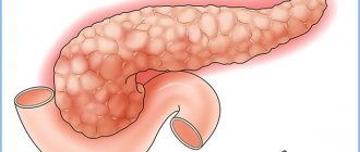
Pulmonary embolism (PE) is a blockage of the artery that transports venous blood from the heart to the lungs for oxygenation by a blood clot.
A pathological clot (thrombus) can form in the veins of the legs, arms or pelvis, or in the atria and ventricles of the heart. This clot is carried by the blood into the pulmonary circulation and blocks the pulmonary artery or its branch (thromboembolism of the branches of the pulmonary artery, thromboembolism of the small branches of the pulmonary artery). If a pulmonary artery or a large branch is blocked by a massive thrombus, death can occur instantly. When small branches of the pulmonary artery in part of the lung are blocked by a thrombus, blood circulation and gas exchange (oxygen and carbon dioxide) worsen and a pulmonary infarction occurs.
If the damage is extensive, the human body ceases to receive a sufficient amount of oxygen and hypoxia occurs. With critical hypoxia due to pulmonary embolism, the patient may die.
Unfortunately, the development of pulmonary embolism is often so rapid that it is not possible to establish a diagnosis during life. The chances of recovery are significantly increased by early diagnosis and anticoagulant therapy in the first hours of the disease.
Thromboembolism spares neither adults nor children, but the main risk group is people aged about sixty years.
Embolism can be gas (the lumen of the artery is closed by an air bubble), bacterial (the reason for blocking the blood flow is a clot of microorganisms), mechanical (clogging with fatty deposits, bone marrow particles).
General information
Pulmonary embolism (abbreviated as PE) is a pathological condition in which blood clots suddenly clog the branches of the pulmonary artery.
Blood clots appear initially in the veins of a person’s systemic circulation. Today, a very high percentage of people with cardiovascular diseases die precisely due to the development of pulmonary embolism. Quite often, pulmonary embolism becomes the cause of death of patients in the period after surgery. According to medical statistics, approximately one fifth of all people with pulmonary thromboembolism die. In this case, death in most cases occurs within the first two hours after the development of embolism.
Experts say that it is difficult to determine the frequency of pulmonary embolism, since about half of cases of the disease go unnoticed. The general symptoms of the disease are often similar to those of other diseases, so the diagnosis is often erroneous.
Classification
To make a correct diagnosis, establish the severity of the pathology and select effective treatment tactics, a detailed classification of pulmonary embolism is used, reflecting all aspects of the manifestation of the pathology.
Depending on the location, pulmonary embolism is divided into left-sided, right-sided, and bilateral.
Blockages can occur at the level of small, large or intermediate blood vessels.
The course of pulmonary thromboembolism is chronic, acute or recurrent.
Doctors, based on the clinical picture of the development of the disease, distinguish:
- Infarction pneumonia, representing thromboembolism of small branches of the pulmonary artery.
- Acute cor pulmonale, in which the disease affects the large branches of the blood vessels of the lungs.
- Recurrent pulmonary embolism of small branches.
Depending on the volume of affected pulmonary blood vessels, the disease can take a massive or non-massive form. This characteristic directly affects the severity of the pathology.
Causes of pulmonary embolism
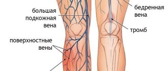
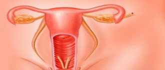
Most often, pulmonary embolism occurs due to blood clots that initially appeared in the deep veins of the legs. Consequently, the main cause of pulmonary embolism is most often the development deep vein thrombosis In more rare cases, thromboembolism is provoked by blood clots from the veins of the right side of the heart, the abdominal cavity, pelvis, and upper extremities. Very often, blood clots appear in those patients who, due to other ailments, are constantly on bed rest. Most often, these are people who suffer from myocardial infarction , lung diseases , as well as those who have suffered spinal cord injuries or undergone surgery on the hip. with thrombophlebitis increases significantly . Very often, pulmonary embolism manifests itself as a complication of cardiovascular diseases: rheumatism , infective endocarditis , cardiomyopathy , hypertension , coronary heart disease .
However, PE sometimes affects people without signs of chronic diseases. This usually happens if a person is in a forced position for a long time, for example, often travels by plane.
In order for a blood clot to form in the human body, the following conditions are necessary: the presence of damage to the vascular wall, slow blood flow at the site of damage, high blood clotting.
Damage to the walls of the vein often occurs during inflammation, during trauma, and also during intravenous injections. In turn, the blood flow slows down due to the development of heart failure in the patient, with a prolonged forced position (wearing a cast, bed rest).
Doctors identify a number of hereditary disorders as the causes of increased blood clotting; a similar condition can also be provoked by the use of oral contraceptives and AIDS . A higher risk of blood clots is found in pregnant women, people with a second blood group, and obese .
The most dangerous are blood clots, which at one end are attached to the wall of the vessel, and the free end of the blood clot is located in the lumen of the vessel. Sometimes only small efforts are enough (a person can cough, make a sudden movement, strain), and such a blood clot breaks off. The blood clot then travels through the bloodstream and ends up in the pulmonary artery. In some cases, the blood clot hits the walls of the vessel and breaks into small pieces. In this case, blockage of small vessels in the lungs may occur.
Diagnostics
Diagnosis of PE begins with a clinical assessment by a physician and may then include specialized tests that can confirm or rule out the diagnosis.
Clinical assessment.
The first step in diagnosing PE is for the doctor to assess whether the person is likely to have PE. The doctor makes this assessment by performing a thorough medical history, assessing risk factors for deep vein thrombosis (DVT), performing a physical examination, measuring the oxygen concentration in the blood, and possibly performing an ultrasound to detect DVT.
Non-invasive tests
After a clinical evaluation by a doctor, special tests, such as blood tests or imaging studies, may be needed.
- D-dimer assay . If the likelihood of thromboembolism is considered low, your doctor may order a D-dimer test. A D-dimer test is a blood test that measures the presence of abnormal levels of clotting activity in the blood, which is expected if a person has DVT or PE. If the clinical likelihood of PE is low and the D-dimer test is negative, PE can be ruled out and the doctor will begin to consider other possible causes of the symptoms.
If the likelihood of PE is assessed as high, or if the D-dimer test is positive, then either a V/Q scan (ventilation/perfusion scan) or a computed tomography (CT) scan of the chest is usually performed.
- V/Q scan : A V/Q scan is a scan of the lungs that uses a radioactive dye injected into a vein to evaluate the flow of blood into lung tissue. If a pulmonary artery is partially blocked by an embolus, less radioactive dye will be delivered to the corresponding portion of the lung tissue and will be visible on the screen.
- Computed tomography (CT) : A CT scan is a non-invasive computerized x-ray procedure that allows the doctor to visualize the pulmonary arteries to see if there is obstruction caused by an embolism.
- Pulmonary angiogram : The pulmonary angiogram has long been considered the gold standard for detecting PE. If the diagnosis is unclear after performing the above tests, your doctor may order a pulmonary angiogram.
Treatment of pulmonary embolism
Once the diagnosis of pulmonary embolism is confirmed, therapy begins immediately. If there is a very high probability of pulmonary embolism, medical therapy may be started even before the diagnosis is confirmed.
Blood thinners - anticoagulants .
The main treatment for pulmonary embolism is the use of anticoagulant blood thinners to prevent further clotting.
Symptoms of pulmonary embolism
Experts define three types of pulmonary embolism, depending on the extent of pulmonary vascular damage observed. With massive pulmonary embolism, more than 50% of the lung vessels are affected. In this case, the symptoms of thromboembolism are expressed by shock, a sharp drop in blood pressure , loss of consciousness, and there is insufficiency of right ventricular function. The consequence of cerebral hypoxia during massive thromboembolism sometimes results in cerebral disorders.
Submassive thromboembolism is determined when 30 to 50% of the pulmonary vessels are affected. With this form of the disease, a person suffers from shortness of breath , but blood pressure remains normal. The dysfunction of the right ventricle is less pronounced.
With non-massive thromboembolism, right ventricular function is not impaired, but the patient suffers from shortness of breath.
According to the severity of the disease, thromboembolism is divided into acute , subacute and recurrent chronic . In the acute form of the disease, pulmonary embolism begins abruptly: hypotension, severe chest pain, and shortness of breath appear. In the case of subacute thromboembolism, there is an increase in right ventricular and respiratory failure, signs of infarction pneumonia . The recurrent chronic form of thromboembolism is characterized by recurrence of shortness of breath and symptoms of pneumonia.
Symptoms of thromboembolism directly depend on how massive the process is, as well as on the condition of the patient’s blood vessels, heart and lungs. The main signs of the development of pulmonary thromboembolism are severe shortness of breath and rapid breathing . The onset of shortness of breath is usually abrupt. If the patient is in a supine position, then it becomes easier for him. The occurrence of shortness of breath is the first and most characteristic symptom of pulmonary embolism. Shortness of breath indicates the development of acute respiratory failure. It can be expressed in different ways: sometimes a person feels like he is a little short of air, in other cases shortness of breath is especially pronounced. Also a sign of thromboembolism is severe tachycardia : the heart contracts at a frequency of more than 100 beats per minute.
In addition to shortness of breath and tachycardia, pain in the chest or a feeling of some discomfort may occur. The pain can vary. Thus, most patients note a sharp dagger pain behind the sternum. The pain can last for several minutes or several hours. If an embolism of the main trunk of the pulmonary artery develops, the pain can be tearing and felt behind the sternum. With massive thromboembolism, pain can spread beyond the sternum. Embolism of small branches of the pulmonary artery can occur without any pain at all. In some cases, coughing up blood, blue or pale lips, ears, and nose may occur.
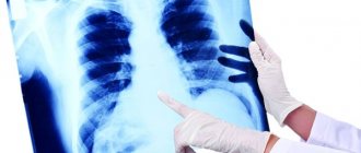
When listening, the specialist detects wheezing in the lungs and systolic murmur over the heart area. An echocardiogram reveals blood clots in the pulmonary arteries and right side of the heart, and there are also signs of impaired right ventricular function. The x-ray shows changes in the patient's lungs.
As a result of the blockage, the pumping function of the right ventricle is reduced, resulting in insufficient blood flowing into the left ventricle. This is fraught with a decrease in blood in the aorta and arteries, which provokes a sharp drop in blood pressure and a state of shock. Under such conditions, the patient develops myocardial infarction and atelectasis .
Often the patient experiences an increase in body temperature to subfebrile, sometimes febrile levels. This is due to the fact that many biologically active substances are released into the blood. The fever may last from two days to two weeks. A few days after pulmonary thromboembolism, some people may experience chest pain, coughing, coughing up blood, and symptoms of pneumonia.
How to treat
Treatment for pulmonary embolism may be:
- conservative;
- minimally invasive;
- operational.
It pursues the following goals:
- emergency removal of a patient from a life-threatening condition;
- elimination of blood clots in the arteries;
- relieving symptoms of the disease;
- restoration of the functionality of the lungs and heart.
The tactics and type of treatment are selected by the doctor taking into account the severity of the disease, concomitant diseases, and the individual characteristics of the patient.
Drug treatment
Drug treatment of pulmonary embolism is carried out using anticoagulants - medications that actively influence blood clotting factors. These agents dissolve existing blood clots and reduce the risk of their formation.
The most common anticoagulants are Warfarin and Heparin. The latter is administered to the patient subcutaneously or intravenously. Warfarin is taken orally. But their long-term use can cause serious consequences - bleeding, cerebral hemorrhage, nausea, vomiting, etc. When taking these drugs, blood clotting should be monitored using a coagulogram.
Today, PE can be treated with safer, effective drugs. These include Apixaban, Dabigatran, Rivaroxaban.
Important information: How to treat chronic idiopathic myelofibrosis and its symptoms
Surgical intervention
In severe forms of pulmonary embolism, conservative treatment becomes ineffective. To save the patient's life, radical measures are required. Indications for surgical intervention for pulmonary embolism should include:
- massive form of the disease;
- ineffectiveness of therapy;
- violation of general circulation;
- relapse, etc.
Pulmonary embolism is eliminated using the following types of surgical interventions:
- embolectomy, which involves removing a blood clot;
- thromboendarterectomy, when the inner wall of the blood vessel is removed along with the plaque.
The operations are complex, involving the opening of the patient's chest and the transition to temporary artificial blood supply to the body.
These interventions are time-consuming and require the participation of high-class specialists - thoracic surgeons and cardiac surgeons.
Today, gentle surgical interventions are often used to eliminate a blood clot:
- catheter embolectomy;
- catheter thrombolysis using medications - streptokinase, alteplase, urokinase.
Manipulations are carried out using a special catheter through small punctures in the skin. The catheter is brought through the main veins to the site of the thrombus, where it is removed under constant computer monitoring.
Installation of a vena cava filter
A vena cava filter is a special mesh-shaped trap designed for detached blood clots. The device is installed in the inferior vena cava and serves for preventive purposes to protect against emboli of the pulmonary artery and heart.
When installing a vena cava filter, minimally invasive treatment methods are used in the form of endovascular intervention. A specialist, through a small puncture in the skin using a catheter through the veins, delivers the mesh to the required location, where it straightens and secures it. The catheter is withdrawn. When installing a trap, the main veins are considered to be the great saphenous, jugular or subclavian veins.
The manipulations are carried out under light anesthesia and last no more than an hour. After this, the patient is prescribed bed rest for 2 days.
Diagnosis of pulmonary embolism
During the diagnostic process, a physical examination of the patient is performed to identify certain clinical syndromes. The doctor can determine shortness of breath, arterial hypotension, and determine body temperature, which increases already in the first hours of the development of pulmonary embolism.
The main methods of examination for thromboembolism should include an ECG, chest x-ray, echocardiogram, and biochemical blood test.
It should be noted that in approximately 20% of cases, the development of thromboembolism cannot be determined using an ECG, since no changes are observed. There are a number of specific signs determined during these studies.
The most informative research method is a ventilation-perfusion scan of the lungs. A study using angiopulmonography is also carried out.
In the process of diagnosing thromboembolism, an instrumental examination is also indicated, during which the doctor determines the presence of phlebothrombosis of the lower extremities. Radiocontrast venography is used to detect venous thrombosis. Carrying out Doppler ultrasound of the vessels of the legs allows us to identify violations of the patency of the veins.
Prognosis for pulmonary embolism
Pulmonary embolism is a life-threatening disease. The prognosis of patients depends on several factors - the presence of concomitant diseases, timely diagnosis and correct treatment.
Approximately 10% of patients with pulmonary embolism die within an hour of the onset of the disease, 30% die later from repeated pulmonary embolism.
Mortality rates also depend on the type of PE. Life-threatening pulmonary embolism, which is characterized by a drop in blood pressure, has a mortality rate of 30–60%.
Prevention of pulmonary embolism
To prevent thromboembolism, it is important to know which conditions predispose to the occurrence of venous thrombosis and thromboembolism. People who suffer from chronic heart failure, are forced to remain in bed for a long time, undergo massive diuretic treatment, or take hormonal contraceptives for a long time should be especially attentive to their own condition. In addition, a number of systemic connective tissue diseases and systemic vasculitis , diabetes mellitus . The risk of developing thromboembolism increases with strokes , spinal cord injuries, prolonged stay of the catheter in the central vein, the presence of cancer and chemotherapy. with varicose veins of the legs , obese people, and cancer patients should be especially attentive to their own health Therefore, to avoid the development of pulmonary embolism, it is important to get out of postoperative bed rest in a timely manner and treat thrombophlebitis of the leg veins. For people at risk, preventive treatment with low molecular weight heparins is indicated.
To prevent manifestations of thromboembolism, it is periodically relevant to take antiplatelet agents : this may include small doses of acetylsalicylic acid .
Prevention
Preventing pulmonary embolism is preventing deep vein thrombosis; The need for it depends on the patient's risks, including:
- type and duration of surgery;
- concomitant diseases, including cancer and hypercoagulability disorders;
- presence of a central venous catheter;
- History of DVT or PE.
Bedridden patients and patients undergoing surgery, especially orthopedic surgery , have an advantage, and most of these patients can be identified before a clot forms. Preventative recommendations include low-dose unfractionated heparin, low-molecular-weight heparins, warfarin, fondaparinux, oral anticoagulants (rivaroxaban, apixaban, dabigatran), and the use of compression devices or elastic compression stockings.
The choice of drug or device depends on various factors, including patient population, perceived risk, contraindications (such as risk of bleeding), relative costs, and ease of use.
Healthy people who simply want to protect themselves from this disease need to undergo constant diagnostics (once every 6 months), exercise, keep their weight under control and be sure not to smoke.
Diet, nutrition for pulmonary embolism
Diet for deep vein thrombosis of the lower extremities
- Efficacy: the therapeutic effect is achieved after 2 months
- Terms: 2-6 months
- Cost of products: 1600-1700 rubles. in Week
Diet for coronary heart disease
- Efficacy: therapeutic effect after 30 days
- Timing: constantly
- Cost of products: 1700-1800 rubles. in Week
What is TELA
Pulmonary embolism - what is it? There is a disruption in the dynamic balance, and blood clots begin to form on the walls of the bloodstream . They grow, becoming large, mobile, loose.
Over time, the blood clot is rejected from the bloodstream and begins to migrate throughout the circulatory system.
Most often, the formation of such a clot occurs in the lower extremities or in the capillaries of the small pelvis.
Attention! Initially, a dangerous clot is transformed through a large blood circle along the vena cava from the lower sections to the right atrium. From there it moves to the right ventricle of the myocardium. Later it is transported along with the blood to the pulmonary artery.
If the embolus has not reached a large size, then it can be transported through the veins for a long period of time until it dissolves in the lymph . Otherwise, when the thrombus is large in size, blockage of a large artery or one of its branches is a matter of time.
So, TELA – what is it? A life-threatening condition in which a broken blood clot clogs a blood vessel in the lungs. The consequences of such blocking are disastrous.
Reasons for the development of the disease
During the development of pathology, clots form and block blood vessels. Among the causes of pulmonary embolism are the following:
- Impaired blood flow. This can be observed against the background of the development of: varicose veins, compression of blood vessels by tumors, previous phlebothrombosis with destruction of vein valves. Blood circulation is disrupted when a person is forced to remain immobile.
- Damage to the wall of a blood vessel, resulting in the process of blood clotting.
- Vein prosthetics.
- Installation of catheters.
- Vein surgery.
- Infectious diseases of a viral or bacterial nature that provoke damage to the endothelium.
- Disruption of the natural process of fibrinolysis (dissolution of blood clots) and hypercoagulation.
The combination of several causes increases the risk of developing pulmonary embolism; clinical pathology requires long-term treatment.
How is the treatment carried out?
In case of thromboembolic syndrome, the patient is sent to the intensive care unit for emergency procedures. Considering the severity of the disorder, the necessary further therapy is prescribed. It is possible to eliminate the symptoms of thromboembolism using conservative and surgical methods. In the first case, treatment is carried out with low severity of ischemia. Pharmacy medications are also prescribed if the operation is impossible. In case of violation, the drugs presented in the table are used.
| Medicines | Action | Name |
| Anticoagulants | They thin the blood, causing the clot to slowly dissolve | "Heparin" |
| "Warfarin" | ||
| Thrombolytic drugs | Dissolving a blood clot | "Streptase" |
| "Urokinase" | ||
| Antiseptics | Eliminate spasms in blood vessels | "Memorel" |
| "Memantine" |
When large vessels are blocked, the clot is removed surgically.
It is strictly forbidden to use blood thinners for symptoms of hypertensive crisis or internal bleeding. If conservative methods do not give a positive result, then surgery is performed. During surgery, the blood clot is removed using a scalpel, after which blood flow is normalized. Open surgery is required for thromboembolism of large arteries.
Treatment
Treatment of thromboembolism is an attempt at a complex and rather aggressive effect on various body systems. The main objective of such measures is to minimize irreversible changes in ischemic organs and tissues, as well as create the prerequisites for recanalization of the thrombus and restoration of adequate blood supply to the affected area. Operations to remove a blood clot themselves can cause further development of thrombosis as a postoperative complication, so their use is limited. The medications used to provide assistance can be represented as follows:
- anticoagulants;
- disaggregants;
- thrombolytics;
- angioprotectors;
- drugs for the correction of cardiac and respiratory disorders;
- solutions for infusions;
- metabolic agents.
Anticoagulants are the gold standard in the treatment and prevention of various thrombotic diseases and complications. Heparin remains the very first representative of this group of medications, but due to many circumstances, today it is used extremely rarely. The modern pharmacological industry produces a large number of derivatives of this drug, with a more pronounced therapeutic effect and minimized side effects.
Thrombolytics are medications that have the ability to dissolve fresh blood clots. The effectiveness of their use is limited by strict time frames. Antiplatelet agents can significantly reduce blood viscosity. Thereby reducing the risk of the formation of fresh clots and improving blood flow in the microvasculature.
The main methods of therapy should be aimed at providing medical care to patients with the causative pathology that causes thromboembolism. People with diseases of the veins of the lower extremities should not forget about the prevention and treatment of their illness. Even the use of compression garments (stockings, knee socks) can play an important role. Heart valve pathology must be treated effectively and promptly.
Treatment of thromboembolism is a complex process and not always successful. The use of folk remedies in an attempt to cure the pathology will only take away precious time. You cannot influence a blood clot by using stockings made of “special” fabric and so on. Diet and nutritional regimen create a minimal background impact on the pathology, except during the rehabilitation period. Alternative medicine can be used to combat the consequences of chronic leg ischemia, for example, if a fungal infection occurs.
Diagnostic measures
In diagnosing this disease, it is important to establish the location of blood clot formation in the pulmonary arteries, as well as to assess the degree of damage and the severity of the disorders. The doctor is faced with the task of determining the source of thromboembolism in order to prevent relapse.
Given the difficulty of making a diagnosis, patients are sent to special vascular departments, which are equipped with technology and have the ability to conduct comprehensive research and therapy.
If PE is suspected, the patient undergoes the following examinations:
- Taking anamnesis and assessing all risk factors.
- General blood and urine analysis.
- Blood test for gas composition, determination of D-dimer in plasma.
- Dynamic ECG to rule out heart attack or heart failure.
- X-ray of the lungs to exclude pneumonia, pneumothorax, malignant tumors, pleurisy.
- Echocardiography is performed to detect high pulmonary artery pressure.
- Lung scintography will show decreased or absent blood flow as a result of the development of pulmonary embolism.
- To detect the exact location of the thrombus, angiopulmonography is prescribed.
- Doppler ultrasound of the veins of the lower extremities.
- Contrast venography to detect the source of pulmonary embolism.
After an accurate diagnosis is made and the cause of the disease is discovered, therapy is prescribed.
What treatment is prescribed?
Urgent Care
If you suspect a blood clot in the lungs, you must urgently call an ambulance; any bodily movements and transportation of the patient with your own transport are strictly prohibited. The standards for resuscitation measures are as follows:
- immobilization of the patient, minimal physical activity;
- installation of a catheter in the main vein, through which medications will be introduced into the bloodstream;
- intravenous injection of Heparin;
- administration of oxygen using a catheter or mask;
- dropper with “Dopamine”, “Reopoliglyukin”;
- use of antibiotics, if necessary.
Emergency care is aimed at normalizing blood circulation in the lungs, preventing the development of sepsis and chronic pulmonary hypertension. After first aid has been provided, it is important for the doctor to urgently establish an accurate diagnosis, then carry out a differential diagnosis. In addition, the etiology of the development of thromboembolism is simultaneously clarified.
Drugs
Fraxiparine is prescribed to dissolve a blood clot.
Drug therapy is aimed at resolving the blood clot and preventing relapse. For these purposes, the following medications are used:
- "Fraxiparin";
- "Heparin";
- "Streptokinase";
- "Urokinase."
Thrombolytic therapy is strictly prohibited after surgery or in the presence of pathologies that can cause heavy bleeding, because drugs that dissolve blood clots thin the blood, which increases the risk of bleeding.
Surgery
Before the operation, the patient is provided with psychological support and prescribed a course of sedatives and hypnotics. Surgery is prescribed if PE affects more than 50% of the lungs. During surgery, the surgeon removes the clot and frees the vessel. Complex surgical intervention is necessary if there is a blockage of the artery trunk or large branches, because blood flow is disrupted throughout the entire area of the lungs. The operation will be followed by rehabilitation and prevention of pulmonary embolism.
Other methods
Catheter fragmentation is a procedure that prevents a blood clot from entering the heart.
If for some reason surgical treatment is contraindicated, there is an alternative method of removing the blood clot - catheter fragmentation. The essence of the method is that a special filter is installed into the affected vessel through a micropuncture, which prevents the detached blood clot from reaching the heart muscle.
Epidemiology
PE is a relatively common cardiovascular pathology (approximately 1 case per thousand population per year). In the United States, PE affects approximately 650,000 people and results in 350,000 deaths per year. PE occurs more often in the elderly (In the United States, venous thromboembolism occurs for the first time in about 100 out of 100,000 people per year and increases exponentially from less than 5 cases per 100,000 people in children under 15 years of age to 500 cases per 100,000 people at age 80 years) . Data on the association of gender with PE vary, but an analysis of a US national database found that men have a 20% to 30% higher mortality rate than women. The incidence of venous thromboembolism among the elderly is higher in men, but in patients under 55 years of age it is higher in women.
Diagnostic methods
MRI is a fast and effective method for diagnosing vascular blockages.
Arterial and venous thromboembolism is confirmed through diagnostic procedures. A phlebologist or surgeon deals with such violations. The doctor examines the damaged area and prescribes an emergency examination. The patient's blood is taken for laboratory testing, MRI and venography with a contrast agent are performed. An effective diagnostic method for thromboembolism is ultrasound examination with Doppler.
Risk groups for pulmonary embolism
Antithrombotic therapy is indicated for the following categories of patients:
- with prolonged bed rest - physical inactivity leads to the formation of unstable blood clots, which are prone to destruction and detachment from the wall;
- in the case of a planned operation, stress and pain stimulate the formation of blood clots; a long operation, especially in the pelvic cavity, lower extremities increases the risk of venous thrombosis. Any tissue damage also triggers the release of clotting factors. Blood loss and centralization of blood circulation increase blood viscosity and thrombophilia;
- when using general anesthesia, too strong or prolonged anesthesia causes hypercoagulation and provokes stagnation of blood in the legs;
- after administration of whole blood or red blood cells;
- elderly - after 40 years, anticoagulant activity decreases, the structure of the vascular wall is disrupted, cardiac output and the return of venous blood to the heart decrease, the risk of thrombosis increases with previous physical inactivity;
- with malignant neoplasms – blood clotting is activated;
- with excess body weight – thrombosis is associated with impaired fat metabolism;
- history of venous thrombosis, thromboembolism, varicose veins.
Additional risk factors include heart failure, atrial fibrillation, bacterial endocarditis, myocarditis, and cardiomyopathy. PE is often detected in patients with diabetes mellitus, sepsis, congenital thrombophilia, and antiphospholipid syndrome.
Pregnancy, especially complicated by gestosis, the labor and postpartum periods are also included in the list of risk factors. Accelerated formation of blood clots can be provoked by the use of hormones for contraception, treatment of menopause, kidney and intestinal diseases, heart attack and stroke, antitumor radiation or chemotherapy.
Complications
An organ infarction may be a consequence of late care provided to the patient.
If a person is not given first aid or treatment is not timely, the following consequences develop:
- pulmonary infarction;
- paradoxical embolism of large vessels;
- chronic hypertension in the pulmonary vessels.
Statistics show that if the pathology develops at lightning speed, the survival rate is very low.
Urgent Care
Emergency care measures for pulmonary embolism can be divided into three groups:
1) maintaining the patient’s life in the first minutes of pulmonary embolism;
2) elimination of fatal reflex reactions;
3) elimination of the embolus.
Life support in cases of clinical death of patients is carried out primarily by resuscitation. The priority measures include the fight against collapse with the help of pressor amines, correction of the acid-base state, and effective oxygen barotherapy. At the same time, it is necessary to begin thrombolytic therapy with native streptokinase drugs (streptodecase, streptase, avelysin, celease, etc.).
The embolus located in the artery causes reflex reactions, due to which severe hemodynamic disorders often occur with non-massive pulmonary embolism. To eliminate pain, 4-5 ml of a 50% analgin solution and 2 ml of droperidol or seduxen are injected intravenously. Drugs are used if necessary. In cases of severe pain, analgesia begins with the administration of drugs in combination with droperidol or seduxen. In addition to the analgesic effect, the feeling of fear of death is suppressed, catecholaminemia, myocardial oxygen demand and electrical instability of the heart are reduced, and the rheological properties of blood and microcirculation are improved. In order to reduce arteriolospasm and bronchospasm, aminophylline, papaverine, no-spa, and prednisolone are used in normal doses. Elimination of the embolus (the basis of pathogenetic treatment) is achieved by thrombolytic therapy, started immediately after the diagnosis of PE. Relative contraindications to thrombolytic therapy, which exist in many patients, are not an obstacle to its use. The high probability of death justifies the risk of treatment.
In the absence of thrombolytic drugs, continuous intravenous administration of heparin at a dose of 1000 units per hour is indicated. The daily dose will be 24,000 units. With this method of administration, relapses of pulmonary embolism occur much less frequently, and rethrombosis is more reliably prevented.
When clarifying the diagnosis of pulmonary embolism, the degree of occlusion of the pulmonary blood flow, and the localization of the embolus, a conservative or surgical treatment method is selected.
Diagnosis of the disease and treatment method
It is not always possible to accurately and timely diagnose the pathology due to its atypical course. Some people have a fever, a cough, a rise in blood pressure, and often pain in the abdomen, which is accompanied by diarrhea. In most cases, these symptoms are not differentiated from thrombophlebitis at all, which is the main mistake.
In people with cardiovascular diseases, differential diagnosis is required. With a massive form of pulmonary embolism, the patient's upper pressure does not exceed 90 mm Hg. Art., and in addition, there is a fainting state and loss of consciousness.
Submassive pulmonary embolism is characterized by improper functioning of the right ventricle of the heart, and the myocardium may be damaged. The non-massive form is the worst for diagnosis, as it is confused with heart attacks and lung diseases. With this type, special attention should be paid to shortness of breath, which accompanies a person constantly. When the heart is listened to, with the form described, murmurs occur in the lung area.
Among the instrumental diagnostic methods for thrombophlebitis the following can be noted:
- Computed tomography, thanks to which blood clots can be found in the branches of the pulmonary artery.
- Ultrasound of the veins allows you to diagnose the initial stages of embolism formation.
- Doppler ultrasound makes it possible to detect segments of arteries where the speed of blood flow is impaired. If there is a decrease in blood flow, a clot is likely blocking the lumen.
- X-ray - thanks to contrast agents, the method allows you to accurately see the location of the blood clot and even its size.
- ECHO-CG of the heart - allows you to see the expansion of the right ventricle, which is a typical symptom of PE, makes it possible to detect other abnormalities and completely examine the vessels of the heart.
Based on laboratory tests and instrumental diagnostic techniques, the doctor prescribes an individual treatment regimen for the patient. The person’s age, body weight, the nature of the manifestation of the disease and concomitant pathologies that are present in the body are taken into account.
A conservative form of treatment for thromboembolism is carried out if the patient has a non-massive form of pathology. The patient needs to be provided with complete rest and a supine position with his head elevated, unless, of course, there is collapse.
If the latter is present, therapeutic measures are taken to eliminate acute vascular insufficiency. If there is pain in the chest, analgesics and antispasmodics are administered. Regular oxygen inhalations are required. For cardiac weakness, glycosides such as Strophanthin and Korglikon are indicated for use. Antihistamines are mandatory: Diphenhydramine, Pipolfen, Suprastin, etc. Thrombolytic and anticoagulant therapy will be important.
Clinical manifestations of pulmonary embolism
The symptoms of this condition are determined by the volume of the pulmonary arteries involved in the pathological process and their size. They are also influenced by the speed at which thromboembolism develops, the extent of disturbances in pulmonary blood flow and the initial condition of the patient. All this causes a wide range of conditions: from the absence of symptoms to death within a few minutes. If we take the classic development of pathology as an example, the symptoms will be as follows:
| Syndrome | Clinical manifestations |
| Cardiovascular |
|
| Pulmonary-pleural |
|
| Immunological |
|
| Feverish | Body temperature 38–39 °C for two to twelve days. |
| Abdominal | Acute pain symptoms in the right hypochondrium, vomiting. |
Signs of the disease
Symptoms of pulmonary embolism depend on many factors:
- General condition of the patient's body.
- Number of damaged arteries.
- The size of the particles that clog the blood vessels.
- Rate of disease progression.
- Degree of disturbances in lung tissue.
Treatment of pulmonary embolism will depend on the clinical condition of the patient. For some, the disease occurs without causing any symptoms and can lead to sudden death. The difficulty of diagnosis also lies in the fact that the pathology’s symptoms resemble many cardiovascular diseases, but the main difference is the suddenness of the development of pulmonary embolism.
The pathology is characterized by several syndromes:
1. From the cardiovascular system:
- Development of heart failure.
- Reduced blood pressure.
- Heart rhythm disturbance.
- Increased heart rate.
- The development of coronary insufficiency, which manifests itself as sudden intense pain in the chest, lasting from 3-5 minutes to several hours.
- Pulmonary heart syndrome is manifested by swelling of the veins in the neck, tachycardia.
- General cerebral disorders with hypoxia, cerebral hemorrhages and, in severe cases, cerebral edema. The patient complains of tinnitus, dizziness, vomiting, convulsions and fainting. In severe situations, there is a high probability of developing coma.
2. Pulmonary-pleural syndrome manifests itself:
- The appearance of shortness of breath and the development of respiratory failure. The skin becomes gray and cyanosis develops.
- Whistling sounds appear.
- Pulmonary infarction most often develops 1-3 days after pulmonary embolism, a cough appears with the discharge of sputum mixed with blood, the body temperature rises, and when listening, moist fine bubbling rales are clearly audible.
3. Feverish syndrome with the appearance of febrile body temperature. It is associated with inflammatory processes in the lung tissue.
4. Enlarged liver, irritation of the peritoneum, and intestinal paresis cause abdominal syndrome. The patient complains of pain in the right side, belching and vomiting.
5. Immunological syndrome is manifested by pulmonitis, pleurisy, skin rashes, and the appearance of immune complexes in a blood test. This syndrome usually develops 2-3 weeks after the diagnosis of PE.
Clinical recommendations for the development of such symptoms are to begin urgent therapy.
Causes
The root cause of thromboembolism is a thrombus-embolus detached from the vascular wall, which, with the blood flow, enters a particular vessel and clogs it. Subsequently, ischemia develops in the affected area, causing symptoms to develop.
The following diseases or conditions can predispose to the formation of thrombus emboli and the development of thromboembolism:
- prolonged stay in one position and sudden rise (for example, prolonged bed rest, flight or trip);
- taking medications that increase blood viscosity;
- thrombophilia (blood clotting disorder);
- some surgical operations;
- atherosclerosis;
- antiphospholipid syndrome;
- arterial hypertension or hypertensive crisis;
- stroke;
- diseases of the heart and blood vessels;
- varicose veins;
- generalized sepsis;
- fractures of large bones;
- bleeding;
- burns or frostbite;
- malignant tumors;
- smoking;
- dehydration;
- pregnancy and childbirth;
- elderly age.
Prognosis and prevention
With non-massive thromboembolism and timely provision of adequate medical care to the patient, the prognosis for life is favorable. Severe concomitant pathology and late medical intervention significantly worsens the prognosis.
If after the first thromboembolism the patient does not receive anticoagulant therapy, there is a high risk of relapse within the first 3 months. Accordingly, properly administered anticoagulant therapy reduces the likelihood of developing recurrent thromboembolism by more than 2 times.
To prevent the occurrence of pulmonary embolism, provoking factors that are determined in a particular patient should be promptly eliminated: varicose veins, thrombophlebitis and others (read above).
Another method of preventing pulmonary embolism is the installation of a filter – a vena cava filter – in the inferior vena cava. They can be temporary (installed for the period of surgery, childbirth, or in other situations that promote thrombus formation) and permanent (installed for already diagnosed deep vein thrombosis, accompanied by the danger of blood clot rupture). Once in the filter, the blood clot is crushed and then easily dissolved by anticoagulants taken by the patient.
What is pulmonary embolism (PE)?
PE is a pathological, life-threatening condition that occurs as a result of occlusion of the pulmonary artery and its branches by a thrombus. It is accompanied by impaired blood flow to the lungs, which is manifested by decreased blood oxygenation, pain symptoms at chest level, suffocation, bluishness of the face, symptoms of pulmonary infarction and other clinical manifestations.
The symptoms of pulmonary embolism are similar to some other conditions, so differential diagnosis using instrumental methods is mandatory. Treatment is carried out using conservative therapy and surgery.
It is important to understand that PE can develop very quickly and, in some cases, be fatal within minutes.
Pathophysiology of pulmonary embolism
A typical manifestation of pulmonary embolism is the development of pulmonary hypertension. In initially healthy people, it occurs when the threshold value of embolic obstruction is exceeded, i.e. occlusion of 50% of the pulmonary circulation. An increase in this indicator leads to a further increase in total pulmonary vascular resistance, an increase in pressure in the right heart and pulmonary trunk, a decrease in cardiac output and oxygen tension in arterial blood. In the acute stage of the disease, massive embolism can lead to a rise in pressure in the pulmonary circulation no higher than 70 mm Hg. Art. Exceeding this parameter indicates a prolonged nature of embolic occlusion or the presence of concomitant cardiopulmonary pathology causing right ventricular hypertrophy. Until a certain time, a stable value of systemic pressure is maintained due to an increase in vascular resistance of the systemic circulation.
The severity of hemodynamic disorders during thromboembolism is entirely determined by the factor of mechanical obstruction of the pulmonary arterial bed. This pattern should be the determining basis for the surgeon’s tactical and therapeutic actions aimed at restoring the patency of the pulmonary arteries.