Benign tumor – fibroadenoma
As practice shows, in nine out of ten cases, the formed breast tumor is a fibroadenoma. The disease is common to both men and women, although it is most common among the fair sex between the ages of 14 and 35 years. And this is perhaps one of the main differences between a fibroadenoma and a cyst and a fatty lobule.
Breast fibroadenoma at a young age most often develops due to unnatural or abnormal growth of adipose tissue in the chest area. Other causes of diseases can be more accurately determined by puncture of the mammary gland. Among them may be:
- diseases associated with the human endocrine system;
- hereditary predisposition and genetic characteristics;
- early pregnancy or the first months after the birth of a child;
- puberty in girls (young people can be excluded in this case);
- stress, fatigue and constant nervous tension.
Characteristics of breast diseases, or rather, knowledge of it, will help you determine in the best way whether a cyst is found in your breast or one of the types of fibroadenoma. As for the latter disease, it is worth noting that fibroadenoma is a neoplasm of the mammary gland, one of the forms of mastopathy and a type of benign tumor. It has a focal nature of distribution, and cannot form in several places at once in one mammary gland. Moreover, the extensive practice of mammology allows us to determine with almost one hundred percent accuracy the exact location of the formation - the upper right quadrant of the breast.
Please note that breast fibroadenoma rarely causes pain upon palpation. The same cannot be said about such a benign tumor as a cyst. The disease is not associated with the epidermis, which means that puncture can determine not only the nature of the disease, but also its type.
Another nuance that distinguishes fibroadenoma from a fatty lobule or cyst is the absence of any clear contours, which is also determined by puncture.
Upon careful examination of the disease, you will notice that the fibroadenoma itself is enclosed in a capsule. It can roll around inside the mammary gland.
What is glandular tissue of the mammary gland, its functions
Structure of the mammary gland
The mammary gland is divided into small lobules due to connecting layers, which diverge from the skin in different directions. The cavity located between the lobules is filled with fatty tissue. It plays the role of a kind of shock-absorbing pillow on which the gland itself is located.
The glandular tissue itself and its structure consists of a large number of small glands. They are located directly in the lobules of the mammary gland. Each section has tubes, the ends of which are slightly expanded. These small expansions are called alveoli.
Throughout a person’s life, the mammary glands undergo constant changes, for example, during puberty they actively grow. This condition is accompanied by the production of certain substances that stimulate the development and functioning of the breast. During menstruation, pregnancy, lactation and hormonal changes, a woman's breasts increase in volume and become more sensitive.
Every month during menstruation and after the end of regulation, the breasts undergo changes. With the beginning of the second phase of the menstrual cycle, the body begins to actively produce progesterone, which stimulates the growth of alveoli. At the end of the cycle, everything returns to its original state.
With the onset of pregnancy, the continued production of progesterone causes the alveoli to mature. In the last stages of pregnancy, the body begins to produce prolactin, which affects the state of organic structures. A woman’s body is intensively preparing for breastfeeding.
Macrophages in preinvasive lesions
Macrophage-type cells in the mammary gland are the main cells that regulate phagocytosis. The essence of this process is to recognize and destroy foreign particles, cancer cells, toxins and other pathogenic microorganisms. Microphages do not function in the blood, but in the tissues of the mammary gland. If during a medical examination they were detected in high concentrations, it is worth assuming that a pathological process is occurring in the chest.
Hemosiderophages and xanthoma cells in the mammary gland are a type of macrophages that are found in connective tissue.
During a laboratory examination, a laboratory employee is faced with the task of finding and studying the structure of all the types of macrophages found. They always contain a large amount of “garbage”, which is the remains and fragments of the aggressors they have digested. If during the study it was possible to recognize fragments located inside macrophages, an accurate diagnosis of the disease will be made.
There are cases when macrophages are connected to red blood cells and hemoglobin destruction products. This state of affairs indicates that the cavities of the mammary gland contain blood. Such conditions are explained by the development of certain forms of mastopathy, with severe injury to soft tissues.
Cyst as a benign tumor
Breast cysts are equally common in both benign and malignant manifestations. The main difference between a cyst and a fibroadenoma is that the tumor can be single or multiple, and develop simultaneously in both mammary glands. Quite often, a cyst forms and subsequently develops in the milk ducts. The puncture can provide accurate information about the location of the spread of nodular neoplasms.
The main characteristics of cystic formation include the following:
Cystic formations can be the cause of mastopathy that was not diagnosed in time. Doctors also identify a risk group, which consists of young girls under the age of 30 who have not yet been pregnant.
A cyst is almost always accompanied by painful sensations, unlike the same fibroadenoma. If you regularly conduct self-examination of your mammary glands, you will probably be able to detect nodular neoplasms at an early stage in order to get rid of them in time.
Diagnostics
If any inflammatory formations occur in the nipple area, it is necessary to carry out a set of diagnostic measures to confirm or exclude Paget's disease.
After examining and palpating the mammary glands, the mammologist-oncologist will prescribe instrumental and laboratory tests that can detect changes in the tissues of the organ and determine the nature of these changes:
- Ultrasound of the breast,
- mammography,
- MRI,
- scintigraphy,
- cytological examination of biopsy material,
- a swab of nipple discharge.
Paget's disease must be differentiated from the following pathologies:
- eczema,
- dermatitis,
- melanoma,
- psoriasis,
- mycosis fungoides,
- tuberculosis,
- Bowen's disease.
Learn about the symptoms of gestational diabetes during pregnancy, as well as the treatment options for the disease.
What a masectomy is and how to perform surgical intervention is written on this page.Go to https://fr-dc.ru/hormones/progesteron/nedostatochnost.html and see a selection of effective methods for treating progesterone deficiency in women.
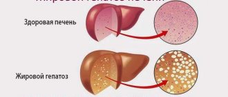
Fat lobule
The fatty lobule of the mammary glands is most often detected by a diagnostic method such as puncture. Quite often, this type of disease is also referred to as fat necrosis, as a more medical and understandable term. Because it is the lobule that causes aseptic necrosis of the mammary glands.
So, a fat lobule is a neoplasm in both or only one mammary gland, which can be directly related to the skin. You can often observe nipple retraction and painful condition of the areola. Fat necrosis, although it is more likely a benign tumor, quite often, as puncture shows, it can develop into a malignant one. Moreover, during the initial diagnosis it is extremely difficult to determine the nature of the fat lobule tumor.
During the development of fat necrosis, a focus of the disease appears, which can be surrounded by a capsule with a dense wall. Note that similar characteristics are observed in both fibroadenoma and cyst.
The presence of a membrane around the filling site is evidence that fat necrosis is a benign tumor. Its absence is the reason for a puncture to exclude the possibility of malignancy.
The most informative diagnostic method is a biopsy. Often it is necessary, although it can be harmful. In advanced stages, the disease is treated with surgery.
Having examined the three most similar types of benign tumors, you can see that they have a lot in common, for example, the characteristics of each type of disease. This is often the reason for an erroneous diagnosis, and, consequently, incorrect treatment. Carefully study the characteristics of each tumor and then the likelihood of error will be reduced to a minimum.
The mammary glands are modified sweat glands with an apocrine type of secretion. Glandular tissue
is of ectodermal origin.
By the time of puberty, the mammary glands reach full development, which reaches its maximum after the first birth of a full-term pregnancy. Under the influence of hormonal stimulation during pregnancy, there is a gradual increase in the number of glandular lobules
.
During the growth and development of the mammary gland, four types of glandular lobules
.
The first type of lobules
are the least differentiated and are known as
virgin lobules
, as they represent the immature female breast before menarche.
Lobules of this type have from 6 to 11 ducts.
Lobules of the second type
evolve from lobules of the first type, the glandular epithelium in them acquires extensive morphological differentiation, characteristic of glands during reproductive age outside of pregnancy. The number of ducts also increases, correspondingly about 47 per lobule.
Lobules of the third type
evolve from lobules of the second type, have an average of 80 ducts or alveoli per lobule. These lobules are already formed under the influence of hormonal stimulation during pregnancy.
And finally, the fourth type of lobules
is presented in women with lactation and reflects the maximum differentiation of the glandular component and the development of the mammary glands during lactation. There are about 120 ducts in the lobules of this type. These lobules are not found in women who have not been pregnant. After the end of lactation, the fourth type lobules regress into the third type lobules. After the onset of menopause, involutional changes occur in the mammary gland in both parous and nulliparous women. This is manifested by an increase in the number of lobules of types 1 and 2. At the end of the fifth decade of life, the mammary gland of parous and nulliparous women consists mainly of type 1 lobules.
Normally, the main tissue elements of the mammary glands, with the help of which their role in reproductive function is realized, is represented by a combination of epithelial and stromal
fabrics.
Epithelial elements
are represented by branching ducts that are associated with the functional units of the gland - lobules and nipple.
Stroma
consists of varying amounts of adipose and fibrous connective tissue that form the volume of the gland itself outside of lactation periods.
At birth, the epithelial component of the mammary gland is represented by a small number of rudimentary ducts located deeper than the nipple-areola complex. During the prepubertal period, these ducts slowly grow and branch, accompanied by an increase in the stromal component. In the postpubertal period, the endings of the ducts form sacular buds, with accompanying growth of the stroma, which increases the volume of the gland during this period. During pregnancy, many glands develop from each bud.
By the end of pregnancy, the glandular component increases to such an extent that the mammary gland consists entirely of glandular tissue, with a small amount of stroma.
After the end of lactation, atrophy of the glandular tissue is noted and the stroma again becomes the dominant component of the mammary gland
.
After menopause, atrophy of glandular components occurs
with a pronounced decrease in the number of lobules to such an extent that in some areas of the glands the lobules disappear completely and only the ducts remain. The connective tissue component of the stroma also decreases, while the adipose tissue of the stroma increases in its content.
From this brief description of changes in the epithelial and stromal elements of the mammary glands depending on the periods of the reproductive cycle, it clearly follows that all these rearrangements are based on physiological, but multidirectional processes of proliferation and apoptosis
, providing the final result with adequate changes in the structure and function of the glands in accordance with the tasks in each age period of the reproductive cycle.
, which in the predominant number of cases are based on cellular hyperplasia, form a rather heterogeneous group of disorders.
In relation to this pathology, the doctor usually solves two diagnostic problems:
firstly, to exclude a malignant neoplasm in a palpable formation, and secondly, when conducting a histological examination (according to indications), to obtain useful information regarding the morphological characteristics of the observed changes (Semiglazov V.F. et al., 1992).
In this regard, the tendency to consider clinically benign changes in the mammary glands in terms of assessing the possible risk of developing a malignant process in the future is indicative (which seems quite correct).
To illustrate what has been said here, it is appropriate to cite the jointly developed decision of the “Conciliation Commission,” which included forty prominent specialists from the American College of Pathologists on the problem of benign breast processes (October 3–5, 1985, New York, USA). The adopted document was based on the results of prospective observations performed by WD Dupont and DL Page (1985) in a large group of patients (1500 people). They underwent a biopsy for clinically benign breast tumors, and their fate was followed over a significant period of time.
In accordance with the results obtained, all benign changes in the mammary glands were divided into three groups according to the relative risk of developing cancer.
1st group.
Non-proliferative processes
(no risk of malignancy).
Types of breast diseases
All pathologies of the mammary glands can be divided into three large groups: diseases of an inflammatory nature (mastitis), benign tumors (mastopathy) and oncological diseases (breast cancer).
Mastitis
Mastitis is an inflammation of the mammary gland that usually appears during breastfeeding. The causative agents of the disease are infections that entered the gland through cracks in the nipple (streptococci, enterobacteria, staphylococci, etc.). And the appearance of stagnation of milk in the glands further intensifies the inflammatory process.
The disease is accompanied by acute pain in the chest, redness of the skin, swelling and distension of the mammary glands, the appearance of a lump in the chest, chills and high fever. There may also be discharge from the nipples and enlarged lymph nodes in the armpits.
The first “pre-mastitis” level of the disease is lactostasis. It is characterized by stagnation of milk in one of the lobes of the mammary glands. If lactostasis is not treated for several days, then after some time the disease turns into mastitis.
Mastopathy
Mastopathy is a benign pathology of the mammary gland. According to medical statistics, every second woman of childbearing age suffers from mastopathy. The appearance of the disease is mainly associated with hormonal disorders.
In older women, a painful lump in the breast may indicate the appearance of a malignant tumor.
General symptoms of mastopathy:
- dull, aching pain in the mammary glands on the eve of the menstrual cycle (mastodynia or mastalgia);
- transparent, whitish, greenish, much less often bloody discharge from the nipples;
- painful lump in the right or left breast.
There are several types of disease:
- diffuse mastopathy (appearance of nodules and compactions in the mammary gland);
- nodular mastopathy (the formation increases in size, can be the size of a pea or a walnut, the lump in the breast hurts regardless of the menstrual cycle);
- fibrocystic mastopathy (manifests in the form of small formations and cystic nodes filled with liquid contents: cyst, breast fibroadenoma, etc.).
1 Alpha ST (General Electric) - X-ray mammography machine
2 Diagnosis of breast diseases
3 Diagnosis of breast diseases
Apocrine metaplasia.
These changes in the mammary gland epithelium are characterized by the transition of cuboidal cells to cylindrical ones, in which round nuclei are defined, with abundant eosinophilic cytoplasm and apocrine secretion.
Moderate
hyperplasia of the epithelial lining of the ducts.
Characterized by an increase in the number of epithelial cells in the ducts to more than two cells in the thickness of the duct, but not more than four. In this case, epithelial cells do not block the lumen of the duct.
Ductal atypical hyperplasia.
This type of epithelial structure has some, but not all, of the features of ductal carcinoma in situ. Near the center of the duct, a population of relatively round identical epithelial cells with regularly spaced nuclei is determined. Closer to the periphery of the duct, epithelial cells retain their orientation.
Variations in the size and shape of the remaining intraductal spaces are noted, as features intermediate between carcinoma in situ and ductal hyperplasia persist. These changes are referred to as " atypical ductal hyperplasia"
».
Lobular atypical hyperplasia.
This lesion is characterized by the proliferation of small identical cells in the acini, which are not stretched by them. Because this type of proliferation has some, but not all, of the features of lobular carcinoma in situ, these changes qualify as “atypical lobular hyperplasia.”
Good afternoon About 4 years ago, fibroadenoma was discovered. She was checked regularly, according to the ultrasound everything was stable, she was not growing, but at the next ultrasound a new formation of unknown origin was discovered in the other breast. And lots of cysts. Tell me what it could be and what to do. Ultrasound conclusion: in the right breast there are 2 hypoechoic formations with clear uneven boundaries, size 10x4x6 and nearby 6x4x4. In the left breast, in an annachogenic inclusion of 6x2.8 mm, a formation of increased echogenicity of 3.4x1.4 mm is determined. Increase or pathologist. No changes in lymph nodes were detected. Conclusion. An echographic picture of focal changes in the right breast with signs of benign. Character (fibroadenoma), focal formation in the left breast (intraductal papilloma? Cyst with dense contents?), simple cysts in both breasts
Ellona, Armavir
ANSWERED: 06/03/2017
With the results of the ultrasound, you need to contact a mammologist in person for an examination and further examination. In terms of additional examination, an ultrasound of the pelvic organs, a study of blood hormone levels, a puncture biopsy and cytology of discharge (if any) are required. Further tactics after receiving the results.
Clarification question
Related questions:
| date | Question | Status |
| 15.09.2018 | Good afternoon. A couple of weeks ago I discovered an enlarged subclavian lymph node on the right. I did an ultrasound - subclavian: single 10*3.2 mm, hypoechoic, CMD erased, without signs of hypervascularization. (in conclusion - signs of lymphadenopathy of the subclavian lymph node on the right). The ultrasound specialist said that yes, it was slightly enlarged and the structure was slightly changed - nothing criminal. But I started looking on the Internet for information about hypoechogenicity and erased CMD - I only find that these are bad signs (metastases or lymphomas). Tell... | |
| 11.01.2014 | Ultrasound of the mammary glands: in the upper outer quadrant on the right, a formation measuring 1.83-1.27 cm is visualized; the contour is not smooth, not clear with a sign of local shadow; in the upper internal quadrant on the left, anechoic formations measuring 0.73-0.51 cm are visualized; 0.52-0.38 cm the contour is smooth, clear; in the lower outer quadrant on the right, a formation measuring 0.96-0.95 cm is visualized; the outline is smooth and clear; Conclusion: Cysts of the left breast. Lesions of the right breast Cytological… | |
| 28.01.2015 | Today I went for an ultrasound of the thyroid gland because I was worried about a tickling feeling, a lump in my throat, and difficulty swallowing food. On the right side of the thyroid gland everything is normal, it is not enlarged, but on the left there is a suspicion of a cyst measuring 2.4 by 1.5 mm. In conclusion, they write an anechoic formation, avascular. What to do? Could this be cancer? Do I need to have surgery? And what to do about difficulty swallowing? I haven’t seen an endocrinologist yet. | |
| 26.10.2015 | DECODE the ultrasound. IN THE RIGHT MUMMY GLAND THE LIQUID FORMATION IS 13, AT 8.6, 6, IN THE LOWER QUADrant THE FORMATION OF MEDIUM ECHO DENSITY. CONCLUSION: signs of fibrocystic mastopathy, probably fibroadenoma. | |
| 19.01.2014 | Hello. I'm very worried. In November 2012, I went to a mammologist with shooting pain in my right breast; an ultrasound showed a cyst measuring 14x7 mm with an internal septum. Nervous cyst. The doctor prescribed mastodinon and B vitamins. After two months, the cyst decreased to 13x6 mm. For a year, I drank Mastodinon periodically. I saw a doctor only in November 2013, the result was a cyst of 15.6x7.4 mm. The doctor prescribed me to take Mastodinon again, and if after three months the cyst does not shrink, then I will need to do... |
A fatty lobule in the mammary gland is easily detected by ultrasound. In other words, it is a fibroadenoma (benign breast tumor). A fatty lobule can manifest itself in the form of nipple retraction and painful sensations in its area.
Usually, the woman herself discovers the fatty lobule during a self-examination in the form of a small pea. Fibroadenoma consists of 2 deformed tissues - fibrous and glandular. The state of the tumor is influenced by hormonal levels; under its regulation, the fat lobe can decrease and increase in size. Typically, during pregnancy and breastfeeding, the tumor becomes larger, and during menopause, on the contrary, it becomes smaller.
When should you see a doctor?
If during a self-examination you find a lump or notice discharge from the nipple, contact a mammologist. The accuracy of diagnosis by an experienced specialist is 90-95%. When examining the mammary glands, the doctor usually uses a “diagnostic triangle”: examination, visualization of the condition (mammography and ultrasound) and, if necessary, a biopsy (a tiny piece of tissue is taken and examined under a microscope).
If during the examination the mammologist finds discharge from the nipple, he can take a smear and send it to the laboratory for examination. The second stage of the preventive examination is ultrasound and mammography. By the way, the last method is much more effective. Mammography is an x-ray examination that can detect even a small formation that is not detectable upon examination and palpation.
Taking care of your breasts is important not only from a beauty point of view, but also from a health point of view. You should visit a mammologist once or twice a year, and the rest of the time you can conduct the examination yourself.
Self-examination is simply necessary, even if you visit a mammologist, because sometimes tumors develop in a matter of months. You can carry out the procedure every month from the 5th to the 12th day of the menstrual cycle. The main thing is to imagine what is a sign of normality and what is a reason to see a doctor. So…
Everything is normal: healthy female breasts and should not be completely homogeneous to the touch. Inside the gland there are 15-20 “lobules”, consisting of glandular tissue, separated by connective septa and adipose tissue. They are what you feel under your fingers during self-examination.
You should consult a doctor if you feel something like a small lump or pronounced lump. And in this matter it is better to play it safe, since some neoplasms in the tissues of the mammary glands cannot be palpated. For example, tumors and cysts less than 1 cm in size and often microcalcifications (calcium deposits).
You shouldn’t delay going to the doctor, because the sooner treatment is started, the better and faster you will cope with the problem.
Breasts suspiciously ache. Everything is normal: for most women, once a month the breasts swell, ache (mastalgia) and become more sensitive than usual (mastodynia). Thus, every time before the start of menstruation, the body prepares for a possible pregnancy.
To minimize discomfort, you can reduce your salt intake a few days before the onset of PMS symptoms - this will reduce the likelihood of swelling. Gentle self-massage of the chest helps a lot, dispersing blood and fluid in the tissues.
You should consult a doctor: if pain appears around or under the nipples, severe chest pain occurs, and hardening of the nipples begins against the background of a high temperature. All these signs can be symptoms of mastitis - inflammation of the mammary glands.
You should consult a doctor: such pain indicates possible mastopathy (one or more benign tumors). To alleviate the condition, avoid products that contain methylxanthines, such as tea, coffee, chocolate, and carbonated drinks. Non-steroidal anti-inflammatory drugs also reduce pain. Other medications - iodine preparations, diuretics and means to improve blood circulation - should be prescribed by a doctor.
You should consult a doctor: Enlarged lymph nodes in the axillary, subclavian and supraclavicular areas are especially alarming signals. Most often, malignant tumor (cancer) cells spread through the lymph nodes. If you notice thickening of the skin in the area of the tumor or a change in the shape of the breast or nipple, you should know that in this case, visiting a mammologist as soon as possible becomes vital.
Milk appeared
Everything is normal: Even a completely healthy woman may experience whitish discharge on her nipples on the eve of menstruation.
You should consult a doctor if there is profuse discharge from the mammary glands and you are not pregnant or breastfeeding. This happens with hormonal changes. If the discharge has a yellowish or greenish tint and there is blood in it, immediately see a mammologist! Let us emphasize once again that any problems associated with the condition of the breast should be resolved as quickly as possible.
Knowing what a healthy mammary gland should look like will help you identify changes and breast diseases in a timely manner. You should study and remember what the mammary gland looks like on different days of the month. A healthy female breast is not uniform to the touch; inside it there are about 20 parts, consisting of glandular tissue and a fatty layer.
These tissues are felt primarily when examined with the fingers. A healthy breast can be felt all the way to the ribs. There should be no hard formations or bumps along the way. Most women experience breast tenderness before and during menstruation. This is due to changes in hormonal levels and is not a pathology.
At the same time, healthy breasts may increase slightly in size. Severe, persistent chest pain should be a concern. The skin of the breast should not peel or peel off. Externally, there should be no sudden appearance of folds, bumps, pimples or lumps on the chest. The nipple skin should be soft and smooth.
Some women have nipples that are slightly retracted. This is also not a pathology. The color of the areolas should be uniform and not change, and there should be no discharge from the nipples. Axillary lymph nodes can be palpated, but should not change shape or consistency. Breast glands are very rarely the same size.
Slight asymmetry observed after breast growth has completed is normal. It is normal to have noticeable stretch marks caused by rapid growth, sudden weight loss or weight gain. The structure of the mammary glands is affected by pregnancy, lactation, taking oral contraceptives, and menopause.
You should know the characteristics of your breasts in order to notice negative changes in them in time. For this purpose, you need to conduct a self-examination once a month. Warning should be caused by discharge from one or both nipples, the appearance of wrinkles and pits on the skin, enlarged axillary lymph nodes, persistent pain in any part of the chest or armpits, a sharp change in the size of one of the mammary glands, a change in the color of the nipples and the skin around them.
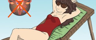
All these changes should alert the woman and require contacting a specialist. To maintain breast health, follow a few simple rules: protect the mammary glands from injury, choose the right bra, do not sunbathe without a swimsuit top, eat right, give up bad habits, take extra vitamins A and E, monitor your body weight and health. Visit a mammologist or gynecologist once a year. What does a healthy mammary gland look like?
- blow to the chest;
- strong squeezing by underwear during massage or sex;
- bruise from a fall;
- subcutaneous administration of pharmaceuticals.
In case of injury, capillaries are damaged, which leads to loss of blood supply to the local area of fatty tissue. The cells die, forming necrotic lesions, which are then replaced by scar tissue. If the outcome is unfavorable, purulent inflammation develops.
If you have a lump, you should make an appointment with a doctor if it does not go away on its own after menstruation and does not decrease in size. For an initial examination, you need to make an appointment with a gynecologist. Palpation and history taking can be carried out either by a doctor or an examining nurse.
A mammologist has the right to diagnose, observe and treat patients with benign neoplasms. When breast cancer is detected, women are sent to a breast oncologist.
Despite the fact that in most cases, tumors found in the breast are benign, women should definitely consult a doctor if new formations are detected.
This is especially true for those women who, along with the tumor, observe the following symptoms of breast cancer:
- hard lumps in the chest, collarbone, or armpit;
- swelling, redness, or rash on the skin of the breast;
- development of pitting or wrinkling in the skin of the breast;
- change in breast shape or size;
- changing the shape of the nipples;
- inversion or retraction of the nipples;
- unusual discharge from the nipples;
- new lumps that do not go away within a full menstrual cycle;
- unintentional weight loss.
Normal breast ultrasound
There are 3 types of tissues in the mammary gland - connective, adipose and glandular epithelium. Normally, the skin should be represented by a hyperechoic area, the glandular epithelium should be an echogenic zone with narrow ducts, and adipose tissue should be a hyperechoic area. Any neoplasms can be clearly identified in these tissues if you know their norm. The fat lobule has a reduced echogenicity compared to other tissues. But, sometimes it happens that fibroadenoma can be a heterogeneous echostructure.
On ultrasound examination, the fatty lobule has a round shape with fairly clear contours. If you look closely, you can find small areas of calcification in the fibroadenoma. There is also a leaf-shaped form of fibroadenoma, which differs from the usual fatty lobule on ultrasound only in its larger size. For the best diagnosis of a neoplasm in the mammary gland, an ultrasound examination should be performed on the 4-5th day of the menstrual cycle.
Symptoms indicating a lump in the breast
Doctors most often treat benign breast lumps when they become large and there is a risk of bust deformation. Some of the neoplasms can go away on their own if they are small in size. These include fat necrosis and cysts. Breast tumors should not be treated using traditional methods.
The main symptom of the disease is neoplasm. But to make a diagnosis, you need to know additional symptoms that help distinguish between breast diseases.
So additional symptoms may be:
- change in color of nipples or breasts,
- enlargement of the mammary gland, changes in its structure,
- increasing the density and rigidity of glandular lobules.
- Nipple discharge
- nipple retraction
- breast deformation.