A biopsy is a diagnostic procedure in which a piece of tissue or organ is removed for subsequent microscopic examination. .
If cancer is suspected, a biopsy is required, since without it the diagnosis is not considered definitively established .
A biopsy is also performed for some non-oncological processes. For example, with autoimmune thyroiditis, some types of hepatitis, Crohn's disease, etc.
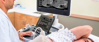
In this situation, it is an additional research method and is carried out when data from non-invasive diagnostic methods (CT, MRI, ultrasound, etc.) is not enough to make a diagnosis
Get Israeli Ministry of Health prices
Enter your details and get a price list on Viber, WhatsApp or Telegram
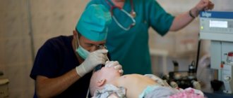
Excisional and incisional biopsy
These types of biopsies are quite painful, so they are performed under general anesthesia or local anesthesia in an operating room (the exception is endoscope-guided biopsy), and require suturing afterward. Excisional biopsy is often performed not only for diagnostic purposes, but also for treatment purposes, while incisional biopsy is performed only for diagnostic purposes. Sometimes during surgery for cancer it is necessary to urgently perform an incisional biopsy to clarify the scope of the operation.
Kinds
In case of prolonged inflammation or suspected oncology, it is necessary to perform a biopsy to exclude or confirm the presence of an oncological process. It is first necessary to perform a general analysis of urine and blood to identify the inflammatory process, and implement instrumental diagnostic methods (ultrasound, CT, MRI).
The collection of biological material can be carried out in several informative ways, the most common and popular among them are presented below:
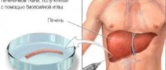
- Trephine biopsy. It is carried out using a thick needle, which in modern medicine is officially called a “trephine”.
- Needle biopsy. The collection of biological material is carried out by puncturing the pathogenic neoplasm using a thin needle.
- Incisional biopsy. The procedure is carried out during a full-fledged operation under local anesthesia or general anesthesia and involves the productive removal of only part of the tumor or affected organ.
- Excisional biopsy. This is a large-scale procedure, during which a complete excision of an organ or malignant tumor is performed, followed by a rehabilitation period.
- Stereotactic. This is a diagnosis carried out by preliminary scanning for the further construction of an individual scheme for the purpose of surgical intervention.
- Brush biopsy. This is the so-called “brush method”, which involves the use of a catheter with a special brush for collecting biopsy material (located at the end of the catheter, as if cutting off the biopsy material).
- Loop. Pathogenic tissues are excised using a special loop (electric or radio wave), in this way a biopsy sample is taken for further research.
- Liquid. This is an innovative technology for identifying tumor markers in liquid biopsy, blood from a vein, and lymph. The method is progressive, but very expensive, and is not carried out in all clinics.
- Transthoracic. The method is implemented with the participation of a tomograph (for more careful control) and is necessary for collecting biological fluid mainly from the lungs.
- Fine needle aspiration. With such a biopsy, the biopsy material is forcibly pumped out using a special needle to conduct exclusively cytological examination (less informative than histology).
- Radio wave. A gentle and absolutely safe technique, which is carried out using special equipment - Surgitron in a hospital setting. Does not require long-term rehabilitation.
- Preskalennaya. This biopsy is used to diagnose the lungs and consists of taking a biopsy sample from the supraclavicular lymph nodes and lipid tissues. The session is carried out with the participation of a local anesthetic.
- Open. Officially, it is a surgical procedure, and tissue collection for examination can be done from an open area. It also has a closed diagnostic form, which is more common in practice.
- Core. Soft tissue sampling is performed using a special trephine with a harpoon system.
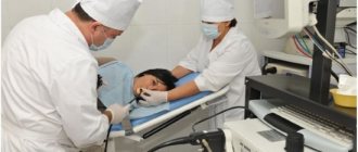
Needle biopsy
A minimally invasive method is a puncture biopsy. Its principle is that a hollow needle is inserted into the pathological formation or organ that needs to be examined. Pieces of tissue through which the needle passed get into it. After the needle is removed, these areas are sent for examination. If you need to examine an organ that is located deeply (that is, it cannot be seen and “palpated”), then the puncture is done under ultrasound or X-ray control.
For greater accuracy and to reduce trauma, a biopsy can be done under the control of ultrasound, endoscope, or x-ray.
In practice, two types of puncture biopsy are used:
- fine-needle (aspiration, classic);
- thick needle (cutting, trephine biopsy).
The advantage of a puncture biopsy is that this procedure is minimally painful. It is done without general or local anesthesia .
Why is a core needle biopsy performed?
In some cases, a local anesthetic is injected into the skin puncture site. But this type of biopsy also has its disadvantages. Firstly, the needle may not penetrate the pathological formation. Secondly, the material remaining in the needle cavity may not be sufficient for examination.
These factors significantly reduce the reliability of the method. The doctor’s experience and the quality of the equipment under whose control the manipulation is performed can compensate for the first drawback. To compensate for the second, modified techniques are used, in particular, core biopsy.
Core needle biopsies use threaded needles that are screwed into tissue like a screw. In this case, tissue areas that are much larger in volume remain in the needle cavity than with a fine-needle biopsy.
Biopsy guns make the procedure much easier for both the doctor and the patient.
This is the name of the devices that are used for fine-needle aspiration biopsy of various organs: pancreas, thyroid and prostate glands, liver, kidney, etc. A sterile needle is attached to the gun, consisting of a trephine (a tube with a very sharp edge) and a harpoon.
When fired, the trephine cuts tissue with great speed, and the harpoon fixes the tissue in the tube. As a result, a large column of material ends up in the needle cavity, which is sent for microscopic examination.
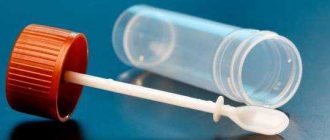
Taking swabs and swabs
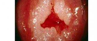
In fact, taking smears and swabs is not a type of biopsy, but they, like biopsies, are used to determine the type of tissue and cells. Fingerprints are taken from accessible research objects. Thus, taking smears for atypical cells is widely used in gynecology for the early diagnosis of cervical cancer.
To obtain swabs, the lumen of a hollow organ is washed with saline; for example, during bronchoscopy, swabs from the bronchi can be obtained. Fluid from a cyst (for example, breast cysts if breast cancer is suspected) or any body cavity, for example, pleural effusion, ascitic fluid, etc., can also be examined for malignant cells.
Reviews
On forums dedicated to medicine, you can find many references to biopsy, patient reviews of this procedure, and stories about impressions. Here are a few such reviews:
- They did a liver biopsy . The procedure itself went quickly, I didn’t feel anything in particular. But the pain came a little later, when the biopsy had already been done. I had to give a painkiller injection. I also read that there are complications due to bile that pours out of the bile ducts;
- We did a kidney biopsy twice . An unpleasant procedure, although quite tolerable. There were no complications, no need to be afraid;
- We did a muscle biopsy , I can say the following: it must be taken from the thigh under local anesthesia. It is very good that pain relief helps to completely eliminate any sensation. No rehabilitation is needed.
Study of the obtained material
Depending on the purpose of the biopsy and the amount of tissue obtained, the following is carried out:
- histological examination of the material;
- cytological examination of the material.
Histological examination examines tissue sections under a microscope..
To do this, pieces of tissue obtained from a biopsy are placed in a fixing liquid (formalin, ethanol, Bouin's fluid) to compact their structure, and then filled with paraffin. After hardening, they are cut into thin layers with a thickness of 3 micrometers using a microtome (a very sharp cutting tool). The sections are placed on a glass slide, paraffin is removed from them and stained with a special substance. After this, the drug is sent for microscopic examination.
In a cytological study, it is not the tissue that is studied, but the cells.
This type of microscopic examination is considered less precise, but requires less material. In addition, the preparation of a cytological preparation does not require lengthy preparation and special equipment.
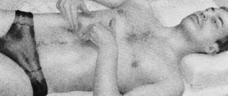
Muscles
Muscle tissue is also taken for biopsy in case of muscle diseases. The procedure is performed through an incision in the skin, which is made with a scalpel. A piece of muscle is simply cut out, so the biopsy requires taking material from the largest muscles.
These can be muscles such as the quadriceps femoris or deltoid muscle . If tissue is taken from a smaller muscle, its function may be impaired.
However, there is another method that allows you to take cells from small muscles using a game that simply pierces the skin and then the muscle. Thus, the damage is minimal and does not affect the functioning of the muscle.
Indications for muscle biopsy are muscle inflammation, atrophy, degeneration of muscle tissue, myopathy, and the like.
Leading Israeli oncologists
- Professor Aron Sulkes
Oncologist - Professor Moshe Inbar
Oncologist
- Professor Ofer Merimsky
Specialist in the treatment of sarcomas
- Dr. Yair Bar
Oncologist
Cytological examination is usually carried out after aspiration biopsy, swabs and smears . It is used to urgently solve diagnostic problems during surgery (establishing the nature of the tumor process, identifying tumor growth into surrounding tissues and metastases, the presence of tumor cells at the edges of the surgical incision, etc.), as well as when biopsy of a tissue area for histological examination is impossible or undesirable (for example, if melanoma is suspected).
Living cells are visible here - an experienced diagnostician quickly distinguishes leukemic (let's say) leukocytes and other atypical elements.
The significance of this method is great when it is necessary to analyze calcified and bone tissues, loose, crumbling masses and very small foci that are not suitable for histological examination.
When biopsy of tumors, it is most rational to carry out both histological and cytological examination of the biopsy specimen. But the results of histological examination are still decisive for establishing a diagnosis of cancer.
Reliability of biopsy results
The reliability of histological examination exceeds 90%. Its positive result serves as the basis for making a final diagnosis and prescribing a treatment regimen, including surgery for cancer.
If a tumor is suspected, it is better to do a biopsy in specialized government medical institutions, where experienced doctors and experienced pathologists work. This will significantly reduce the likelihood of a diagnostic error. If the result of the histological examination is negative, but the doctor has good reason to believe that the patient still has cancer, repeat biopsies are performed.
Cytological examination is a screening (intermediate) diagnostic method. Its results largely depend on the amount of material and its preservation, as well as on how precisely it is taken. If the result of a cytological examination is positive, then this serves as the basis for a more complex histological examination.
A negative result does not reject the suspected diagnosis of cancer.
Contraindications
Biopsy is not permitted for all patients according to indications; there are absolute and relative medical restrictions that are important not to violate.
Medical contraindications affect the following clinical pictures:
- blood clotting disorder;
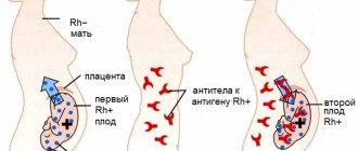
- periods of pregnancy and lactation;
- diseases of the reproductive system;
- inflammatory and infectious processes of the acute stage;
- systemic, somatic diseases;
- high threshold of pain sensitivity;
- after extensive blood loss.