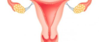
The uterus consists of three parts;
- The fundus is the apex of the uterus, above the entry point of the fallopian tubes.
- Body - for blastocyst implantation.
- The cervix is the lower part of the uterus and connects it to the vagina. This part is structurally and functionally different from the rest of the uterus.
The wall of the uterus consists of three layers of muscle tissue. Muscle fibers run longitudinally, circularly and obliquely. They are intertwined between the connective tissue of blood vessels, elastic and collagen fibers. This strong muscle wall expands and becomes thinner as the baby develops inside the uterus.
After childbirth, the dilated uterus returns to its normal size in about six to eight weeks. Its dimensions, however, become approximately 1 cm larger in all directions than before childbirth. The uterus also becomes a little heavier.
Uterus-structure
https://youtu.be/Fgo_e1vd-EE
Operations[ | ]
Soviet poster campaigning against criminal abortion
| This section is missing references to information sources. Information must be verifiable, otherwise it may be questioned and deleted. You may edit this article to include links to authoritative sources. This mark was set on September 13, 2012 . |
- Abortion
(not to be confused with the term "spontaneous abortion", meaning "miscarriage") is an operation aimed at terminating a pregnancy. It is usually performed in the first 20 weeks (sometimes up to 9 months under circumstances permitted by law or the criminal nature of abortion) in a hospital setting. - Vacuum aspiration
or the so-called “mini-abortion” is an intervention aimed at terminating pregnancy at an extremely early stage - from twenty to twenty-five days without expected menstruation. It is a minimally invasive operation and can be performed on an outpatient basis. - Caesarean section
(Latin: caesarea “royal” and sectio “incision”) - childbirth using abdominal surgery, in which the newborn is removed not through the natural birth canal, but through an incision in the abdominal wall of the uterus. - Hysterectomy
- (Greek hystera uterus + Greek ectome ectomy, removal; it is possible to write hysterectomy; another common name is hysterectomy) - a gynecological operation in which a woman’s uterus is removed.
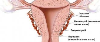
Histological structure of the uterus
The fundus and body of the uterus consist of three layers of tissue;
The peritoneum is a two-layer membrane, continuous with the abdominal peritoneum. Also known as perimetry. The myometrium is a thick, smooth muscle layer. Cells in this layer undergo hypertrophy and hyperplasia during pregnancy in preparation for expulsion of the fetus at birth. The endometrium is the inner lining of the uterus. This can be further divided into 2 parts: Deep basal layer : changes little during the menstrual cycle and is not lost during menstruation. Functionality of the superficial layer : Proliferates in response to estrogens and becomes secretory in response to progesterone. It is shed during menstruation and is regenerated from cells in the stratum basale.
How does the structure of the uterus change during pregnancy?
The anatomical structure of the uterus does not change during pregnancy, unlike its size and position in the pelvic cavity and abdominal cavity. Due to the complexity of attaching the uterine ligaments, a pregnant woman should not raise her arms too high. This is explained by the fact that the uterine ligaments are overstrained, which causes overstrain and displacement of the organ as a whole. During long periods of gestation, there is a risk of fetal displacement. The structure of a woman’s uterus during pregnancy is such that it can not only accommodate a child with water, but also the muscle layer can work and contract to expel the fetus.
Ligaments of the uterus
The tone of the pelvic floor provides primary support for the uterus. Some ligaments provide additional support, holding the uterus in place.
This:
Broad ligament: This is a double layer of peritoneum that attaches the sides of the uterus to the pelvis. It acts as a mesentery for the uterus and helps maintain it in position.
Round ligament: a remnant of the gubernaculum extending from the uterine horns to the labia majora through the inguinal canal. It functions to maintain the position of the uterus.
Ovarian ligament: attaches the ovaries to the uterus.
Cardinal ligament. Located at the base of the broad ligament, the cardinal ligament extends from the cervix to the lateral walls of the pelvis. It contains the uterine artery and vein in addition to supporting the uterus.
Uterosacral ligament: Extends from the cervix to the sacrum. Provides support to the uterus.
The structure of the uterus: photo with a description of its location
In the photos of information and medical sites you can see that the uterus is located in the pelvis, namely between the rectum and bladder. In the normal position, the location of the uterus should be tilted anteriorly. Its maintenance and mobility is ensured by ligaments located on both sides.
As a result of its anatomical features, the uterus reacts to any changes in nearby organs. It moves backward if the bladder is full or forward if the rectum is full. She is also able to rise up if she is pregnant.
Body of the uterus
The body of the uterus is the upper, wider lobe of the uterus. It consists mainly of muscles. A thick layer of muscle is surrounded on the outside by a connective tissue membrane.
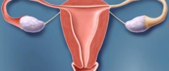
Inward, towards the uterine cavity, it is lined with a gland-rich mucous membrane (endometrium). The lining of the uterus cyclically thickens under the influence of female sex hormones and prepares for implantation of an embryo.
If fertilization and implantation of the egg stops, the outer layers of the uterine lining are shed by monthly bleeding ( menstruation ). The fallopian tubes flow into the upper right and left corners of the uterine body. They establish a connection between the uterine cavity and the ovaries - the female germinal glands.
Diagnostic methods
Depending on the nature of the pathologies of the female reproductive system, the doctor may prescribe studies:
- ultrasound (ultrasound);
- X-ray;
- hormonal;
- colposcopy (microscopic examination of the cervix);
- hysteroscopy (visual examination of the uterine cavity);
- fertiloscopy (examination of fallopian tube patency);
- cytological (cellular);
- histological (tissue section);
Infertility is one of the consequences that occurs when there is an abnormality in the structure or development of the female genital organs. Statistics say that every second woman facing infertility has connective tissue adhesions or disturbances in the peristalsis of the fallopian tubes, as a result of ongoing or past inflammatory processes in the pelvic organs.
If you notice problems conceiving a child, remember that in most cases, failures and disorders of sexual function can be treated and corrected. The main thing is to consult a doctor in a timely manner.
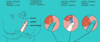
Cervix - what is it?
The cervix is the lower part of the uterus that extends into the vagina. It has a thick layer of mucous membrane and closes the uterus from the outside with the outer cervix, and from the inside with the inner cervix.
The length of the cervix is individual for each woman. On average it ranges from three to five centimeters.
When viewed from the side, the cervix is tilted forward against the uterus and against the axis of the vagina.
The cervix occupies approximately the lower third of the uterus and protrudes at the top of the vagina. The cervix is composed of connective tissue and muscle and has a hollow duct in the longitudinal direction, the cervical canal (cervical canal). It is lined with a mucous membrane, the glands of which form viscous mucus.
Mucus has the task of closing the outer cavity of the uterus and, thus, protecting it from germs from the vagina. Only during fertile days and during menstruation does the mucus become thinner and the canal opens a few millimeters.
During pregnancy, a closed cervix closes the fetal cavity downwards. The mucous membrane lining the cervix in the birth area is flatter than the mucous membrane inside the uterus. There it resembles a mucous membrane, as in the oral cavity (squamous epithelium).
The cervix is the connection from the uterine cavity to the vagina. At the upper end is the inner cervix, at the lower end is the outer cervix. Certain glands in the cervix produce cervical mucus : its amount and consistency changes during the menstrual cycle.
What is the uterus and where is it located?
The uterus is an organ of a woman’s reproductive system in which the fetus develops from the moment the fertilized egg leaves the fallopian tube until the child is born. Its shape resembles an inverted pear.
The uterus is located in the pelvis between the bladder and rectum. Its position can change during the day: when the organs of the urinary and digestive systems are filled, it moves slightly, and after urination or defecation it returns to its original place. But the most noticeable change in the position of the uterus is observed simultaneously with its growth during pregnancy, as well as after childbirth.
Structure of the uterus
Using an ultrasound of the uterus, you can see that it consists of three structural parts. The upper convex side is called the fundus, the middle widened part is the body, and the lower narrow part is the cervix.
The cervix consists of an isthmus, an elongated cervical canal and a vaginal part. The inside of the uterus is hollow. Its cavity communicates on the lower side with the lumen of the vagina, and on the sides with the canals of the fallopian tubes.
The organ wall is three-layered:
1 The outermost layer, facing the pelvic cavity, is called the perimeter . This membrane is closely connected with the outer covers of the bladder and intestines and consists of connective tissue cells.
2 The middle, thickest layer - myometrium , includes three layers of muscle cells: outer longitudinal, circular and inner longitudinal - they are named after the direction of the muscle fibers.
3 The inner lining, endometrium , consists of a basal and functional layer (facing the uterine cavity). Contains epithelial cells and many glands in which uterine secretions are formed.
The cervix has more connective dense collagen tissue and fewer muscle fibers than other parts of the organ.
The wall of the uterus is penetrated by numerous blood vessels. Arterial blood, saturated with oxygen, is brought by paired uterine arteries and internal branches of the iliac artery. They branch and give rise to smaller vessels that supply blood to the entire uterus and its appendages.
The blood that has passed through the capillaries of the organ is collected in larger vessels: uterine, ovarian and internal iliac veins. In addition to blood vessels, the uterus also contains lymphatic vessels.
The vital activity of uterine tissue is controlled by hormones of the endocrine system, as well as the nervous system. The wall of the uterus includes branches of the pelvic splanchnic nerves associated with the inferior hypogastric nerve plexus.
Ligaments and muscles of the uterus
In order for the uterus to maintain its position, it is held in the pelvic cavity by connective tissue ligaments, of which the most famous are:
Interesting! Is gluten harmful: who needs a gluten-free diet?
1 Paired broad ligaments of the uterus (right and left) are attached to the peritoneal membrane. Anatomically, they are connected to ligaments that fix the position of the ovaries.
2 The round ligament contains both connective tissue and muscle cells. It starts from the wall of the uterus, passes through the deep opening of the inguinal canal and connects with the tissue of the labia majora.
3 The cardinal ligaments connect the lower part of the uterus (near the cervix) to the urogenital diaphragm. This fixation protects the organ from displacement to the left or right.
Through ligaments, the uterus is connected to the fallopian tubes and ovaries, which ensures the correct relative position of the organs of the female reproductive system.
In addition to the ligaments, the correct location of the pelvic organs, including the uterus, is ensured by a set of muscles called the pelvic floor. The composition of its outer layer includes the ischiocavernosus, bulbospongiosus, superficial transverse and external muscles.
The middle layer is called the urogenital diaphragm, it contains the muscle that compresses the urethra and the deep transverse muscle. The internal pelvic diaphragm unites the pubococcygeus, ischiococcygeus and iliococcygeus muscles. The pelvic floor muscles prevent deformation of the organs, which would impair their blood supply and function.
Uterus dimensions
When a girl is born, the length of her uterus is about 4 cm. It begins to increase at the age of 7. After the final formation of the reproductive system during puberty, the uterus reaches dimensions of 7-8 cm in length and 3-4 cm in width. The thickness of the walls in different parts of the organ and in different phases of the menstrual cycle varies from 2 to 4 cm. Its weight in a nulliparous woman is about 50 g.
The most significant changes in the size of the uterus occur during pregnancy, when in 9 months it increases to 38 cm in length and up to 26 cm in diameter. Weight increases to 1-2 kg.
After childbirth, a woman’s uterus shrinks, but does not return to its original parameters: now its weight is approximately 100 g, and its length is 1-2 cm greater than before conception. These sizes remain throughout the entire childbearing period; after the second and subsequent births, no noticeable increase occurs.
When a woman's reproductive period ends and menopause occurs, the uterus decreases in size and weight, the wall becomes thinner, and the muscles and ligaments often weaken. Already 5 years after the end of menstruation, the organ returns to the size it was at birth.
What function does the cervix perform?
The cervix produces viscous, sticky mucus (cervical mucus). This forms an effective barrier against sperm and germs rising from the vagina during the days of a woman's infertility. On fertile days (around ovulation , the mucus formed by the cervix becomes transparent and spinny. This mucus becomes accessible to sperm, but also to microbes.
During fertilization, the cervix moves slightly forward and thus absorbs sperm through the outer cervix. Sperm enters the uterus through the cervix and then into the fallopian tubes. During menstruation , blood flows from the uterus through the cervix into the vagina.
During pregnancy, the uterus not only grows, but also changes its shape. It stretches and contracts to one or two centimeters, but should still be about two and a half centimeters in length before delivery. A decrease in cervical length is an important indicator of pregnancy progression.
Pathologies[ | ]
Developmental anomalies[ | ]
- Aplasia (agenesis) of the uterus
- extremely rarely, the uterus may be completely absent. There may be a small infantile uterus, usually with a pronounced anterior incursion. - Duplication of the uterine body
is a defect in the development of the uterus, which is characterized by duplication of the uterus or its body, which occurs due to the incomplete fusion of two Müllerian ducts at the stage of early embryonic development. As a result, a woman with a double uterus may have one or two cervixes and one vagina. With complete nonfusion of these ducts, two uteruses with two cervixes and two vaginas develop. - Intrauterine septum
- incomplete fusion of the embryonic rudiments of the uterus in various variants, can lead to the presence of a septum in the uterus - a “bicornuate” uterus with a clearly visible sagittal depression at the bottom or a “saddle-shaped” uterus without a septum in the cavity, but with a notch at the bottom. With a bicornuate uterus, one of the horns may be very small, rudimentary, and sometimes unlaced. - Hypoplasia is underdevelopment of this organ in a woman. In this case, the uterus is smaller than it should be normally.
Links[ | ]
- Media files on Wikimedia Commons
- BSE.sci-lib.com (unspecified)
. — The meaning of the word “Uterus” in the Great Soviet Encyclopedia. Retrieved September 2, 2008. Archived August 24, 2011. - Reference-anatomia.ru (unspecified)
. — Article “Uterus” in the Handbook of Human Anatomy. Retrieved September 2, 2008. - Golkom.ru (unspecified)
. — Article “Uterus” in the Concise Medical Encyclopedia. Retrieved September 2, 2008.