A gastric biopsy is a method of examining lesions in the stomach. The human body is made up of microscopic structural units called cells.
Monitoring changes at the cellular level allows you to establish an accurate diagnosis and identify the presence or absence of pathology with 100% accuracy.
Tissue collection for analysis is carried out through a biopsy. During the procedure, the doctor takes a small part of tissue from the patient in order to then study the material under multiple magnification.
Indications for prescribing a gastric biopsy
A gastric biopsy is prescribed if the following indications exist:
- suspicion of a precancerous condition;
- gastritis in acute and/or chronic form;
- peptic ulcer with the threat of tissue degeneration into a malignant tumor;
- in case of damage to the gastric mucosa, to clarify the area of resection;
- to identify Helicobacter bacteria;
- suspicion of metaplasia;
- to monitor the condition of the organ after radiation treatment or surgery (especially after removal of polyps).
The indication for a biopsy is the presence of any atypical areas on the body of the stomach.
Gastrobiopsy of the stomach
The manipulation is carried out by introducing a special biopsy probe (gastroscope) to take a biopsy from the inner gastric wall. To perform the selection, special knives or vacuum tubes can be used to suction (aspiration biopsy) tissue particles or a suspension of cells. Gastrobiopsy is rarely accompanied by complications.
Sighting
The procedure is performed with a special reusable device - a fibrogastroscope. The device is equipped with a device for multiple targeted sampling. Advantages of the device:
- special flexibility, which facilitates insertion of the device into the lumen of the organ;
- minimizing discomfort;
- high quality and clarity images;
- the ability to examine the distal end of the stomach through controlled bending of the device.
The process is negatively affected only by severe deformation and severe stenosis of the organ cavity.
Blind
The manipulation is performed using a probe without visual control. This technique is also called search technology. The method must be performed by a high-level specialist, as there is a risk of severe injury to the mucous membrane.
Contraindications
Not all patients can undergo intervention, since gastric biopsy can cause complications in the presence of a number of diseases.
Contraindications to using the method are:
- pathologies of the cardiovascular system;
- state of shock and disruption of the central nervous system;
- diseases of the ENT organs, inflammatory processes in the pharyngeal region;
- gastritis of erosive and phlegmonous type;
- infectious lesions of the body;
- narrow esophagus;
- obstruction of the intestinal tract;
- a condition in which the patient cannot breathe through the mouth (nasal congestion due to infections, damaged septum or the presence of growths of various nature);
- destruction of the gastric epithelium;
- chemical burns of the digestive system;
- unsatisfactory condition of the patient (according to general indicators);
- mental disorders.
In addition to direct contraindications, it is necessary to take into account the emotional state of a person. If the patient is not ready for the procedure and experiences severe fear, the study is not prescribed.
ATTENTION! Mental disorders are not absolute contraindications. If necessary, an unstable patient may be prescribed a biopsy under intravenous anesthesia.
Preparing for the study
Your attending physician will tell you about proper preparation for FGS.
Preparation for EGD of the stomach with biopsy does not require special measures, however, there are several recommendations for patients. The attending physician must prepare the patient psychologically for the procedure - explain the risks and possible complications, describe the course of the study and the necessary measures.
Regardless of the duration of the procedure, a number of general tips can be identified:
- A few days before the study, stop drinking alcoholic beverages, spicy and hot foods.
- Do not take food or liquid 3-4 hours before the test.
- The evening before the test, the patient may take sedatives to reduce stress levels.
- Do not take medications that can reduce the effectiveness of hemostasis (Aspirin, etc.).
- If the patient has an increased gag reflex, local anesthetics may be used.
How to prepare for a gastric biopsy
If the patient has received a referral for a gastric biopsy, it is advisable to switch to inpatient treatment.
It is possible to remove a biopsy specimen in a clinic setting. But if complications develop, the doctor will not be able to provide timely assistance to the patient.
Before the procedure, an x-ray of the stomach is required to confirm the absence of contraindications.
Food interferes with an objective examination of the gastric cavity and can complicate the process of collecting material. Therefore, two days before the biopsy, it is necessary to exclude chocolate, seeds and nuts, as well as alcoholic beverages from the diet.
The last meal should be no later than 12 hours before the scheduled biopsy.
The patient comes to the procedure on an empty stomach.
You are allowed to drink water no later than two to four hours before the start of biopsy sampling.
ATTENTION! Immediately before the biopsy, it is forbidden to brush your teeth or use chewing gum.
Possible complications
FGDS, like biopsy, are routine procedures in the practice of an endoscopist. However, the risk of adverse consequences must always be considered. Due to various reasons, complications such as:
- Perforation of the esophagus.
- Stomach injury.
- Bleeding.
- Damage to teeth.
- Panic attack.
- Allergic reaction to anesthetics or anesthetic drugs.
Symptoms of complications do not always appear immediately, so you should definitely consult a doctor if you experience any of the following symptoms:
- severe weakness;
- pain in the neck, throat, stomach and/or chest;
- intense nausea;
- vomiting blood;
- fever, chills;
- dizziness;
- dark stool;
- widespread itchy skin rash;
- swelling of the eyelids, lips, cheeks;
- severe difficulty breathing.
Remember that normally, EGDS with a biopsy does not cause any severe pain. The patient may feel soreness and discomfort in the throat due to the administration of the drug, sometimes belching with air (it is injected into the digestive canal for a better view of the mucous membrane). A marked deterioration in health is a clear reason to seek medical help.
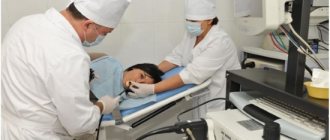
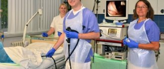
Progress of the procedure
- The patient will be placed on the couch, on his left side (bending in the back is unacceptable, the spine must be straight).
- The working area (throat and larynx) is treated with lidocaine to minimize discomfort.
- Using endoscopy, an instrument (endoscope) is inserted into the stomach, which the patient pushes through the throat.
- The doctor corrects the patient's breathing technique.
- A biopsy sample is taken.
- The endoscope is removed.
Samples are taken not only from atypical areas. Material from the areas that are the subject of the study and material from the border zone, at the junction of healthy and atypical tissues, are sent for research.
Upon completion of the procedure, the doctor reports any abnormalities found. The extracted material is sent for laboratory analysis, where a fragment of the stomach is degreased and treated with paraffin, cut into layers and examined under a microscope.
Description of methods
Gastroscopy of the stomach, called FGDS, involves a visual examination of the lumen and mucous membrane of the digestive tract (esophagus, stomach, duodenum). For this purpose, a special flexible probe in the form of a tube is used, equipped with a camera and an optical system. Gastroscopy can be of a purely diagnostic nature or used for the purpose of performing a biopsy - taking samples of affected tissue for subsequent research. Advantages of the method:
- A thin elastic tube, equipped with a modernized control system, allows you to study in detail the entire surface of the gastrointestinal mucosa.
- Detection of all superficial wall destructions not visualized by radiography.
- Determining the cause of gastrointestinal bleeding.
- Detection of malignant and benign growths in the stomach.
- Tracing malignancy (malignancy) of tumors.
- Tracing the development of peptic ulcer disease.
- Taking a biopsy sample while performing a biopsy.
Biopsy is a diagnostic technique that involves taking a small piece of tissue from the mucosa or a suspension of cells from a tumor. The biopsy is sent for analysis, which is carried out under a microscope to detect malignant cells in the epithelium, determine their etiology, and the degree of damage to the organ. Based on the results, a decision is made on the need for surgical intervention. There is a general classification for gastrobiopsy into 2 types, differing in technique:
- sighting;
- blind.
What to do after the examination
Immediately after the procedure, the patient can go home. Eating is allowed only two hours after the biopsy. In the future, the person can return to normal life, eliminating the intake of hot food. The risk of complications is minimal.
ATTENTION! You can drink alcohol only one day after the biopsy is taken.
Mild bleeding is possible, which does not require medical intervention. The sensitivity of the larynx and the swallowing reflex return gradually.
Preparation at different times
The following general recommendations are expected to be implemented:
- For three days, do not eat spicy foods or drink alcohol.
- Do not eat anything for 10 hours before the procedure.
- Do not take blood thinning drugs (Aspirin, Paracetamol), NSAIDs.
- Immediately before the procedure, you should rinse your throat with an antiseptic.
During gastroscopy in the morning, you should:
- do not eat from 22:00 the night before;
- do not drink in the morning;
- do not use medications.
When diagnosing in the evening:
- do not eat 8 hours before the procedure;
- Drink water for the last time 2 hours before the procedure, and take sip of still water throughout the day;
- no smoking for 3 hours.
General recommendations, regardless of the time of FGDS of the stomach:
- cleaning the stomach from secretion, mucus, blood;
- diet;
- minimal water consumption;
- refusal of exercise.
How is it carried out?
The collection of biopsy material is carried out using special endoscopic equipment - a fiber gastroscope. The procedure is called fibrogastroscopy, or FGS for short. Gastroscopes can be flexible or rigid. Each gastroscope has an optical system and a video camera that transmits the image to the screen. In addition, gastroscopes have instrumental channels that allow various manipulations. There is also such a study as FGDS - in this case, the antrum and lumen of the duodenum are examined.
Previously, the biopsy was blind - performed using a special gastric tube that did not have an optical system. Visual control over the progress of the procedure is impossible.
Using an endoscope and a scalpel or a special knife, the doctor cuts out several pieces of mucous membrane from different parts of the stomach. The muscle layer of the organ is not affected, so no special anesthesia is required. The pieces are immediately placed in a sodium chloride solution and sent to the laboratory.
A special gastroscope knife allows you to carefully cut off pieces of the gastric mucosa
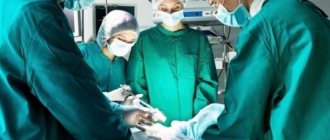
A biopsy can also be performed during surgery, when it is necessary to decide on the volume of tissue to be removed. Then the resulting material is urgently taken to the laboratory, and a specialist gives an opinion within 10-15 minutes. The operating team does not perform any actions at this time. After confirmation of the diagnosis, the operation continues in one capacity or another.
To prepare histological material, pieces of mucous membrane are poured with paraffin and kept for a certain time. Then they are cut with a special tool into the thinnest plates. Then these plates are painted with special dyes. A specialist examines the material under a microscope and gives an opinion.
This is what a finished histological specimen of the gastric mucosa looks like
Types of biopsies
Modern surgery has many options for endoscopic biopsies. With fibrogastroduodenoscopy, the specialist has the opportunity to take samples not only from the stomach - the duodenum is additionally examined, which means it is possible to take scrapings of its walls. In some cases, a partial puncture of the pancreas is taken. The study is carried out under mandatory further ultrasound control, since pancreatic tissue is restored slowly and is most susceptible to transformation into oncology.
Colonoscopy allows you to take additional examination of the mucous layer of the colon and rectum.
An important place is occupied by the timely detection of liver cancer, as the worst complication after hepatitis or cirrhosis.
Biopsy for prostate cancer
If prostate cancer is suspected, a biopsy provides the most reliable information and, based on its results, treatment tactics are determined. Modern biopsy technology is performed using a TRUS (transrectal ultrasound) ultrasound device with a probe into the lumen of which special short puncture needles are inserted.
The probe is inserted through the wall of the rectum into the prostate, the gland is scanned and, under the control of an ultrasound screen, the required number of puncture needles are “shot” into different parts of the prostate - from 10 to 30 needles. Plunging into the prostate, they collect “columns” of its tissue and are pulled back through the probe.
To prevent infection from the rectum, the patient is prescribed antibiotics. In Israel, a method of percutaneous prostate biopsy has been developed, when the introduction is performed not through the intestine, but through the skin of the perineum, and the number of samples from different parts of the prostate has been increased to 50, which increases the accuracy of the study.
Decoding the results
How long does a biopsy take? If it is urgent, carried out during the operation, then almost immediately, but in standard situations you have to wait 2-3 days for an answer. If samples are sent to another city or country, then the waiting time for a response extends to 1.5-2 weeks.
In the case of a gastric biopsy, deciphering the results obtained is of great importance. In this case, the following parameters are assessed:
- thickness of the mucous membrane;
- epithelium - its character, the degree of its secretion;
- presence of inflammation;
- signs of atrophy, metaplasia, dysplasia;
- degree of H. pylori contamination.
When deciphering the histology of the stomach, one should remember that:
- Sometimes the results may be questionable or unreliable if the amount of material was insufficient, and the study needs to be repeated.
- Gastric cytology is especially important for identifying atypical cells.
- It is the doctor who must ultimately interpret the data obtained.
In general, the results of histological examination can be divided into the following groups:
- Malignant tumors. The type of tumor, the type of cancer cells and the nature of their differentiation (for example, high-, low-differentiated) are determined.
- Benign tumors. The type of tumor and cell type are indicated.
- Gastritis. Its type and the nature of changes in the mucous membrane are described.
- Norm. The stomach tissues are not changed.
- (-) – negative result, normal;
- (+) – weak contamination, up to 20 H. pylori bacteria in the microscope field;
- (++) – average, moderate contamination, 20-40 bacteria in the field of view;
- (+++) – high contamination, more than 40 H. pylori in the field of view.