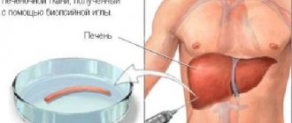
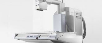
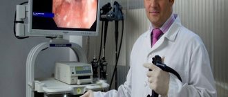
A research method used for accurate diagnosis of diseases, examination of the mucous membrane and detection of inflammatory changes in the gastrointestinal tract using a special device - an endoscope (a flexible tube with fiber optics, equipped with a special channel and the ability to place instruments in it for performing a biopsy) is called FGS of the stomach. The decoding of FGS sounds like “fibrogastroscopy” (from the Greek “stomach” and “I observe”, “I look”). This method of examining the gastrointestinal tract is the most reliable, fastest (carried out under local anesthesia within a few minutes) and allows you to establish an accurate diagnosis, and, if necessary, makes it possible to perform a biopsy (taking a tissue sample for a more detailed study in a bacteriological laboratory).
How to prepare for FGS
Preparing for the FGS is not particularly difficult. The main aspect for a good result is the presence of a positive psychological attitude of the patient and his complete trust in the endoscopist. The doctor must know about all the patient’s concomitant diseases and the medications he is currently taking.
FGS is an examination that is carried out on an empty stomach in a specially equipped room in a hospital or clinic. On the day of the procedure, smoking, eating, drinking and chewing gum is prohibited. A couple of days before the examination, it is advisable for the patient not to eat foods that are difficult for the stomach and cause gas formation (legumes, a variety of dairy products, fish, meat). The last dinner on the eve of the examination day should be no later than 18:00.
Methods for painless passage
In the last decade, techniques have begun to be actively used that can significantly reduce discomfort or even painlessly undergo gastroscopy of the stomach:
- Sedation. Sedation will help you survive FGDS calmly without fear. The patient is injected intravenously with a drug from the group of benzodiazepines, after which he falls asleep. The procedure is widely carried out and makes it easier to endure unpleasant sensations and painlessly perform FGDS of the stomach for adults and children. A few minutes after completion of the study, the patient wakes up. In an hour he can go home. The risk of side effects is low.
- Anesthesia (general anesthesia). Used more rarely under the supervision of an anesthesiologist. The patient is given several drugs for pain relief, but mechanical ventilation is not required. Usually performed before planned surgery.
Capsule endoscopy. This is an innovative technique in which the patient does not have to swallow the tube. Instead, it swallows a small capsule-shaped chamber that travels all the way through the digestive system. The image is recorded and then viewed by a doctor without any problems. Among the disadvantages of capsule endoscopy is the impossibility of a more thorough examination of the changed area of the mucosa, conducting a Helicobacter test or biopsy. Helps those who cannot undergo the standard FGDS procedure.
Insertion of a probe through the nose (transnasal technique) is also used. This technique provides a more comfortable FGDS, since it causes nausea much less often and does not affect hemodynamic parameters (which is important for patients with cardiovascular diseases). A probe with a significantly smaller diameter is used - 5.4 mm (standard - 11 mm). When asked whether it is painful to do gastroscopy of the stomach in this way, many patients answer negatively.
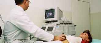
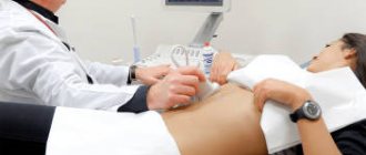
Some diseases can be diagnosed using less unpleasant procedures: x-rays of the stomach and ultrasound of the abdominal cavity.
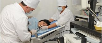
How does the FGS procedure work technically?
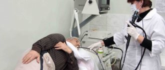
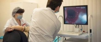
The prepared patient's larynx is frozen with a special local anesthetic. After this, in order to prevent the jaw from closing during the procedure, he clamps a special mouth opener with his teeth, through which the endoscope will pass.
In a sitting position, the patient, under the guidance of the doctor, makes swallowing movements until the device reaches the desired depth of the area being examined. After this, the patient is placed on his side and oxygen is introduced into the stomach under slight pressure (to straighten the walls). The doctor, examining the mucous membrane, can take an analysis of the stomach contents (stomach juice), remove the tumor, carry out treatment or perform a biopsy.
Is it painful to have a gastroscopy?
Most patients who have undergone FGDS in the traditional way, if asked to describe their feelings from the procedure, call it unpleasant. To reduce the severity of these sensations, a local anesthetic is used, which is applied to the back wall of the oropharynx. However, it does not always save the patient from discomfort. The most common complaints from patients:
- nausea;
- a feeling of discomfort in the neck, behind the sternum or in the abdomen;
- increased salivation;
- belching;
- feeling of lack of air;
- panic attacks;
- aching or cutting pain in the neck or abdomen.
Oddly enough, pain syndrome is relatively rare. It is possible in the presence of acute pathology (for example, exacerbation of peptic ulcer disease in young patients), complications (wall perforation) or careless examination, however, swallowing the intestine to check the stomach is unpleasant. The main thing is to behave correctly during the procedure and listen to the doctor’s recommendations.
What do FGS results show?
Endoscopy is the only method by which structural changes in the mucosa can be seen at a very early stage. The attending gastroenterologist, prescribing such a procedure, clarifies the diagnosis using additional methods of studying the patient’s body. The results of FGS of the stomach will help confirm or refute the presence of diseases such as stomach or esophageal cancer, esophagitis, benign tumor, gastritis of high or low acidity, gastric ulcer, polyps, gastric dysfunction and other gastrointestinal ailments. With the help of FGS, you can monitor the dynamics of treatment of the disease.
Based on the results of the biopsy, we can talk about the essence of the processes occurring in the tissues of the body, the presence of cancer cells or ordinary inflammation in them. Histological examination helps to detect pathologies in the early stages of development.
Main indications
The procedure in question is prescribed to patients who are suspected of having diseases affecting the gastric mucosa. The following phenomena may indicate this:
- heartburn and sour belching, signs of ulcer development;
- pain in the upper abdominal cavity, symptoms of ulcers, erosion or inflammation;
- deterioration or lack of appetite;
- difficulty swallowing, occurs with diseases of the cardiac part of the stomach and cardiac sphincter;
- nausea and regular belching of air after eating;
- weight loss without objective reasons (a sign of cancer pathology;
- anemia, in the absence of other causes, can occur with gastritis;
Fibroscopy is performed to make a diagnosis and monitor the effectiveness of treatment, and if cancer is suspected, patients are prescribed a biopsy.
FGDS of the stomach is not recommended if the patient:
- hypertensive crisis, a significant increase in blood pressure (200/100 or more);
- there was an acute myocardial infarction;
- acute phase after stroke;
- poor blood clotting;
- mental disorders;
- bronchial asthma.
What to expect during FGS
The process of inserting a tube into the gastrointestinal tract may be accompanied by gag reflexes or regurgitation. There is no need to be ashamed of this, because such a reaction is an indicator of a normally functioning organ. Such responses of the body to FGS occur due to air, which is pumped into the stomach using an endoscope and straightens its walls for a better view. The subsequent decoding of the FGS depends on this. The doctor thoroughly examines the mucous membrane and, if there is a need to remove a polyp, eliminates it, and if it is necessary to take part of the material for a biopsy, he takes it absolutely painlessly.
Carrying out FGDS with biopsy: indications and contraindications, preparation for the procedure
Stomach diseases occur in almost every third person. First of all, these are various types of gastritis and peptic ulcers. However, more serious diseases can also occur, for example, tumor damage to the organ wall with the development of metastases. The gold standard in diagnosing stomach diseases is fibroesophagogastroscopy, which allows the doctor to visually assess the condition of the mucous membrane of the organ and draw a conclusion about the necessary treatment. But such a procedure cannot always give an accurate answer about damage to the stomach. Then gastroscopy with biopsy is used - a method of morphological examination of the organ after preliminary staining of the obtained gastric sample.
Possible complications after FGS
The use of this serious diagnostic procedure has established itself in medical practice as one of the safest studies. Complications after FGS are reduced to zero. An endoscope is a very expensive, reusable instrument, and before each manipulation it is processed according to medical standards, so infection with any disease or infection is excluded.
But, as you know, in medicine there is always a small percentage of complications even from the most harmless procedures. In this case, this indicator is very small and depends directly on the patient himself. The patient must properly prepare physically and psychologically, and during FGS, strictly follow the recommendations of the endoscopist. Possible complications as a result of FGS are perforation of the wall of the organ being examined or minor bleeding due to pinching off a small portion of the wall of the esophagus, stomach or duodenum for biopsy.
After the procedure, the patient may experience discomfort when swallowing for some time. The normal state occurs within 24 hours. A full transcript of the FGS and the necessary recommendations for adjusting nutrition in the first few days will be given to the patient immediately after the study.
Treatment
Treatment must be prompt. The main method is surgical intervention. The patient undergoes gastropancreatoduodenal resection. This type of treatment is difficult for the body and is allowed for patients after checking their level of exhaustion, the amount of protein in the blood and other indicators.
We invite you to read: Demodicosis of the scalp: treatment of subcutaneous hair mites on the head in people, symptoms, photos
If cancer treatment begins at stage I or II, the survival rate is 80–90%. At stage III, it also makes sense to start treatment: the five-year life expectancy in this case reaches 5–10%.
If the patient's health condition does not allow radical therapy, treatment consists of conditionally radical operations, for example pancreaticoduodenectomy.
If there is no hope for the patient's recovery, palliative therapy is used, which is aimed at alleviating symptoms. In particular, they ensure the outflow of bile using various types of anastomoses. Such treatment not only alleviates suffering, but in some cases can prolong the patient’s life.
After cancer of the papilla of Vater is found, therapy is prescribed that corresponds to the stage of the pathological process. The main treatment method is surgery. Only removal of areas affected by malignant neoplasm allows one to cope with the disease.
The prognosis for the life of such patients is not very comforting. About 40% of patients who undergo resection survive the next 5 years. Statistics show how serious a pathology cancer of the large duodenal nipple is.
The volume of tissue that will be removed during surgery depends on the stage of the tumor process. Surgeons can perform resection:
- large part of the pancreas;
- nearby lymph nodes;
- parts of the small intestine;
- gallbladder and part of its duct;
- pyloric part of the stomach.
Sometimes the surgical method is combined with radiation therapy. Chemotherapy is rarely prescribed, since statistics show that it is ineffective for this type of cancer.
Vater's nipple cancer most often occurs in people over 55 years of age. Timely diagnosis and surgical intervention can stop the process and save the patient’s life.
How does FGS differ from other methods of studying the stomach?
Each research method has features depending on the area being analyzed. Some names seem to be similar, but in practice the procedures are very different. The endoscopist doctor is well versed in what FGS shows and what FGDS or EGDS shows. All these methods can be given one definition - fibrogastroscopy. Even if the endoscopist does FGS with an emphasis on the gastric cavity, he will still look at both the duodenum, as is done with FGDS (fibrogastroduodenoscopy), and the esophagus, which is examined with EGDS (esophagogastroduodenoscopy). Also, when examining all parts of the gastrointestinal tract, the video gastroscopy method can be used, during which video recording occurs using an endoscopic camera.
Every patient should understand what the endoscopy procedure is, how important it is, and that it depends only on him whether he will be healthy or not.
This research method, due to its high efficiency and low likelihood of possible complications, is widely practiced and used in medical work. Deciphering FGS by experienced specialists will help make the correct diagnosis and prescribe appropriate treatment.
General information
Cancer of the large duodenal papilla is a malignant neoplasia of the large duodenal (Vater) papilla, localized in the descending part of the duodenum and representing the anastomosis of the main pancreatic duct and the common bile duct.
It accounts for 40% of the total number of oncological lesions of the pyloroduodenal zone, 5% of the total number of gastrointestinal neoplasias and 1-2% of the total number of cancers of various localizations. Cancer of the major duodenal papilla is the third most common cause of obstructive jaundice. Typically affects older patients, with an average age of 54 years. Very rarely detected in children. Women suffer less often than men. Treatment is carried out by specialists in the field of oncology, gastroenterology and abdominal surgery.
Major duodenal papilla cancer
FGDS - when not to do it
The FGDS procedure also has contraindications.
This examination cannot be carried out if the patient has a sore throat or has an acute respiratory disease. When is it necessary to sign up for an FGDS procedure:
- Body temperature remains at 38 degrees Celsius.
- Constant nausea, as well as vomiting, with which blood clots come out (in this situation, the color resembles dark chocolate).
- Defecation is difficult, and the stake is black (that is, there is a trace of blood in it).
- My stomach hurts all the time.
BDS pathologies
Diseases of the major duodenal papilla are very diverse. With the development of modern diagnostic methods, conclusions about functional disorders in this structure are much more common than previously thought. However, due to untimely and rather difficult diagnosis, medical practice is often faced with a huge number of unsatisfactory results in the treatment of patients with cholelithiasis or pancreatitis, which developed against the background of disorders in the structure of the obstructive system.
Tumor-like neoplasms are considered a common pathology of BDS - hyperplastic polyps account for up to 87% of benign neoplasms. Polyps, as a rule, do not degenerate into malignant tissue. Adenomas are a rare disease; BDS cancer accounts for up to 25% of all malignant neoplasms. OBD stenosis is diagnosed in 4-40% of patients. As a rule, the pathologies of BDS are interrelated with cholelithiasis (GSD), which occurs in every tenth resident.
Spastic stenosis of the abdominal joint
BDS stenosis is a pathology with a benign course, which is caused by obstruction of the bile and pancreatic ducts due to inflammatory changes and cicatricial narrowing of the papilla. How is everything going? The passage of the stone causes injury to the papilla, and an active infectious process in the folds leads to the development of fibrous tissue and stenosis of the areas of the ampulla of the ampulla.
As is known, the structure of the BDS is directly influenced by a person’s age. Elderly people with cholelithiasis suffer from an atrophic-sclerotic form of chronic papillitis. The contingent whose age has not reached the age of sixty is susceptible to hyperplastic changes in the BDS (adenomatous, adenomyomatous).