Liver hemangioma - what it is, treatment and causes
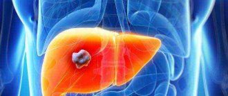
Liver hemangioma is a benign tumor, the formation of which occurs somewhat unusually and, in essence, is a ball of vessels huddled together, incorrectly developed and formed in the embryonic period. In this regard, the disease is considered congenital.
Usually, as a result of the examination, the formation of one such tumor is revealed and its size generally does not exceed 3-4 cm. Most patients do not have any symptoms, so they may not suspect its presence for a long time. It is extremely rare that situations arise that can provoke an increase in hemangioma up to 10 cm in size, which will be clearly expressed by characteristic symptoms. Such patients require mandatory treatment.
General information
Hemangioma is essentially a benign tumor of blood vessels that contain certain pathologies. Such vessels are arranged randomly, grow uncontrollably and do not fulfill their main role, which is to transport oxygen and nutrients to the organs. Such neoplasms can occur in any part of the body and in any organ - in the liver, kidneys, ovaries. They can even appear in the brain and spinal cord.
Here are a few more interesting facts that will help you understand what it is - liver hemangioma. Treatment and preventive measures for this pathology are carried out only in isolated cases, which will be discussed below. Basically, doctors take a wait-and-see approach, since in 90% of children, hemangiomas resolve on their own by the age of 10, leaving no traces.
In girls, such formations are observed approximately 7 times more often than in boys (according to some sources, 4 times), and the lower the baby’s weight, the larger the tumor.
A pleasant and very important feature of these formations is that they never develop into cancer.
Ultrasound contrast Dopplerography
For the differential diagnosis of hemangiomas, the use of ultrasound contrast Dopplerography seems very promising. The pharmacokinetics of contrast enhancement and the interpretation of the resulting image with ultrasound are identical to those with bolus contrast enhancement with CT: this is intense peripheral staining of tortuous and lacunar dilated vessels in the arterial phase and prolonged staining of the central part of the tumor. In small formations, point accumulation of contrast agent is more often observed. A convincing differential diagnostic feature is the visualization of the feeding vessel, which is easier to detect with V3 angiography.
Liver hemangioma is a benign vascular neoplasm localized in the liver parenchyma. This tumor is 6 times more common in women than in men, which is due to the influence of the female sex hormone estrogen on its growth and development. Liver hemangioma (ICD code) is a tumor that does not degenerate into malignant and in rare cases requires treatment or resection. Very often, this disease is found in pregnant women or those who have recently given birth, which is also caused by an increase in the level of female sex hormone during gestation.
Causes of liver hemangioma in children
The etiology of this pathology is not fully understood. In principle, this can be said about all benign and malignant tumors. The reasons for their occurrence are in the realm of speculation. If we talk about liver hemangioma observed in infants, experts associate its appearance with adverse events during pregnancy (up to 12 weeks) that can affect the formation of fetal blood vessels. This includes all known causes that negatively affect the development of the embryo:
- Hereditary factor.
- Transfer of diseases by the expectant mother. Regarding hemangiomas, influenza and ARVI are especially dangerous, since the viruses that cause them release toxins that negatively affect the formation of blood vessels.
- Constant stress in the first trimester.
- Use of certain medications.
- Lack of vitamins in a pregnant woman's diet.
- Smoking and drinking alcohol.
Why is hemangioma dangerous?
The main danger of an angioma, especially a large tumor, is its rupture and massive bleeding into the abdominal cavity. A rupture can occur due to heavy physical exertion, a blow to the liver area, or increased intra-abdominal pressure. Without timely medical care, this condition ends in death.
Other complications of the tumor are:
- thrombosis of hepatic vessels;
- leg torsion;
- transition to a malignant form.
Complications are rare, and the tumor usually grows very slowly. But in women, liver hemangioma grows faster during pregnancy, which increases the risk of complications.
Symptoms
Not only the causes of liver hemangioma are not entirely clear. And the treatment of this pathology also does not have a clear algorithm. As noted above, most doctors only monitor the condition of the tumor if it happens to be discovered. The fact is that liver hemangioma does not manifest itself in any way in most people. People live with it until they are very old and do not suspect that they have such a pathology. This mainly applies to small single tumors, which have virtually no effect on the functioning of the liver.
If there are several hemangiomas in the liver or there is only one, but very large in size (from 5 cm and above), a person may develop symptoms that are difficult not to pay attention to:
- Enlarged liver.
- Pain or unexplained discomfort in the right side.
- Nausea, worse after eating fried, spicy foods.
In rare cases, when liver hemangioma reaches very large volumes (weight - 1500 grams or more), it can compress the bile ducts. Then the patient may additionally experience the following symptoms:
- Vomit.
- Dark urine.
- Yellowing of the skin and sclera of the eyes.
- Problems with blood pressure.
- Fatigue quickly (a person cannot run or walk quickly, as he immediately develops painful sensations in the right side).
- Swelling of the legs if the inferior vena cava is compressed by the tumor.
Nutrition for liver hemangioma
If the liver hemangioma is small in size, then treatment in this case comes down to regular visits to the doctor and proper nutrition. The diet for this disease comes down to:
- reducing alcohol consumption or completely abstaining from it;
- consumption of low-fat meat and fish;
- reducing the amount of fatty foods consumed per day;
- limiting smoked meats and pickles in the diet;
- refusal of fried foods;
- consuming sufficient, but not excessive amounts of carbohydrate-rich foods.
Meals must be fractional. That is, a patient with liver hemangioma needs to eat food in small portions, but often. This will make it easier for the body and, in particular, the diseased organ to cope with the load that arises during the digestion process.
What is it - liver hemangioma in adults
This pathology is found mainly in young and middle-aged women (from 30 to 50 years old). Doctors attribute this to the peculiarity of female hormones. Hemangioma in adults is the same as in children, that is, a benign vascular tumor. It is formed from intertwined abnormal vessels filled with blood. The difference may be that over the years in adults, the accumulation of blood can increase significantly, sometimes reaching 5 kg or more. An incredible case was recorded when the tumor weighed 35 kg!
The causes of liver hemangioma in adults can be:
- Incorrect development of blood vessels at the embryonic stage.
- Liver injuries.
- Endocrine diseases.
- Hormonal disorders.
- Taking hormonal contraceptives.
Of course, too large a tumor has a negative impact on neighboring tissues, compressing the bile ducts and normally developed blood vessels. This explains why the symptoms of hemangioma in adults are often more obvious than in children. In adults there are:
- Enlarged liver.
- Unpleasant sensations, pain, pressure, heaviness in the right side.
- Nausea after eating.
- Sometimes vomiting occurs (especially after eating fatty and spicy foods, large doses of alcohol).
- Swelling of the legs.
- Yellowness of the sclera and skin.
In adults, other pathological formations are often detected simultaneously with hemangioma - cysts, diseases of the bile ducts, gallbladder, hernia in the esophagus. Each of these deviations from the norm adds its own symptoms. For example, with problems with gall, a bitter taste in the mouth, flatulence, irritability, itchy skin, and stool disturbances are observed, and with a hernia, heartburn, rotten or sour belching, regurgitation of food, and difficulty swallowing are observed. These symptoms do not indicate hemangioma, but if they are present, you should definitely check the condition of the liver.
Classification
There are four types of liver hemangioma:
- Capillary. Small vessels take part in its formation. For this reason, such tumors are very small (up to 2 cm), and they are much more difficult to identify.
- Cavernous. It is formed by the fusion of larger vessels in diameter. Such a tumor can be 5 or more centimeters in size. Often it is a cavity filled with blood, due to the addition of which the tumor grows.
- Membrane. It differs in that multiple partitions are observed in the tumor.
- Mixed. This type is observed very rarely and represents both capillary and cavernous hemangiomas present in the liver. This often leads to liver failure, and subsequently to cirrhosis.
Note that in the vast majority of cases, people have either capillary or cavernous hemangioma, and it is most often located in the right lobe of the liver.
Complications
Can a liver hemangioma cause harm to human health, since it does not develop into cancer, causes almost no trouble, plus it resolves itself? It turns out it can. Under some circumstances, this tumor, a cavity filled with blood, can burst, causing massive bleeding. Moreover, mortality is observed in 80% of cases.
Hemangiomas can cause rupture:
- Liver injuries (impact, bruise).
- Sudden movements.
- High loads (for example, lifting a barbell).
The symptoms of this phenomenon are very characteristic:
- A sharp stabbing pain that appears suddenly and spreads throughout the peritoneum.
- Decreased pressure.
- Impaired functioning of the heart and lungs.
- Cold sweat.
- Loss of consciousness.
- Coma.
In view of such a deadly complication, liver hemangioma is desirable to detect and observe its growth dynamics in a timely manner.
Diagnostics
Even if there are no obvious symptoms of the pathology we describe, it is advisable to diagnose and treat liver hemangioma. The reasons for its rupture can be the most ordinary, from which no one is immune (for example, a person tripped or slipped on a slippery road and fell unsuccessfully), and the consequences of such an injury are always serious. Even if it is possible to save the patient’s life, internal bleeding will require long-term treatment and rehabilitation.
Most often, this tumor in the liver is discovered during routine (prophylactic examination) or other examinations due to any diseases of the internal organs.
You can notice a tumor in the liver with the following types of hardware diagnostics of the abdominal cavity:
- Ultrasound.
- MRI.
- CT.
- MSCT.
If a liver hemangioma is detected, the following is additionally performed:
- Celiacography (angiography of the celiac trunk).
- Hepatoscintigraphy (to make sure that the detected tumor is benign).
An effective test in the presence or absence of symptoms of pathology in the liver is a biochemical analysis of plasma and blood, better known as liver tests. Blood is drawn from a vein. A person must take this test on an empty stomach. A few days before the test, you should not drink alcohol or eat fatty, fried, or spicy foods. On the day of donating blood, you should not smoke until you visit the laboratory.
Liver tests are performed to examine both children and adults, but the norms for all indicators are not the same for different age categories.
If a hemangioma is suspected, the amount of albumin in the plasma is determined. If it is less than normal, this may indicate many pathologies in the liver, including hemangioma.
The amount of bilirubin, transaminases AlT and AST, GGTP, and prothrombin time are also determined.
Since doctors call a hereditary factor one of the reasons for the development of hemangioma in the liver, genetic markers are studied during diagnosis. Scientists suggest that this pathology more often develops in people with blood group 2.
Hemangioma puncture is not done, as this can cause bleeding.
Causes
Although hemangioma is one of the most common benign tumors, the etiological basis for its development is still missing. There is no clear connection with hereditary factors, although there is a theory about congenital genesis (formation mechanism).
At the same time, researchers were able to establish that the following can provoke the growth of tumors:
- Drugs from the group of steroids.
- Estrogens.
- Stimulation with human chorionic gonadotropin.
These pharmacological agents affect pre-existing vascular lesions, but there is no convincing evidence that they are capable of causing tumor formation. In addition, an increase in the size of hemangiomas can be observed during pregnancy - I always warn patients planning to conceive a child about the need for timely monitoring of the condition and visiting a doctor.
Read more about hardware research
Everyone knows what ultrasound, CT and MRI are, so we won’t dwell on them. Let's talk about other studies that are not yet known to everyone.
Celiacography allows you to recreate the picture of blood flow in the vessels. It is performed by injecting a contrast agent, after which an angiogram is performed. The procedure is expensive. In Moscow, the average price is 19,300 rubles (for comparison, note that the MSCT described below costs from 6,300 rubles).
The MSCT hardware examination method is the same as computed tomography, only spiral. When performing this, the tomograph tube rotates continuously, and the tomograph itself moves. As a result, it is possible to obtain three-dimensional images with a slice of only 0.5 mm. This study is very informative for diseases of the gallbladder, pancreas, spleen, lymph nodes and liver hemangioma.
Hepatoscintigraphy is an effective and relatively inexpensive (from 5,700 rubles) study using a radiotracer injected into the liver. The drug is distributed throughout its volume and begins to emit specific signals. Using them, the operator can accurately determine the presence, location and size of not only liver hemangioma, but also changes in the parenchyma, hepatomegaly, and portal hypertension.
Treatment criteria
Considering the high mortality rate when a hemangioma ruptures, it is logical to assume that if it is detected, a course of therapy is necessary. However, in practice everything is different. In children, such a tumor is not treated in 99% of cases, since it has the excellent property of dissolving on its own with age.
Treatment of liver hemangioma in adults, depending on the clinical indications, is carried out using different methods: conservative, folk, surgical, non-invasive, based on new technologies.
A few words about size. Hemangioma is round only in some, very rare cases, which allows us to talk about its diameter. In most patients it has an arbitrary shape. Its value is usually measured in three projections where its cross-section is maximum.
Regardless of whether the hemangioma is large or small, if it does not bother the patient at all, treatment is not prescribed. Doctors only monitor the dynamics of its growth. For this purpose, the patient undergoes an ultrasound scan of the liver every six months.
Prevention
When the etiology (i.e., the cause of occurrence) of a pathological condition is not sufficiently clear, prevention is difficult, if not completely impossible. Liver hemangioma is no exception, however, if present, it is advisable to periodically examine it.
Hemangioma of the liver is a neoplasm in the organ, the origin of which is little known even to specialists. It more often affects women and develops at any age, but if proper treatment is started in time, getting rid of this problem can be relatively simple and painless.
Conservative and traditional methods of therapy
At the moment, official medicine offers a therapeutic course using agents that correct the presence and amount of hormones. The doctor prescribes the medications individually in each case, based on test results, location and size of the tumor.
Traditional treatment of liver hemangioma is of great help. There are a lot of recipes from healers. Each one uses only natural products.
Drugs have different effects:
- Strengthen the immune system.
- Relieves symptoms (pain, nausea).
- Relieves inflammation of neighboring organs caused by tumor pressure.
- They have choleretic properties, which reduce the load on the liver.
- Restoring the liver after surgery.
They do not resolve the tumor or reduce its size.
We offer several recipes:
- Tea mushroom. Those who have it drink tea infused with it to prevent any diseases. In addition, the drink itself tastes good. A mushroom is a living creature; it needs to be grown in a sweet tea infusion. Drink the drink several times a day. This remedy has a lot of medicinal effects, including choleretic, tonic, and anti-inflammatory.
- Honey. This product has a lot of useful substances. It is used for the prevention and treatment of dozens of ailments. To improve the condition of liver hemangioma, you need to drink a glass of warm water every morning, in which a teaspoon of honey is diluted. After this, you need to lie on your right side for 15 minutes.
- Pumpkin, mint and milk thistle. Take 200 ml of pumpkin oil, 100 ml of milk thistle oil (you can prepare it yourself, from the seeds of the plant, which need to be infused in regular oil for two weeks), 5 drops of mint essential oil. Mix everything thoroughly. Take a spoon half an hour before breakfast for 30 days.
- Oat seeds. Fill a glass with them (a little below the rim), add plain water, and leave for 10 hours. The seeds should swell. After this, they need to be boiled over low heat for half an hour, and left to steep for another 12 hours. Strain. Add 1 liter of boiled water to the resulting broth. Drink 100 ml three times a day.
Diet
One of the methods of maintaining a neoplasm in a stable state is organizing proper nutrition for liver hemangioma. This includes traditional contraindications for diseases of this organ - fried, fatty, salty. It is also advisable to exclude:
- alcohol;
- spicy seasonings;
- yolks;
- mushrooms;
- pears;
- chocolate;
- fresh bread;
- carbonated drinks.
Liver hemangioma requires diet. If the tumor is small, you can stop its growth by choosing a menu with suitable products. Nutritionists suggest using in your daily diet:
- bread in the form of crackers;
- vegetables, greens;
- porridge;
- citrus;
- fruits other than pears;
- vegetable oil – sunflower, olive;
- honey;
- dried fruits compote.
Such a disease does not require adherence to a strict diet. It is enough just to exclude “heavy foods” from the patient’s daily diet, and also try to eat fried and fatty foods less often. It is also not recommended to eat canned and salted foods.
Surgical methods
Treatment with folk remedies for liver hemangioma can only be used as an auxiliary treatment to reduce symptoms (if any) and to improve the functioning of all body systems.
Surgical treatment is performed in the following situations:
- Rupture of tumor vessels and bleeding.
- The size of the neoplasm is more than 50 mm.
- Growth dynamics (if the tumor size increases by 50% or more per year).
- Severe symptoms associated with the influence of hemangioma on neighboring tissues.
- Assumption that the tumor is malignant.
There are two types of liver surgery:
- Lobectomy. It involves removing only the segment of the liver where the tumor is located. The cost of such an operation in Moscow is from 6,500 rubles.
- Hemihepatectomy. Consists of removing from the 5th to 8th segments of the liver. The cost of such surgical intervention is from 10,500 rubles.
Operations are not performed if patients have contraindications:
- Cirrhosis of the liver.
- Growth of hemangioma into the main hepatic vessels.
- The location of the tumor is in the two lobes of the liver.
What it is?
Liver hemangioma is a benign neoplasm formed from blood vessels. Usually there is only one tumor in the tissues of this organ, but occasionally there are cases of multiple lesions.
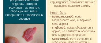
The sizes of neoplasms can vary - from 2 mm to 20 cm. Upon visual examination, a vascular tumor looks like a flat or lumpy spot. An important diagnostic sign is the color of the growth: in hemangioma it is red or reddish-blue. In this case, the tumor is clearly visible against the background of the surrounding liver tissue.
Large growths may have a thin stalk, while small ones are attached to the organ without one.
Invasive methods
Gentle and at the same time very effective treatment of liver hemangioma is becoming increasingly in demand. These methods include:
1. Sclerosis. It is used not only for liver hemangioma, but also for the treatment of vascular diseases in other parts of the body. It involves injecting a substance called “sclerosant” into the vessels with a syringe. Under its influence, the vessels stick together, which prevents blood flow to the tumor and its further growth.
2. Embolization. During this procedure, under local anesthesia, emboli substances are injected into the liver vessels, which clog their lumens. As a result, new blood flows to the tumor stop.
3. Microwave radiation and liquid nitrogen. This method successfully treats hemangiomas in children and adults. It is not traumatic and does not cause bleeding. Vessels are destroyed under the influence of microwave rays and nitrogen supplied at very low temperatures, leaving no scars.
4. Laser therapy. It is based on the fact that the laser beam glues the vessels together, which stops the flow of blood into the hemangioma. The method has contraindications:
- Insulin dependence.
- Epilepsy.
- Infectious diseases.
- The presence of oncology in any organ.
5. Electrocoagulation. Mainly used to remove skin tumors (moles, condylomas, nevi). The method is based on burning out formations with a special element heated to high temperatures. If a device called an electrocoagulator operates in monopolar mode, bleeding in the vessel stops. In addition, doctors began to use an innovative method based on the introduction of specific substances into pathological vessels, which are heated to high temperatures and burn out the tumor, and dead tissue is further broken down.
Opinions of doctors and patients
The overwhelming majority of doctors believe that liver hemangioma, which does not make itself felt and does not interfere with a person’s life in peace, does not need to be treated. At numerous medical forums, experts explain to people that with this pathology you can play sports, give birth to children, and work in any specialty.
Patients in reviews of liver hemangioma express complete optimism that its treatment does not cause any complications. Health is completely restored after surgery or other medical effects.
Some patients note that their liver hemangioma disappeared after gerudotherapy, although doctors do not recommend this method.
Which doctor should I contact?
At an early stage of development, there are no symptoms of liver hemangioma, so the tumor is most often discovered accidentally during ultrasound diagnosis.
It is necessary to visit a hepatologist to make an accurate diagnosis and ensure that the neoplasm is benign. If there is no hepatologist in your city or it is difficult to get to one, you can contact a general practitioner.
The main research method for hemangioma is ultrasound of the liver and gallbladder. The study is available to most patients and does not cause any radiation exposure to the body.
You can see what a liver hemangioma looks like on an ultrasound. It is a round formation with fairly clear contours. The contents of the tumor are heterogeneous.