Enamel hypoplasia is the underdevelopment of the enamel layer of teeth. A similar situation occurs for various reasons and is typical for both milk and permanent organs. Sometimes hypoplasia reaches an extreme degree and implies a complete absence of enamel coating; this form is called aplasia.
Enamel hypoplasia is not difficult to recognize. With it, spots, grooves, grooves, sometimes highly pigmented, are clearly visible on the teeth, contributing to carious lesions of the teeth with the subsequent transition of the disease to pulpitis. Aplasia is characterized by high sensitivity of teeth to external irritants. Both of these dental disorders provoke the formation of malocclusion.
Causes and risk factors
The main prerequisite for the development of hypoplasia is the defective processes of mineralization of dental tissues due to the pathology of protein and mineral metabolism. During the formation of tooth buds in conditions of disturbed metabolism, the functions of enameloblasts, the cells that form enamel, suffer. The result of incorrect functioning of these cells is the appearance of various defects in dental tissue.
Enameloblast deficiency can occur both in the prenatal period and in the first year of a child’s life, when active processes of formation and mineralization of the rudiments of milk and permanent teeth occur.
Causes of mineral and protein metabolism disorders leading to enamel and dentin hypoplasia:
- diseases of the central nervous system accompanied by impaired calcium and phosphorus metabolism;
- endocrine pathology (diseases of the thyroid and parathyroid glands);
- acute infectious processes in a newborn (for example, osteomyelitis);
- intoxication with digestive disorders and loss of significant volumes of fluid and minerals (dysentery, etc.);
- intrauterine infections (syphilis, TORCH infections), respiratory viral diseases suffered by the mother during pregnancy, influenza, etc.;
- rickets;
- hemolytic anemia;
- toxicosis, gestosis in the mother;
- hypovitaminosis C, D, E in a newborn;
- complicated allergy history, atopic dermatitis;
- eating disorders.
Risk factor for dental hypoplasia – diseases of the thyroid or parathyroid glands
In addition to metabolic disorders, hypoplasia of enamel and dentin can be caused by injuries to the upper and lower jaws. Also, a number of studies indicate the possibility of genetically determined damage to dental tissue.
A risk factor for congenital hypoplasia is prematurity.
Causes of development of tooth enamel hypoplasia
Enamel hypoplasia develops in two cases:
- Initially incorrect laying of embryonic cells;
- Maternal diseases and extremely severe toxicosis affecting the development of the fetus in utero.
These circumstances become sources of abnormal metabolism in the emerging new organism. The consequences become visible later, when the baby begins to teethe.
Many expectant mothers believe that the lack of enamel in a newborn baby is explained solely by a lack of essential minerals, and as soon as their intake into the child’s body is increased, the teeth will be restored. Unfortunately, areas of hypoplasia cannot be eliminated in this way. The disorder implies its dependence on the influence of pathogenic factors that occurred at the stage of fetal formation, that is, long before the birth of the baby. An analysis of registered diseases of pregnant women and children born with dental enamel hypoplasia shows a direct dependence of the pathology on the diseases and manifestations of toxicosis suffered by the expectant mothers. The health of children's teeth is affected by outbreaks of acute respiratory viral infections, rubella, and toxoplasmosis. If the baby was injured during childbirth or was born prematurely, subsequent enamel abnormalities are also possible.
The occurrence of hypoplasia cannot be associated with other systemic or chronic diseases diagnosed in the child. 40% of children included in health groups 1 and 2 are susceptible to this disease.
On baby teeth, hypoplasia appears if harmful factors affected the fetus during pregnancy. Teeth erupt and immediately look unhealthy. But much more often, pathology is recorded when it affects permanent teeth. This is considered a dangerous situation, because enamel defects invariably mean an almost one hundred percent likelihood of caries and other dental and orthodontic diseases.
The occurrence of enamel hypoplasia in permanent teeth is influenced by the metabolism and illnesses of a child aged 5-6 months. Therefore, during this period it is extremely important to monitor the baby’s health, provide him with adequate nutrition and healthy sleep. Hence the explanation for the frequency of occurrence of anomalies on permanent teeth - after all, newborn children get sick more often than pregnant mothers.
At the same time, the nature of the manifestations of hypoplasia has been studied and correlated with the time when the child suffered the disease. For example, if the baby was ill at six months, the anomaly will appear on the central incisors in the area of the cutting edge and on the surface of the tubercles of the first molars. Reason: at 5-6 months the formation of these permanent teeth occurs. The disease, which occurs in the ninth month of the child’s development, will be reflected in hypoplasia of the enamel of the lateral incisors, including the edge of the canines.
Diseases affecting the metabolism of a child’s body later result in large areas of defective enamel on the teeth. Almost the entire crown part of the tooth is covered with uneven, lumpy, pigmented enamel. However, quickly treated or minor pathologies leave behind only white spots on the enamel.
Forms of the disease
In accordance with the prevalence of the process, the following forms of dental hypoplasia are distinguished:
- systemic – all teeth or their absolute majority are affected (develops in the prenatal period);
- local – single teeth are involved in the pathological process (the result of inflammatory processes or injuries);
- focal - damage to several adjacent teeth (can be observed in absolutely healthy children).
With systemic hypoplasia, all teeth or most of them are affected
According to morphological manifestations:
- spotted;
- wavy;
- cup-shaped;
- grooved;
- erosive;
- edge;
- "tetracycline teeth";
- teeth of Hutchinson, Fournier, Pfluger, Turner.
The main complications of dental hypoplasia are the progressive destruction of enamel and dentin, and the addition of the carious process.
Depending on the condition of the surface of the defect, hypoplasia occurs:
- smooth;
- dim;
- shiny.
By severity:
- light;
- moderate severity;
- severe (up to complete absence of enamel - aplasia).
Based on the presence of concomitant pathology and complications, the following are distinguished:
- hypoplasia in the spot stage;
- hypoplasia in the stage of usurs and defects;
- hypoplasia complicated by caries;
- hypoplasia combined with caries;
- hypoplasia, complicated and combined with caries;
- enamel aplasia.
Diagnostic methods
To diagnose the disease, you need to visit a dentist. During the initial examination, the specialist will indicate the presence of pathology. The testing algorithm for hypoplasia is simple:
- determining the nature of a woman’s pregnancy,
- studying the life history of a small patient,
- examination of the oral cavity using an instrument,
- clarification of the time of appearance of the first signs of the disease.
After diagnosis, the doctor draws up a treatment plan. For each patient it is developed individually, as it depends on the type and degree of neglect of the disease.
Symptoms
The main symptoms of the disease are:
- the appearance of spots, grooves, irregularities and other defects with a clear or blurred outline on the enamel of the tooth (teeth);
- roughness and pigmentation of enamel;
- shortening of tooth roots;
- symmetrical nature of the lesion, often multiple;
- change in bite;
- shortening of tooth crowns;
- thinning of dentin;
- expansion of channels;
- increased sensitivity to temperature effects;
- depressions on the surface of the teeth with thinning or absence of enamel at the bottom of the defect.
Hypoplasia is characterized by increased tooth sensitivity
“Tetracycline teeth” is a pathology that develops when a mother during pregnancy or a child in the first year of life is treated with tetracycline drugs. Hypoplasia in this case is manifested by a specific green-brown, brown or yellow-brown staining of the enamel (and in severe cases, dentin).
Hypoplasia is much more common in childhood, on non-permanent teeth. If a child has a chronic somatic pathology, the risk of hypoplasia is approximately 50%.
The sign of Hutchinson's (Hutchinson's) teeth is a characteristic barrel-shaped shape (the width of the neck of the tooth exceeds the width of the crown), combined with moon-shaped notches on the free edge of the incisors, atrophy of their chewing surface and (often) absence of enamel.
Fournier's teeth have a similar appearance to Hutchinson's teeth; the main difference is the absence of a semilunar notch on the cutting edge - it is absolutely smooth.
Pflueger teeth are a consequence of improper formation of dentin, which is manifested by the cone-shaped shape of the first molars (large molars).
All three listed types of dental hypoplasia are characteristic of a congenital syphilitic process.
Tourneur's teeth are characterized by the presence of chalky spots on the enamel, which after eruption can undergo pigmentation; Large molars are more often affected. Sometimes crown deformation with partial or complete absence of enamel is detected. This pathology is characterized by the single nature and asymmetry of the lesion.
What is hypoplasia?
- Enamel hypoplasia is a pathology, the main symptom of which is the partial absence of tooth enamel. In the early stages, it manifests itself in the form of pigment spots on the surface of the tooth, grooves, pits, and chips. The last stage of this disease is considered to be aplasia or the complete absence of enamel. In all cases, this pathology is congenital in nature and is a consequence of metabolic disorders in the embryo. Dental hypoplasia is a frequently diagnosed abnormality. Currently, about 40% of clinically healthy children suffer from this disease. The reverse process to this disease is enamel hyperplasia - the appearance of excess tooth tissue.
- The disease can be detected both in a preschooler who has only baby teeth, and in older children who have already developed molars. The disease can be identified during the first examination by a specialist. Based on the location of the spots, the dentist can determine at what point in the development of the fetus this disease began and what led to it.
- When hypoplasia of the enamel of baby teeth is diagnosed, the child is registered with a dentist, and in the future he will have to visit the doctor’s office several times a year and undergo prophylaxis. If the disease is detected in the early stages, it is possible to quickly cope with aesthetic defects, as well as prevent further development of the disease.
- Clinical manifestations of the disease depend on its stage and type. If metabolic processes have mild disturbances, there is mainly a change in the color of the enamel. They usually appear as single yellowish spots. Unlike carious formations, they do not cause discomfort and are not stained with food coloring. With deeper processes, the formation of grooves and depressions in the tissues is usually observed, and in the last stages of the disease the enamel layer may be absent altogether.
Diagnostics
Hypoplasia of enamel and dentin is diagnosed based on:
- characteristic clinical picture;
- X-ray examination (changes in tooth roots, canals, hard tissues are revealed);
- fluorescent stomatoscopy (there is no quenching of the glow in places where spots are localized);
- results of vital staining (no staining of enamel defects with dyes);
- assessing the dynamics of the process (the stability of pathological changes is established).
Dental hypoplasia has a clear clinical picture, diagnosis is not difficult
Symptoms of hypoplasia
The clinical manifestation of the disease depends on its type. Symptoms of the systemic form include:
- spots of different shapes and shades,
- complete absence of the enamel layer,
- defects of a wavy or grooved appearance,
- point change in the pigmentation of the dental covering,
- smoothness and shine of the enamel at the site of the lesion,
- shrinkage of the tooth crown,
- complete absence of pain in places where the integrity of the enamel is damaged.
Also, with hypoplasia, teeth can change their shape. There are only 3 variants of pathology with the following categorical changes:
- Hutchinson's teeth - the central incisors become thinner in the lower part, and take on the appearance of a screwdriver or barrel,
- Pflueger teeth - the crowns of the first molars near the cheek are thicker than those at the chewing surface, the teeth are cone-shaped,
- Fournier's teeth - the clinical picture is similar to Hutchison's symptom, there is no semilunar recess.
Symptoms of local hypoplasia:
- white or yellow spots on the coating,
- the appearance of pinpoint grooves on the surface of the tooth,
- thinning of the tooth body due to the absence of the enamel layer (entirely, partially).
Treatment
The choice of treatment for hypoplasia in each specific case depends on the depth and extent of the defect, the severity of the disturbance of mineral metabolism, and the degree of deterioration in the aesthetic appearance of the teeth.
Conservative treatment of enamel and dentin hypoplasia consists of saturating tooth tissues with minerals (remineralizing therapy).
Microabrasion or bleaching of hypoplastic areas is used when the affected area is small and allows you to eliminate small superficial cosmetic defects of the enamel.
Replacement of affected tissues is carried out if it is impossible to eliminate the defect in a low-traumatic way. The main directions in this case are restoration of hypoplastic areas with composite material, installation of veneers and lumineers, metal-ceramic, ceramic, metal-plastic crowns.
Treatment and prevention of enamel hypoplasia
- Measures for the treatment of enamel hypoplasia, as well as enamel hyperplasia, are chosen in accordance with the stage of the disease, as well as the rate of its progression. If it manifests itself exclusively in the form of pigment spots on the teeth, is localized and does not lead to noticeable destruction of the enamel, the doctor may limit himself to preventive measures and prescribe tooth remineralization to the patient.
- If the stains are pronounced, the dentist will decide to grind the damaged part of the tooth. This method prevents further development of the disease and gives a good aesthetic effect.
- In case of erosive depressions or lesions of a mixed form, a specialist can use composite materials to fill the foci of the disease or an alternative to them: veneers and lumineers - special inlays that cover the affected teeth.
- In severe stages of the disease, when the patient is missing significant areas of tooth enamel, the doctor will prescribe prosthetic crowns. This measure will prevent further development of caries, and will also help achieve the desired aesthetic effect.
It is important to understand that all of the above therapeutic measures are aimed mainly at eliminating the consequences of the disease, but they cannot completely stop the processes of enamel destruction.
Patients, especially those with a hereditary tendency to hypoplasia, are advised to consult a doctor from time to time, undergo examination and eliminate new foci of the disease.
As for the prevention of hypoplasia, it is based on a set of measures aimed at preventing metabolic disorders, primarily during the formation of the fetus (in pregnant women), also in children before adolescence. Such measures include:
- formation of a healthy balanced diet with the necessary content of vitamins and minerals;
- prevention of childhood injuries;
- timely treatment of various infectious diseases;
- a set of measures aimed at increasing immunity and ensuring proper metabolism;
- timely and productive treatment of childhood caries and other diseases of the teeth and gums.
At an older age, the prevention of child enamel hypoplasia should also be given due attention. Here, the most important aspect of disease prevention can be considered remineralization of teeth. It must be carried out every six months in a clinical setting. Enamel hyperplasia also provides similar measures to prevent the disease.
Danger and consequences
Disorders associated with hypoplasia are often attributed to other vascular diseases, which makes the diagnosis of pathology much more complicated. If not treated in a timely manner, its consequences may include deterioration in vision, hearing and functioning of the vestibular apparatus, changes in the composition of blood and other biological fluids , as well as atherosclerosis and thrombosis.
Therefore, at the first suspicion of hypoplasia, the patient should consult a doctor as soon as possible.
Prevention
It is impossible to return damaged enamel to its original structure, but there is always a chance to prevent the development of this disease. Every mother should take care of the child’s dental health during pregnancy, namely:
- treat diseases in a timely manner;
- maintain proper mineral metabolism in the body;
- eat well;
- To avoid negative effects on the fetus, take medications only as prescribed by your doctor.
Proper nutrition during pregnancy is the key to your baby’s health
Measures to prevent hypoplasia of permanent teeth:
- regular visits to the pediatric dentist, mandatory treatment of baby teeth;
- prevention and timely treatment of infectious and other diseases in children;
- providing a nutritious diet;
- taking measures to prevent injury.
A visit to the pediatric dentist is a mandatory procedure for children over 1 year of age.
Disease prevention
The most effective prevention is to pay attention to your health during pregnancy. You should beware of infectious diseases, and if you fail to protect yourself, then treatment should be timely and complete. From the first days of pregnancy, you should pay attention to a nutritious diet, enriched with essential vitamins. After the birth of a child, it is also worth monitoring the baby’s proper and varied diet. After the introduction of complete complementary feeding, in accordance with the child’s age, his diet should include:
- sources of calcium: dairy and fermented milk products (milk, yogurt, kefir, cottage cheese, cheese);
- sources of vitamin C: spinach, broccoli, citrus fruits, sweet peppers and rosehip decoction;
- sources of vitamin B: fish, rice, greens, legumes, mushrooms (after 3 years);
- sources of vitamin A: seaweed, offal (liver, heart), egg yolks.
Food products for pregnant women should be as fresh as possible and should not contain preservatives or excess salt. The balance between such essential nutritional components as proteins, fats, carbohydrates, as well as vitamins and minerals is of great importance.
An important part of preventing enamel pathology is proper hygiene. After the first tooth appears, it is necessary to accustom the baby to regular cleaning of the oral cavity. First, use silicone brushes, and then switch to full-fledged toothbrushes with soft bristles. In addition, any healthy child needs to visit the dentist’s office twice a year, who will be able to notice the development of the disease at a very early stage.
Symptoms and signs
The defect may not produce symptoms for a long time and only appear in adulthood or old age.
At a young age, the functions of the vertebral arteries are taken over by their thinner branches, which provides a compensatory effect. But over the years, the body’s resources begin to be depleted, and it can no longer compensate for blood flow disturbances on its own - during this period, the first unpleasant manifestations of hypoplasia appear in a person.
Common symptoms of the pathology include:
- frequent and causeless dizziness;
- dysfunction of the vestibular apparatus;
- weakness and decreased performance;
- lethargy and drowsiness;
- numbness and loss of sensation in the extremities (especially the fingers);
- distorted perception of space.
Over time, the symptoms increase and those associated with a defect in the right or left artery come to the fore.
Thus, right-sided hypoplasia is manifested by a disturbance in the emotional background : irritability, frequent mood swings, lethargy, fatigue and drowsiness, which can give way to insomnia. The person becomes weather dependent, may feel as if they have been on a merry-go-round for a long time, stumble when walking, and constantly bump into objects.
The main feature of left-sided hypoplasia is stagnation of blood in the vessels , which is why severe pain in the cervical spine, as well as increased blood pressure, joins the general symptoms of the pathology.
Familiarize yourself with the symptoms of aortic dissection, a dangerous disease that requires competent medical care. Aortic stenosis - is it scary? Find out what the disease can mean and how to prevent it from this article.
What is cavernous sinus thrombosis and how it manifests itself, ways to prevent the disease - all this is described in our publication.
Damage to the enamel layer of permanent teeth
The formation of the rudiments of permanent teeth occurs from six months to 1.5 years. Many somatic diseases suffered by the child at this time can give impetus to the development of enamel pathologies.
Among them:
- diseases of the digestive and endocrine system;
- rickets;
- dystrophy;
- syphilis;
- severe form of infectious diseases;
- brain dysfunction;
- Iron-deficiency anemia.
Diseases suffered by a child can become a prerequisite for the development of enamel hypoplasia
Note: through scientific research it has been proven that the location and nature of erosion of the dentition depends on the time and severity of the diseases suffered.
In children whose illness occurred at the time of the beginning of the formation of permanent teeth (6-7 months), damage to the cutting edges of the 1st central incisors and the cusps of the 6th teeth is observed; if this happened at 8-9 months or later - 2 and 3 teeth on both sides, as well as large chewing incisors in the crown area. With a prolonged recurrent course of the infectious disease, the enamel coating may acquire a lumpy structure. The light form leaves only small dark spots on it, while the heavy form can lead to the complete disappearance of the protective coating.
Enamel hypoplasia in children
Where and when does pathology begin?
The disease begins to develop in the womb. When a baby is born with normally developed organs and body parts, which subsequently, due to various reasons, stop developing and growing, this cannot be called hypoplasia.
Hypoplasia in a child is not always detected immediately after birth. There are cases when pathology is revealed only after some time, for example, during puberty - this is hypoplasia of the genital organs or mammary glands. It appears only during their development and formation.
How is the treatment carried out?
Taking tetracycline during pregnancy leads to damage to the tooth buds of the fetus.
If the pathology is weakly expressed and manifests itself only in the form of pigment spots that are insignificant in color and progression, the doctor can take preventive measures, in particular tooth remineralization.
In other cases, when there is severe damage to the enamel, developing at a rapid pace, modern dentistry only offers grinding of the surface of the affected area. This procedure helps prevent the spread of the disease and provides an excellent aesthetic effect.
If there are erosive depressions or mixed types of damage, the doctor may recommend the use of composite technology for restoring tooth enamel. Another option is veneers or lumineers.
In advanced forms of pathology, when the enamel becomes very thin, experts recommend using prosthetic crowns. This procedure will help prevent the development of caries and give your teeth the desired appearance.
It is important! All the methods described above only help eliminate the consequences of the pathology; they do not allow it to be completely stopped. Patients who have a genetic predisposition to the development of hypoplasia are recommended to visit their dentist at least once every six months.
When should you see a doctor?
Hypoplasia of the vertebral arteries is treated by a neurologist, who should be contacted immediately after the first symptoms appear . Delay in this case can be extremely dangerous, since signs of hemodynamic disturbances indicate that the body can no longer compensate for blood flow disturbances on its own. After listening to the patient’s complaints and collecting anamnesis in order to clarify the diagnosis, the doctor must prescribe additional tests and studies.
Classification of dental hypoplasia
Depending on the signs of manifestation, there are different types of dental damage:
- Cup-shaped (erosive). It appears as the formation of oval-shaped holes on the surface of the tooth. The enamel thins unevenly, its minimum thickness is located in the very recess of the hole. At the bottom of the formation, enamel may be completely absent.
- Spotted form. Symptoms of this form are a change in the color of the enamel and the appearance of spots of white and yellow shades.
- Furrowed shape. It is characterized by the appearance of defects in the form of grooves, which are located parallel to the cutting edge. The grooved form also includes wavy hypoplasia, when the grooves are located in such a way that the surface becomes wavy.
- Mixed hypoplasia. It is diagnosed when one patient has different forms of manifestation of the disease.
- Aplastic hypoplasia. It is a severe form of the disease, characterized by deep damage to the enamel up to its complete absence.
Severe hypoplasia leads to changes in the shape of the teeth. These pathologies are named after the doctors who studied them:
- Pflueger's teeth. Change in the shape of permanent molars. They take on the shape of a cone, while the tubercles and crown of the cervical region are underdeveloped.
- Hutchinson's teeth. This type of pathology is distinguished by screwdriver-shaped or barrel-shaped incisors, with a characteristic crescent-shaped notch along the cutting edge.
- Fournier's teeth. They present themselves as the same form of the disease as described by Hutchinson, but without the semilunar notch.
All three forms are included in the triad of symptoms of congenital syphilis, however, they are not always a mandatory marker of the disease. Sometimes they occur against the background of other conditions.
Consequences
Many doctors consider abnormal variations in the structure of the circulatory system of the brain as predictors of epileptiform disorders. Consequences of a severe disease:
- Aneurysm formation.
- Rupture of the vascular wall with subsequent formation of a hemorrhage focus.
- Deterioration in cognitive abilities, general health and well-being.
- Stroke.
The development of a stroke is associated with deterioration of blood supply to a region of the brain or intracranial hemorrhage. After a stroke, only 20% of patients regain the ability to self-care. In other cases, the patient faces disability with a stable, pronounced neurological deficit. The mortality rate is 35-40%.
Hypoplasia of blood vessels in the brain is a pathology that in most cases does not pose a threat to the health and life of the patient. The need for drug or surgical treatment occurs if the abnormal structure of the elements of the circulatory system leads to cerebral circulatory failure, occurring in acute or chronic form.
"Tetracycline" teeth
“Tetracycline” teeth also belong to systemic dental hypoplasia, but they should be considered separately.
The coloring of such teeth is changed as a result of taking the drug tetracycline during the period of formation, as well as mineralization of the hard tissues of the tooth. Tetracycline tends to be deposited in the dentin and enamel of teeth that are at the developmental stage and in the bones of children in the prenatal period (if a woman takes tetracycline during pregnancy) or after birth (when tetracycline is introduced into the child’s body to achieve a therapeutic effect for various diseases).
Taking tetracycline is undesirable not only because of the color change, but also because this drug can cause hypoplasia. The nature of the changes depends on the type of drug and the doses in which it was taken. Small doses only affect the color of teeth, but when taking significant doses, along with a change in color, underdevelopment of the enamel layer occurs.
Treatment of a pregnant woman with tetracycline causes discoloration of her child's front teeth. The third part of the crown of the incisors, starting from the cutting edge, and the chewing surface of multi-rooted teeth acquire a yellowish color. This is due to the ability of the drug to penetrate the placental barrier (the placenta protects the child from many harmful factors).
Taking tetracycline drugs, starting from the sixth month of a child’s life, leads to staining of permanent teeth, which are at the stage of formation at this time. As a rule, the color does not change in the entire crown, but only in its part, which is still being formed at the moment when the above-mentioned medicine enters the body.
The type of tetracycline and its amount directly determine whether the teeth become light yellow or acquire a dark shade.
An interesting feature of tetracycline teeth is that they can emit fluorescent radiation when exposed to ultraviolet light. This method can be used to diagnose this type of hypoplasia or if there is doubt when making a diagnosis.
Based on the fact that teeth stained with tetracycline cannot be whitened, it should be prescribed only in vital circumstances.
Causes of hypoplasia
Disorders of intrauterine development of the fetus lead to the occurrence of hypoplasia. And these violations can be caused by a variety of factors:
- Anomalies of embryonic cell division
- Oligohydramnios (reduced amount of amniotic fluid)
- Radiation
- Injuries of a pregnant woman
- Exposure to high temperature
- Work in hazardous industries
- Smoking
- Consumption of alcoholic beverages, drugs and certain medications
- Infectious diseases (rubella, toxoplasmosis, influenza, etc.)
Gnomik.ru recommends Rmh24.ru Urgent repair of refrigerators We repair refrigerators of all brands. Free visit of a specialist and diagnostics!
Use promo code: Gnomic.ru
and get a guaranteed 10% discount
We work in Moscow and the region.
Call
Call a specialist
Classification of the disease
Hypoplasia affecting the cerebral artery is a pathology that is characterized by a change in the normal shape and size of the vessel, which affects its patency and the speed of blood movement along the channel.
The diagnosis of hypoplasia of cerebral vessels almost always correlates with such disorders as TIA (transistor ischemic attack) in 46% of cases, vegetative-vascular dystonia, dyscirculatory encephalopathy, vertebrobasilar insufficiency, hypoxia, ischemic infarction in 38% of cases, necrosis.
Hypoplasia of blood vessels in the brain is characterized by changes in the structure of the blood flow system. In this case, the elements of the circulatory system differ from normal vessels in the following ways:
- Abnormal tortuosity (loops, kinks).
- Irregular shape.
- The presence of defects - thickening of the wall, pathological expansion of the bed (aneurysm).
As a result of such changes, the transportation of blood through the vascular bed is disrupted, and the blood supply to brain tissue deteriorates. Against the background of pathology, brain dysfunction develops, a change in neurological status occurs, and disruptions in the body’s functioning occur. The nature and severity of the disorders depend on the location of the damaged area of the circulatory system. The blood supply to the brain is primarily through the vertebral and carotid arteries.
If there is a blockage in any part of the bloodstream, the blood moves around the main path along the Circle of Willis. As a result, disturbances associated with imperfections in the main channel are partially compensated. The circle of Willis has a normal anatomical structure in 25% of people. In most cases, it also has an abnormal structure, which impedes the blood supply to the brain.
The abnormal structure of certain sections of the vertebral artery occurs in 10% of healthy people. Often the pathology does not manifest itself and does not cause a person discomfort, but it increases the risk of developing an ischemic stroke. Cerebrovascular pathologies have a major impact on mortality rates. Stroke causes the death of 4.6 million people worldwide every year.
Hypoplasia of the A1 segment of the right or left anterior cerebral artery is a variant of the formation of the circle of Willis, which is a congenital abnormality that occurs with a frequency of 1-13%. The pathology is an increased risk factor for the development of hemispheric infarctions, which are accompanied by a significant deterioration in blood flow due to occlusion (blockage) of the carotid arteries.
Hypoplasia of the posterior right and left communicating arteries, which are located in the brain, is a variant of an incorrectly formed circle of Willis. Hypoplasia of the venous sinuses located in the brain is considered a normal variant. In most cases, it is not accompanied by pathological symptoms and disorders. Hypoplasia of the left or right transverse sinus, located in the brain, is observed in 20-49% of healthy people.
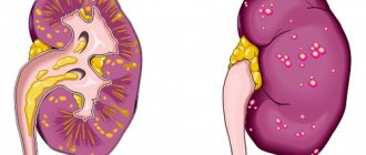
Hypoplasia is a widely used medical term that means underdevelopment or abnormal anatomical structure of an organ. Brain hypoplasia, also known as microcephaly, is a disease in which there is a decrease in the volume of the skull and brain matter. The disease is a severe form of damage to the central nervous system, leading to mental impairment, disability, and death.
Pituitary hypoplasia is a pathology that provokes disruptions in the endocrine system, which negatively affects the growth and increase in muscle mass in a child. In adults, it is manifested by a decrease in bone density, hypoglycemia, anorexia, lack of pigmentation of areas of the skin, amenorrhea (prolonged absence of menstruation), impotence and other disorders of the reproductive system.
What diseases can enamel hypoplasia be confused with?
Differential diagnosis of enamel hypoplasia is carried out with initial and superficial caries.
- With initial caries, the white spot is single, most often located at the neck of the tooth, and with hypoplasia, the spots are formed in the plural and can be located anywhere on the surface of the crown.
When stained with a solution of methylene blue, carious spots change color, while hypoplastic spots remain unchanged.
- The difference between hypoplasia and superficial caries is that when probing caries, the location of the lesion is determined by the roughness to which the instrument clings. With hypoplasia, the enamel remains smooth and dense even in severe forms of the disease.
Complications with hypoplasia
Hypoplasia of primary teeth must be treated, otherwise areas devoid of enamel can become a source of infection, which will have a negative impact on the buds of permanent teeth. Untreated disease leads to increased sensitivity and abrasion of the enamel. Teeth damaged by hypoplasia are more susceptible to caries. Enamel thinned by disease is suitable soil for the proliferation of carious bacteria, which can quickly penetrate into the deep layers of dentin and can lead to the development of pulpitis. Complicated pulpitis, in turn, can lead to tooth loss and infection of neighboring tissues. In addition to the complications that hypoplasia can entail, there are also aesthetic problems. Damaged teeth appear stained, chipped, and uneven, and the enamel changes color and structure. Such a smile can hardly be called beautiful. A qualified doctor using modern aesthetic dentistry techniques will help you get your teeth in order.
Treatment of hypoplasia
In mild cases of underdevelopment of tooth enamel (with small and hardly noticeable stains), treatment is not carried out. If the signs are localized on the front surface of the incisors, clearly visible when talking and smiling, if the deep tissues are affected, treatment is definitely necessary.
Treatment of dental hypoplasia
Untimely help with hypoplasia can lead to unpleasant consequences, such as:
- destruction of tooth tissue;
- rapid abrasion of the tooth edge;
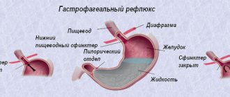
- the development of malocclusion, which in turn provokes diseases of the gastrointestinal tract.
The treatment protocol is selected individually depending on the severity of the lesion.
Whitening
It is carried out for mild forms of hypoplasia (spotty) under the supervision of a doctor at home or in a dental office. Home whitening is more comfortable and costs patients less, but takes longer.
After teeth whitening, remineralization begins
One of the most popular modern technologies is whitening using special trays made to individual sizes and filled with whitening gel. One session of wearing a mouth guard lasts from 3 to 10 hours. Other methods: laser and photo whitening (carried out in an office setting).
Sealing
The method is effective for single depressions, stripes and grooves. Treatment is carried out according to the traditional scheme:
- cleaning the dentition;
- leveling the surface with a boron;
- etching of tooth enamel;
- applying adhesive composition;
- restoration of a damaged tooth through filling.
Treatment of hypoplasia with filling
Careful attention to restored teeth allows you to preserve their functions and aesthetic appearance for many years.
Video - Treatment of enamel hypoplasia
Veneers
The technology involves attaching special plates to the front surface of the teeth. This method allows you to make the visible part of the dentition immaculate, while the palatal part remains affected.
Veneers for teeth
Orthopedic crowns
This method must be resorted to when there is significant deformation of the tooth shape, structure and color of the enamel coating or its absence. Installing crowns involves serious preparation of the hard surfaces of the tooth (grinding with a burr).
Orthopedic crowns
Important: if there is at least a minimal opportunity to restore the dentition in a more gentle way, you should not rush to seek help from orthopedists.
Enamel hypoplasia in young children
Mild hypoplasia of primary teeth occurs in almost every second child.
Without receiving all the necessary beneficial microelements, a child’s body becomes vulnerable, as a result of which the enamel becomes very thin and begins to deteriorate; a tooth can break even with a slight load.
Usually this disease is not transmitted to the permanent teeth, except in cases of long-term serious illness after birth. Therefore, hypoplasia detected at an early age should not cause serious concern, but treatment is still required, since the quality of permanent teeth depends on the health of baby teeth.
Important! Scientific studies have found that rubella disease during pregnancy in 9 out of 10 cases leads to the development of hypoplasia in the unborn child.
Causes
The development of hypoplasia is based on complex disturbances in the process of organogenesis, i.e., the formation of organs at the stage of embryonic development. Such violations can result from:
- genetic abnormalities;
- intrauterine infections (cytomegalovirus, herpes, rubella, syphilis, toxoplasmosis, influenza);
- oligohydramnios;
- smoking, drinking alcohol or using drugs;
- exposure to ionizing radiation or occupational hazards;
- living in a region with an unfavorable environmental situation.
Signs of hypoplasia, despite the congenital nature of the pathology, do not always appear from the moment the child is born.
general information
Hypoplasia of the vertebral arteries is a congenital disease characterized by their insufficient development. The lumen of the vessel at the site of its insertion into the bone canal narrows significantly , which causes a disruption in the blood supply to the brain tissue.
The pathology can be left-sided, right-sided or bilateral , and most often the disorder concerns the right artery - according to statistics, this phenomenon is observed in every tenth person in the world.
Bilateral hypoplasia is considered a fairly rare disease, but if the diagnosis is not timely, the pathological process that occurs in one of the arteries may affect the other in the future.