Our fingers are capable of performing coordinated and very subtle movements that have a significant impact on our daily activities and ability to work. Their fractures, from which no one is immune, can cause significant problems: restrictions on the full functions of the hand, bending of the fingers and pain even with minimal loads. In the future, these consequences of injury can negatively affect professional activities and impose restrictions on daily life, and fractures of the thumb can cause disability. That is why any finger fractures should be a reason to contact a specialist.
Such injuries occur quite often and are observed in 5% of patients with fractures. In this article we will introduce you to the causes, types, signs and methods of diagnosis, first aid and treatment of finger fractures. This information will help you suspect the presence of such an injury in time and make the right decision about the need for treatment from a specialist.
Causes and types of fractures
The main cause of finger fractures is direct trauma: a strong blow or compression, a fall from a height, industrial or road accidents. Such injuries often occur in athletes - especially among volleyball players, basketball players, artistic gymnasts and boxers. And especially dangerous fractures can occur when fingers get caught in complex working mechanisms.
In more rare cases, a fracture of the fingers is provoked by minimal mechanical impact due to osteoporosis, osteomyelitis and, extremely rarely, in the presence of a tumor or its metastases.
According to statistics, a fracture of the little finger occurs more often. This fact is explained by the fact that it is located on the edge of the brush. And the most dangerous thing in terms of restoring further working capacity is a fracture of the thumb. Injuries to other fingers can also be dangerous if they are not fused properly, leading to impaired fine motor skills.
Like all fractures, finger injuries can be open or closed. If the integrity of the skin is compromised, the risk of infection of the fracture area and the development of such a dangerous complication as osteomyelitis increases significantly.
Depending on the location of the fragments, all finger fractures are divided into injuries with and without displacement. Displaced fractures are divided into fractures with divergence or overlap of fragments.
Depending on the number of fragments, fractures are divided into:
- splinter-free;
- single-splintered;
- two-splintered;
- comminuted.
Depending on the nature of the fault line, a fracture can be:
- transverse;
- longitudinal;
- screw;
- oblique;
- T-shaped;
- S-shaped, etc.
Experts classify a subperiosteal fracture of the phalanges as a separate type of injury. As a rule, it can only occur in children and the periosteum remains intact. This is due to the fact that at this age the periosteum remains flexible and soft. Such injuries are more difficult to detect, but respond very well to treatment, do not require reduction and heal quickly.
Types of finger fracture
Because of
- Traumatic fractures are damage to the finger bone due to trauma.
- Pathological fracture - a fracture of a finger in the area of pathological restructuring (affected by any disease - osteoporosis, tumor, osteomyelitis, etc.) Osteoporosis is the most common cause of a pathological fracture.
The nature
- Closed fractures (without breaking the skin)
— Incomplete
— Complete
- Open fractures (with skin damage)
— Primary open
— Secondary open
Based on the presence of offset:
- Fractures without displacement of fragments
- Displaced fractures.
Symptoms
The leading symptoms of a broken finger are severe pain, redness and swelling in the area of injury.
The patient spares the injured finger and tries not to move it. As with other fractures, such finger injuries cause severe pain. Later, the following possible symptoms appear in the area of the fracture:
- redness and swelling at the fracture site;
- the skin at the site of injury is warmer;
- significant limitation of movements of the injured finger;
- the appearance of pain when trying to press on the top of the injured finger;
- gentle finger position.
The presence of all of the above probable signs of a finger fracture in almost 100% of cases indicates a violation of the integrity of the bone and does not require diagnostics to identify reliable symptoms of a fracture:
- detection of cracks by palpation;
- crepitus on palpation;
- identifying pathological mobility in those areas of the finger where it should not exist;
- changing the shape of the brush;
- shortening of the damaged finger (when comparing its length with the same healthy finger on the other hand).
It should be remembered that carrying out manipulations to determine reliable signs of a fracture is always accompanied by pain and can cause progression of the fracture. When palpating an injured finger by a person without medical training, there is a high risk of damage to nerves, tendons and blood vessels. In the future, such injuries will require surgical treatment and can lead to irreversible impairment of hand function.
Fractures of the phalanges of the fingers
Fracture of the distal phalanx of the finger is the most common among household injuries. As a rule, it happens due to a strong pinching of a finger in a drawer or door. Often such an injury is accompanied by a tear off of the nail plate, which is not restored in the future. If the nail remains in place, but is damaged, after some time its complete restoration is observed. In most cases, a fracture of the nail phalanx of the finger is accompanied by a blue discoloration of the nail and a feeling of numbness. Unlike a bruise, the fracture site instantly swells and a hematoma forms.
A fracture of the middle or main phalanx of a finger usually occurs as a result of a fist fight. With such an injury, there are clear signs of joint deformation.
First aid
If you suspect a broken finger, it is important to correctly provide first aid to the victim - the future success of treatment depends on the adequacy of these measures:
- Calm the victim and let him take a pain reliever (Analgin, Ketorol, Nimesil, Ibufen, etc.).
- Call an ambulance.
- If there is a wound, treat it with an antiseptic solution and apply a sterile bandage.
- If there is severe bleeding, stop it by applying a tourniquet, attaching a note to it about the time of application.
- Immobilize the injured finger using available means (branches, wooden sticks, wire, etc.). The position of the finger should be such that the victim feels the least pain. One end of the improvised splint is attached to the injured finger (its end should be 2-3 cm higher than the finger). The other end should rest on your palm and forearm. After this, the splint is fixed with a bandage. Bandaging is performed in the direction from the elbow to the fingers. If it is impossible to apply a splint, the injured finger is bandaged to the adjacent one. After immobilizing the finger, for greater reliability, the hand is immobilized with a scarf.
- Apply ice to the injured area and remove it every 5-10 minutes for 2 minutes to prevent frostbite.
Complications
Compared to traumatic injuries to other bone structures of the upper extremity, complications of finger fractures occur infrequently. It can be:
- hematoma - the formation of a bruise in soft tissues;
- traumatic shock - a critical violation of blood circulation at the tissue level, occurring against the background of severe pain (this can happen due to the patient’s low pain sensitivity);
- improper consolidation - incomplete fusion of the formed bone fragments;
- neurological disorders – paresthesia in the form of numbness, tingling, “pins and needles”;
- restriction of movements, and in difficult cases – their complete absence. May be observed due to injury to nerve endings or improper consolidation;
- infectious and inflammatory complications.
In the latter case it is:
- infection and suppuration in open fractures;
- osteomyelitis – the formation of purulent-necrotic foci in bone tissue, which is accompanied by the formation of fistulas (pathological tracts);
- sepsis - the spread of infection from an infected wound through the blood or lymph throughout the body with the possible formation of secondary foci in organs and tissues.
Possible complications due to self-medication or inadequate treatment
If not treated correctly, a fracture of the fingers can lead to the following complications:
- Formation of pseudarthrosis (false joint). This complication causes the finger to bend in a place where it would not normally bend. It can develop with insufficient immobilization. As a result, over time, the fragments rub against each other, become rounded, and the bone canal in them overgrows, and one bone is divided into two short ones. There is a small gap between them, and the finger acquires pathological mobility. This complication can only be eliminated through surgery. In the future, the injured finger becomes shorter.
- Formation of excessively large callus. If the fragments are incorrectly compared, a giant callus develops, which not only leads to the formation of a cosmetic defect, but also causes restrictions in the movements of the damaged finger.
- The appearance of contractures. With unprofessional treatment, inflammation in the affected area lasts for a long time, and improper immobilization leads to shortening of the tendons. As a result, the injured finger loses its mobility. Elimination of such complications is always lengthy and is accompanied by pain during procedures for stretching the shortened tendon.
- Vicious union. If the fragments are incorrectly compared, one of them is displaced and, after fusion, ends up in a non-physiological position. As a result, the injured finger cannot move fully.
- Formation of ankylosis (complete immobility of the joint). This complication can develop with fractures in the articular surfaces. If treated incorrectly, they completely grow together, become immobile, and the finger completely loses its functions. Ankylosis can lead to disability for the victim, since there are no methods for treating such a complication yet.
- Osteomyelitis. This complication occurs when the bone marrow becomes infected in an open fracture at the time of injury or after surgery. The disease is accompanied by severe pain and often becomes chronic. After the bone heals, the victim periodically experiences exacerbations of osteomyelitis, causing severe suffering. Treatment for this complication can only be surgical.
Classification
For convenience in diagnosis and treatment, a unified classification system for finger injuries has been developed.
She identifies several types of bone integrity disorders:
- Closed fracture - the skin remains unchanged, the muscle tissue is not damaged.
- Open fracture - the bone and its fragments pierce the dermis. An open fracture of the middle finger and the rest is a rare phenomenon and in most cases is associated with domestic trauma, when the finger is subjected to aggressive mechanical action.
- Without displacement - the finger bones do not leave the border of the periosteum.
- Displaced fracture - the periosteum is injured, and the fragments move, touching nearby tissues, joints, and blood vessels. A displaced finger fracture is divided into fractures with divergence or overlap of fragments.
- Compression fracture of a finger - when squeezed, the bone fractures, which requires immediate medical intervention.
- Comminuted fracture - this most often involves injury to the joint and ligaments of the finger due to numerous fragments.
- A direct or transverse fracture of the index finger and others is determined by the shape of the fracture.
- Articular and extra-articular fracture of the finger - the injury occurs outside the joint or in it itself.
- Combined finger fractures are injuries to several bones.
What types of fractures are distinguished by location?
- The most common and unpleasant is a fracture of the thumb. His treatment is complicated by the use of a traction apparatus. The proximal part and closer to the ulnar side can break.
- Quite often, the outermost and smallest finger on the hand, the little finger, is injured. The leading damage factor is a blow to a hard surface.
- A fracture of the ring finger is a complex one and requires a long recovery, during which the fingers should be developed using special exercises.
- Injuries to the middle and index fingers require immediate medical attention and should not be treated at home.
- It happens that the tip or the very edge of the finger is damaged - a marginal fracture, which causes deformation of the nail plate.
- If a fracture of the nail phalanx occurs, this can be seen by the bend that appears at a certain angle.
Any fracture of the phalanx of a finger is dangerous in itself to a greater or lesser extent, since if the bone fragments do not heal correctly, motor activity may be impaired.
Fractures of the phalanges are pathological. They are provoked by chronic diseases that destroy the bone structure - osteoporosis, osteomyelitis.
Based on the nature of the fracture line, a fracture can be:
- Transverse.
- longitudinal
- screw.
- Obliquely.
- T-shaped.
- S-shaped.
A separate type is a subperiosteal fracture. As a rule, it occurs mainly in children on the small finger. With it, the periosteum remains intact.
https://youtu.be/hqTHVs68MmY
Diagnostics
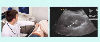
After interviewing and examining the victim and clarifying all the circumstances of the injury, to confirm the diagnosis of a “finger fracture,” the doctor prescribes an x-ray of an individual finger or the entire hand. Pictures are taken in two projections - frontal and lateral. In the future, they make it possible to create a complete picture of the fracture - its shape, exact location and depth - and prescribe the most effective treatment.
After removing the plaster, a repeat X-ray is required to monitor the quality of healing and confirm the correct placement of intraosseous devices for fixing fragments (if any were used).
Rehabilitation after a fracture of the phalanx of a finger
Rehabilitation is a very important stage in restoring full functionality of the finger. If you neglect rehabilitation measures, there is a risk of permanently losing the functionality of the joint.
Rehabilitation after a fracture of the phalanx of a finger includes:
- Therapeutic gymnastics - exercise therapy. To restore range of motion, special expanders or rubber balls are used to help restore the flexion and extension functions of the fingers. To restore fine motor skills, it is useful to transfer small buttons, beads, beads from one container to another.
- Massage. Light massage movements promote blood flow to the damaged area, which helps activate healing processes. The first few days of movement should be very light and soft, and after that the pressure may increase. The duration of the massage is 15-20 minutes.
- Physiotherapy. After removing the cast, physiotherapeutic procedures are recommended. These include magnetic therapy and light therapy, which relieve swelling and reduce pain. Electrophoresis has a stimulating effect and activates the synthesis of bone tissue cells. The number of procedures is prescribed by the doctor on an individual basis.
Are you dealing with an injury or are you worried about chronic pain? Make an appointment with a sports physiotherapist! Make an appointment by calling +7. Detailed information on the website physiotherapist.ru
Treatment
The treatment tactics for finger fractures are determined by their severity. The following techniques can be used:
- closed reduction;
- skeletal traction;
- open reduction.
Closed reduction
This method of treatment can be used for closed simple displaced fractures. Before the closed reduction procedure, a local anesthetic test (Procaine or Lidocaine) is performed. In the absence of allergies, the drug is injected into the soft tissue surrounding the fracture area.
After the onset of local anesthesia, the finger is pulled along its axis and all its joints are bent so that an angle of approximately 120° is formed. Next, pressure is applied to the angle of the fracture, which puts the fragment in the position necessary for healing.
After this, a plaster cast is performed to ensure reliable immobilization. The cast is applied from the upper third of the forearm to the end of the injured finger (while the finger remains partially bent).
When performing such a bandage, be sure to take care of the correct position of the hand. It should be extended at the wrist joint by 30°, and the bent injured finger should lightly touch the palm. This position of the hand helps prevent the development of contractures and repeated displacement of fragments.
After applying the plaster, the victim is advised to keep his arm elevated for 2-3 days. This position prevents the appearance of pronounced edema. Analgesics are prescribed to relieve pain.
Skeletal traction
This method of treatment is used when it is impossible to match displacements by closed reduction or in cases of comminuted fractures. Local anesthesia is administered for pain relief.
The same plaster splint is placed on the arm as for closed reduction, but with one feature. A wire is attached opposite the injured finger (on the palmar side), the end of which is several centimeters longer than the finger and is equipped with a hook. The fragments are repositioned and staples, pins or threads are passed through the soft tissue. When performing such designs on the nail phalanx, the nail plate is covered with several layers of polymer varnish (for strengthening).
After completion of skeletal traction, the victim is prescribed analgesics, anti-inflammatory and antibacterial agents.
Open reduction
Indications for performing open reduction (osteosynthesis) are the following cases:
- open fractures;
- complex comminuted fractures;
- purulent complications due to other methods of treating a fracture;
- improperly healed fracture.
Osteosynthesis surgery is performed under general anesthesia. To fix the fragments in the position necessary for proper healing, knitting needles or screws are used (in rarer cases). After the operation is completed, a plaster cast is applied.
The Ilizarov apparatus can also be used for the same purposes. It promotes more reliable fixation of fragments and does not require wearing a plaster cast. The only disadvantage of such an external fixator is the need for daily treatment.
Osteosynthesis can be:
- one-stage - fixation devices remain in the bones for life;
- two-stage - fixation devices are removed from the bone after 3-4 weeks.
After completion of osteosynthesis, the victim is prescribed analgesics, anti-inflammatory and antibacterial agents.
Treatment of a fractured phalanx of a finger
In the absence of complications in the form of displacement of fragments, a conservative method of treatment is prescribed - immobilization of the hand with the application of a plaster cast. If there is displacement, surgical intervention will be required - repositioning (comparing) fragments of the broken bone and fixing them using knitting needles, screws or a metal plate. The type of construction is selected by the doctor individually.
If possible, the surgeon performs closed reposition of the fragments by percutaneously introducing wires. This method has the advantage of no seam on the skin. The disadvantage of closed reduction is the presence of the wire above the skin and the associated high risk of tissue infection. The plaster cast should fix the hand for a month; early development of the finger is impossible, which leads to additional complications. With open reposition, healing occurs twice as fast - the sutures are removed after 2 weeks. Rehabilitation measures can be carried out a few days after applying the plaster.
How long should the cast be worn?
Depending on the severity of the injury and other factors, the duration of immobilization ranges from 2-3 to 6 weeks.
The duration of immobilization for fractures of the fingers depends on the severity of the injury, age and the presence of concomitant diseases that interfere with bone healing:
- for simple fractures without displacement (after closed reduction), the plaster is worn for 2-3 weeks, and ability to work is restored after 3-4 weeks;
- for simple and comminuted fractures with displacement (after skeletal traction), the plaster is worn for 3-4 weeks, and working capacity is restored after 6-8 weeks;
- for complex fractures (after osteosynthesis), the cast is worn for about 6 weeks, and working capacity is restored after 8-10 weeks.
Duration of wearing a cast for a fracture of the phalanx of a finger
A fracture of the phalanx of a finger heals within two to four weeks. During this time, the patient undergoes repeated radiographs to monitor the correct fusion. However, the duration of wearing a cast directly depends on the complexity of the fracture. For a fracture without complications, the plaster is removed after two to four weeks. With a moderate fracture, bone healing occurs within one and a half to two months. If the fragments were repositioned using metal devices, healing of the injury lasts about 2.5 months.
Rehabilitation
After the cast is removed, the patient is prescribed a rehabilitation program that ensures the most complete restoration of the functions of the injured finger. For this we may recommend:
- therapeutic gymnastics complexes;
- massage courses;
- physiotherapeutic procedures: UHF, salt and soda baths, ozokerite applications, mechanotherapy.
Fractures of the fingers are common injuries. They should not be treated on their own and are always a reason to contact a specialist. Depending on the severity of the injury, the doctor will be able to choose an effective treatment tactic that ensures the most correct healing of the fragments and complete restoration of the functions of the finger in the future.
Recovery and rehabilitation
How long a finger fracture takes to heal depends on its type. This may take from three to ten weeks.
After a broken finger, you need to wear a cast for 2 to 6 weeks, depending on its type.
When the bones heal, the braces are removed, and the person must undergo rehabilitation to fully restore the functionality of the hand. For this purpose they prescribe:
- therapeutic gymnastics exercises,
- massage,
- physiotherapy.
Also today, special fixators - orthoses - are widely used. They are comfortable, but it is important to ensure that the orthosis fits perfectly.
Physiotherapy
https://youtu.be/S7W3op3kHig
Full recovery after a fracture takes quite a long time. To speed up the process, you can perform special exercises:
- Before starting work, rub your hands vigorously to improve blood flow,
- rotate the brush in both directions,
- perform exercises aimed at developing fine motor skills (typing, sorting cereals, putting together puzzles, etc.),
- Exercises with an expander allow you to quickly develop your finger muscles.
It is also useful to read about wrist fractures.
What to do
Having understood the characteristics of the injury, it is necessary to correctly provide first aid to the victim. Without this, the bone may heal incorrectly, and damage to nerves and blood vessels may negatively affect the functions of the limb. After providing first aid, it is advisable to consult a doctor.
This is especially important if the thumb is fractured. It performs a supporting function, and its improper healing will limit a person’s mobility and create problems with choosing shoes. Medical attention is needed when there is doubt about whether a toe is broken. It's better to play it safe and get examined.
Before going to the emergency room, it is important to try to immobilize the injured foot. If your foot was wearing shoes, you should carefully remove them, as swelling will then develop and this will be difficult to do. You should try not to move or step on the injured leg. It is recommended to sit or lie down and raise your foot higher - on a pillow or on the back of the sofa.
Then it is recommended to apply ice to the injury site. This will help reduce pain and swelling. You need to do such compresses for 5-10 minutes with breaks. This will help prevent frostbite. If the pain is severe, you can take a painkiller. Most often it is recommended to take Ibuprofen, Nimesil or Ketanov. You cannot put pressure on the affected area, move your finger, or try to align the bones. Such injuries must be treated by a specialist.
These measures are also used in case of a bruised toe. Sometimes rest and the application of a cold compress can completely eliminate all symptoms of this injury in a few hours. In the event of a fracture, these measures can slightly alleviate the victim’s condition, but then it is still necessary to seek medical help.
Many people believe that if a toe is injured, it is not necessary to do this, especially if the pain is not severe and the fracture is closed. But in fact, even minor injuries are best treated under the supervision of a specialist, this will help avoid complications.
Symptoms and first signs
Many people are concerned about how to recognize an injury in time. In some cases, intense pain appears after several hours or even days. A broken finger can easily be confused with a bruise or dislocation, which can delay a trip to a traumatologist. An incorrect diagnosis leads to unpleasant consequences that will take a long time to eliminate.
To understand at the first stages whether a finger is broken or not, it is enough to know a number of reliable signs of pathology.
Reliable symptoms of an open fracture:
- presence of a wound: damage to the skin with bone fragments;
- bleeding from cut vessels;
- severe pain;
- inability to bend or straighten a finger;
- displacement of fragments relative to each other;
- pronounced swelling and hyperemia of the hand.
An open fracture is characterized by the presence of a wound
Reliable symptoms of a closed fracture:
- crepitus (crunching of bone fragments);
- pathological mobility of the bone: it can bend and unbend in places where there is no joint;
- swelling and hyperemia that does not go away for a long time;
- intense pain when touched, which goes away only after using painkillers.
A distinctive feature of a closed fracture is the deformation of the finger and its swelling
It is quite difficult to distinguish a closed fracture from a severe bruise, guided only by clinical signs. To establish an accurate diagnosis, it is recommended to contact the trauma department.
What can be the consequences of unqualified treatment of a broken finger?
The unpleasant consequences that can arise from incorrect treatment of broken phalanges are quite extensive:
- Pseudoarthrosis is a process in which a false joint is formed, leading to flexion of the phalanx where it would not bend if it were healthy. The complication can be eliminated through surgery, but the finger will become shorter after this.
- A large callus on a bone - such a callus is formed if the bone fragments are not aligned correctly. The callus leads to the formation of a cosmetic defect and limits the motor functions of the broken finger.
- Contractures - if the treatment process is carried out unprofessionally, then healing the inflammation becomes a lengthy process, and improper fixation of a broken finger causes shortening of the tendon. The process of stretching the tendons becomes lengthy and quite painful.
- Vicious union. In this case, the mobility of the finger will not be fully possible - this is the result of the fact that incorrectly aligned fragments have shifted and then fused in an unnatural position.
- Ankylosis is a complication accompanying those fractures that affected the joint area. Medicine does not know how to treat such a complication, so the person may remain disabled - the finger will forever lose its motor ability.
- Osteomyelitis is the penetration of infection into the bone marrow, which can occur at the time of a fracture or in the postoperative period. The disease threatens to become chronic and, in moments of exacerbation, bring severe suffering to a person. It can only be treated surgically.
Prevention
Often, a fracture of the thumb leads to dysfunction of the hand, and therefore takes a person out of his usual way of life for a long time. Therapy takes a lot of time, so it is better to try to avoid such injuries using simple preventive measures:
- A balanced and proper diet is the key to the health of the whole body. The more vitamin D and calcium a person consumes, the less prone his bones are to fractures. You need to include cottage cheese and fermented milk products in your diet; it is useful to eat broccoli, legumes, nuts, white cabbage and sesame seeds. In addition to the presence of calcium, these products contain magnesium, which strengthens and restores bone tissue.
- Bad habits negatively affect the entire body. Nicotine prevents the absorption of many microelements, and alcohol provokes traumatic situations that lead to fractures and bruises. Therefore, maintaining a healthy lifestyle is especially important for human life.
- Many injuries occur due to human negligence. Therefore, you should not neglect safety precautions in the workplace. Automatic tools and equipment must be used carefully.
- Regular exercise helps strengthen your hands. You need to remember that any lesson should begin only with a warm-up. Before loading the hands, the joints and phalanges of the fingers need to be well warmed up, otherwise injury cannot be avoided. To do this, you can use simple exercises:
- rotating the brushes in a circle;
- the same rotations, but the hands should be locked;
- dynamic clenching of the palms into a fist.
Main symptoms of a fracture
The symptoms of a little finger fracture are usually quite obvious and allow you to quickly determine the presence of an injury of this type. The symptoms that the victim himself can easily identify visually are the following:
More to read:
Treatment of a broken toe
- swelling of the injured finger;
- sharp pain that intensifies with flexion and extension of the phalanx, as well as with load on the finger;
- the appearance of a hematoma - blue or redness of the finger;
- inability to carry out normal manipulations with the injured finger.
If we are talking about a displaced fracture, then deformation of the phalanx and the presence of protrusions in places where the bones are displaced will be obvious. Open fractures are characterized by the presence of tears in the skin and soft tissues, and fragments coming out.
There are symptoms that the victim himself is unlikely to notice. Their presence is determined by a specialist. These include:
- curvature or shortening of the finger caused by displacement of the phalanx bone;
- the presence of severe pain when stretching the little finger;
- noticeable mobility of fragments, their crunching when pressed.
In the case of an intra-articular fracture of the little finger, it will be somewhat more difficult to determine. The only symptom that clearly indicates the presence of such an injury is immobilization of the finger and the inability to perform usual manipulations. An accurate diagnosis in this case can be made only after examination and x-rays.
Regardless of whether the victim was able to independently determine the presence of a fracture of the little finger or not, he must consult a specialist for further diagnosis and treatment. A competent approach will ensure that all functions of the damaged finger return to normal.
If a child is injured
Detecting a fracture of the little finger in children is complicated. The reason for this is the presence of uncovered growth zones. As a result, it is quite difficult to determine the specific location of the fracture. Even an x-ray procedure will not bring the necessary results.
In such cases, the doctor has no choice but to make a diagnosis based on the existing signs and his own observations. Usually, a healthy finger is compared with an injured one, and the presence of a fracture is determined by the existing differences (difference in length, presence of bumps on the finger, which are bony protrusions, etc.).
A special immobilizing splint or plaster is applied to the patient’s injured little finger in a certain position: the finger should be slightly bent. Both the plaster cast and the immobilizing splint must necessarily cover the entire finger: from the upper phalanx to the base of the little finger. It takes less time for a child to completely heal an injured finger than for an adult. This period can be only 2-3 weeks.
Diagnostic measures
Radiography is the most common and accessible diagnostic method.
Diagnostic methods and subsequent treatment measures depend on what a fracture of the little finger on the hand looks like. In case of a closed injury, the doctor performs an initial examination of the patient and sends him for radiography. This method is the optimal means of diagnosing pathologies in traumatology; it allows you to record the anatomical structures of the damaged area on film.
How to determine a fracture of the little finger and distinguish it from a severe bruise? It is important to understand the main features that distinguish both injuries.
Characteristic symptoms of a fracture.
With a bruise, damage to the integrity of the skin does not occur; such an injury is internal. Swelling and hemorrhages may appear at the site of the injury. The patient may experience some limitations in mobility.
The video in this article will tell you about the features of little finger injuries.
A fracture is classified as a severe injury in which the integrity of the cartilage or bone is damaged. Vascular rupture often occurs. Injuries to the skin, muscles and nerve fibers.
Why is it necessary to apply plaster?
The following instructions will help distinguish a fracture from a bruise:
- the pain of a bruise subsides after several hours;
- when a bone is damaged, the pain syndrome tends to intensify (it is important to note that painkillers in such cases are ineffective);
- swelling when the integrity of the bone is damaged decreases within 2-3 days;
- In case of a bruise, the little finger can be bent (through pain), but in case of a fracture such movement is impossible.
The described characteristics are also quite subjective, therefore, if you suspect a bruise or fracture, you should contact a specialist. The doctor will conduct an examination, establish an accurate diagnosis and select the correct treatment method.
Symptoms
Symptoms that a person has broken the little finger on his hand are akin to the symptoms of fractures of other limbs:
- Sharp pain that intensifies with movement. A person who breaks his little finger will not be able to move, and over time, swelling will form in the affected area.
- The soft tissue around them changes color to a darker color, and hemorrhage under the nail is possible.
- Shortening or curvature of the finger, excessive mobility, or even crunching of fragments when squeezed.
In children, marginal avulsions of the nail phalanx may occur (more typical for open fractures). In rare cases, a characteristic soft tissue defect is formed.
Injuries in childhood tend to be masked, so even an x-ray does not always give a clear result.
When examining children, doctors have to be especially careful. The most important and indicative sign is severe pain when loading the axis of the finger.
Structure and functions of the hand
If the structure of the hand is considered from the point of view of anatomy, then it will be interesting to know that it is a whole conglomerate of 32 bones and bones, fastened together by layers of muscles and tendons that help the upper limbs withstand heavy loads.
The hand has 3 sections:
- metacarpus;
- wrist;
- fingers.
The metacarpus consists of 5, and the wrist of 8 thin tubular bones that are dependent on each other. They are inextricably linked with the fingers, each of which has three phalanges:
- proximal;
- average;
- distal.
The exception is the thumb, which lacks the middle phalanx.
The fingers are quite mobile, precise in movements and functional. They consist of tubular shortened bones with movable joints and a whole network of medium and small vessels. The joints of the phalanges have a shape that excludes lateral movements, but does not prevent flexion and extension.
Main functions of fingers:
- manipulative;
- tactile.
Due to hypermobility of the fingers, bruises, dislocations, injuries, and various injuries occur quite often.
Any injury is fraught with consequences, and the easiest, in terms of recovery, are cracks or incomplete fractures.
A sudden blow, falling on your fingers, twisting, getting stuck - any of these actions can cause a broken finger. It is dangerous to underestimate this type of damage, especially if the fingers were broken in a child.
More often than others, a fracture of one or more phalanges of the fingers occurs in everyday conditions.