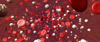
Blood is a functional system that ensures the delivery of oxygen and nutrients to tissue cells and the removal of metabolic products from organs and intertissue spaces. It consists of plasma and formed elements: red blood cells, leukocytes, platelets.
Anisocytosis is a condition in which red blood cells of abnormal size are detected in the blood.
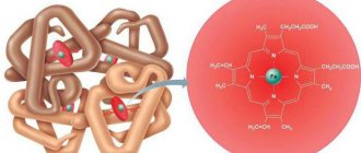
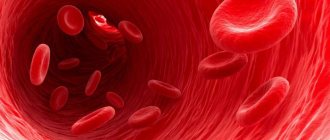
Red blood cells are biconcave disc-shaped cells without a nucleus that provide transport of respiratory gases (oxygen and carbon dioxide), amino acids, hormones, and also maintain blood pH levels.
Red blood cell distribution width by volume (RDW) is a calculated indicator reflecting the degree of heterogeneity of red blood cells by volume, an indicator of anisocytosis, which means the appearance of larger or smaller cells in a blood test, in contrast to the norm.
In human blood, red blood cells can have the following sizes:
- normocytes with an average diameter of 7.5 µm (7.2–7.7 µm): up to 75%;
- microcytes: ~12.5%;
- macrocytes: ~12.5%.
Under various physiological and pathological conditions, the composition of the blood and the percentage of cells in it change. In diagnosing changes in the qualitative and quantitative composition of blood, the morphological characteristics of red blood cells are used. They are assessed using an automatic analyzer (erythrocyte indices: MCV, MCH, MCHC, RDW) or visually - in a blood smear under a microscope. To do this, a clinical (general) blood test is taken from a vein or finger.
To accurately determine the erythrocyte index, modern hematological analyzers must be used
Modern hematology analyzers maintain cell stability, which is difficult to achieve with manual counting. This is due to the smear drying out under the microscope and the diameter of the red blood cells decreasing by 10–20%. Therefore, assessing the degree of anisocytosis under a microscope may be erroneous.
What is RDW
Red blood cell distribution width (RDW) is the change in size (volume) of red blood cells (RBCs). Basically, this test tells you how equal or unequal your red blood cells are in size. The RDW is part of a complete blood count that also measures hemoglobin , hematocrit, and red and white blood cell counts. [, ]
LEFT – ALMOST THE SAME SIZES OF RED CELLS (RDW – NORMAL), RIGHT – SIZES OF RED CELLS VARY GREATLY (RDW – NOT NORMAL)
Low RDW values mean your red blood cells are about the same size, which is normal and desirable. But higher RDW values mean that your red blood cells are produced in different sizes. In other words, there are some problems with the production and survival of red blood cells. [, , ]
value is important , but not specific (not indicating a specific disease) value, therefore the width of the distribution of erythrocytes by volume is used in conjunction with other blood indicators.
Therefore, along with RDW, blood tests also measure such indicators as: MCV (average erythrocyte volume), MCHC (average hemoglobin concentration in an erythrocyte).
RDW calculation methods and units
Depending on laboratory equipment, red blood cell distribution width can be calculated as coefficient of variation (RDW-CV) or standard deviation (RDW-SD).
It is important to understand that when testing the width of the distribution of red blood cells, it is not the actual width or diameter of individual red blood cells that is measured, but the width of the distribution curve of red blood cells (histogram), which is produced by the blood analyzer.
RDW-CV shows how much the red blood cell volume differs from the average value.
RDW-CV is calculated based on the width of the distribution curve as well as the average cell size. The calculation formula is as follows:
RDW-CV = (1 SD/MCV) * 100
Where:
SD – standard deviation of red blood cell sizes
MCV – average erythrocyte volume
The indicator calculated in this way is usually denoted as a percentage (%).
RDW-CV is directly dependent on MCV, so may not always reflect actual changes in red blood cell size. For example, if most red blood cells are small (as in microcytosis), then the RDW-CV will remain within the normal range.
RDW-SD is a direct measurement of the histogram width of the red blood cell size distribution in femtoliters (fL), which is measured by calculating the histogram width at the 20% height level.
1 fl = 10-15 l
Because RDW-SD shows the actual difference between maximum and minimum red cell volume and is independent of MCV, this index more accurately reflects changes in red cell size.
RDW increased: association with diseases
Elevated RDW values may indicate, but do not diagnose or confirm, the following diseases: [, , , ]
- Lack of iron in the body or vitamin deficiency
- Lack of B vitamins, including B12 and folic acid
- Anemia (various types, including sickle cell anemia)
- Inflammation
- Insomnia
- Cardiovascular diseases
- Blood loss due to hemorrhage (including surgery)
- Thalassemia
- Liver disease
- Kidney disease
- Cancer
- Alzheimer's disease
- Alcoholism
- and others.
However, RDW may still be at normal levels in people with leukemia , or with certain types of anemia (such as aplastic anemia ). Therefore, it is important to look not only at the RDW value, but also at the relationship with other markers. []
Why measure RDW in a blood test?
RDW is usually assessed to diagnose anemia of various origins, conditions in which red blood cells cannot carry enough oxygen throughout the body.
RDW is also used to diagnose thalassemia, diabetes, liver disease and cancer.
In addition, the reasons for prescribing a blood test with erythrocyte indices may be:
- health control for insufficient intake of iron and vitamins
- presence of symptoms of anemia (weakness, pale skin, dizziness, etc.)
- significant blood loss
- chronic diseases affecting red blood cells
In recent years, many clinical studies have proven that changes in RDW levels may be associated with many cardiovascular diseases (CVDs). RDW plays a significant role in assessing the severity and progression of CVD. However, the mechanisms of the relationship between RDW and CVD prognosis remain unclear.
Normal RDW values
Normal RDW values are the same for men and women. The range of normal RDW in adults is 11.5 – 15.5% , but may vary slightly depending on the reagents used by different laboratories.
In newborn children, normal RDW values are increased to 14.9 – 18.7% due to stress at birth and adjustment to life outside the mother. But as children grow older, their normal RDW values gradually approach those of adults.
At the same time, with age, red blood cells become more varied in size (volume), which leads to an increase in RDW. []
When is such an analysis prescribed?
The RDW value is determined by a general blood test. It is taken on an empty stomach, from a vein in adults, and from a finger in small children. Such an examination is necessary in order to timely identify possible pathological conditions, clarify the diagnosis or monitor the correctness of the prescribed therapy. Such tests are necessary for pregnant women, as well as for patients before surgery.
Research is being conducted on RDW not only in adults. It is very important for children. From about six months of age, a child’s blood composition should be the same as that of an adult. Determining the RDW level allows you to timely identify various deviations in its development. This analysis is especially indicative for diagnosing iron deficiency anemia, lack of folic acid or vitamin B12. It allows timely detection of congenital pathologies of the hematopoietic system, for example, thalassemia, as well as the presence of malignant tumors.
Such tests in a child are very important, as they allow timely detection of pathologies.
In addition, doctors prescribe this test for adults and children when the following symptoms appear:
- general malaise, weakness, fatigue;
- drowsiness during the day, decreased performance;
- causeless increase in body temperature;
- increased sweating;
- sudden mood swings;
- dryness and pallor of the skin, sometimes slightly yellowing.
RDW reduced
A low RDW value indicates that your red blood cells are uniform in size, which is desirable for health. [] However, it is worth remembering that there is still a possibility of having some kind of disease. []
For example, with reduced RDW values, one may suspect disturbances in the functioning of the spleen, where damaged red blood cells are recycled. In addition, if RDW is low, it is worth thinking about the following possible reasons:
- Recent surgery
- Blood donation
- Severe blood loss (including hidden in the stomach and intestines)
- Hormonal disorders during pregnancy, puberty, and when taking contraceptives
Do you need any preparation for the analysis?
To obtain accurate information, preparation for analysis is required. First of all, blood should be donated on an empty stomach, after a 10-12 hour fast.
Newborns should not eat 30–40 minutes before blood sampling. Children under 5 years old – for 2 – 3 hours.
Avoid stressful situations and physical activity the day before the test.
When taking any medications, it is recommended to consult a doctor to see if this will somehow affect the results of the study.
The RDW blood test in adults involves drawing venous blood as part of a complete blood count. In children, blood is taken from a fingertip for analysis.
When preparing for the procedure, you need to consider the following basic rules:
- do not drink alcohol before taking the test (the day before and immediately before the procedure);
- do not smoke or use nicotine substitutes (patch, spray, tablets, chewing gum) 1-2 hours before blood sampling;
- donate blood strictly on an empty stomach, i.e. the last meal should be at least 12 hours before donating blood;
- Consult with your doctor in advance regarding the possibility of taking medications on the eve of the study.
RDW increased
A high RDW means your red blood cells (RBCs) are unequal in size. This condition is called anisocytosis. [, ] This happens when, for some reason, you have trouble producing red blood cells.
Anisocytosis is determined by RDW and is classified according to red blood cell size along with MCV (mean erythrocyte volume):
- Anisocytosis with microcytosis (small red blood cells). Possible causes: iron deficiency, sickle cell anemia.
- Anisocytosis with macrocytosis (large red blood cells). Possible causes: deficiency of vitamin B12 or folic acid, autoimmune hemolytic anemia, cytotoxic chemotherapy, chronic liver disease, myelodysplastic syndrome.
- Anisocytosis with normal erythrocyte sizes. Possible causes: iron deficiency, vitamin B12 or folic acid deficiency, dimorphic anemia, sickle cell anemia, chronic liver disease, myelodysplastic syndrome.
ABNORMAL SHAPE AND SIZE OF RED CYTES
Changes in test results in children
Although a child's body works a little differently, normal rdw levels are also within fifteen percent. If this indicator increases, the child must undergo additional examinations. It is best to consult several specialists, and only then decide on the choice of treatment.
Any conclusions and diagnosis can only be made by a doctor. Protect yourself from self-diagnosis and even more so from self-medication.
The situation when the erythrocyte distribution index is reduced is very rare and does not have any clinical significance.
When RDW in a blood test is elevated, this indicates that red blood cells are scattered throughout the volume. That is, there can be normocytes, microcytes, and macrocytes.
The reasons that lead to such heterogeneity of erythrocytes may be the following:
- lead poisoning;
- pathology of the cardiovascular system;
- blood transfusion;
- Iron-deficiency anemia;
- liver damage;
- myelodysplastic syndrome.
Hematology analyzers greatly facilitate the work of doctors in the laboratory. Using parameters that can be determined during a blood test, it is possible to diagnose a wide group of blood diseases and assess the severity of the lesion. If there are deviations from the norm, it is better to consult a doctor for a decoding.
Graduate of the Faculty of Preventive Medicine and Medical Biochemistry, Department of Medical Biochemistry of SSMU (formerly ASMA). Diploma specialty: biochemist (clinical laboratory diagnostics doctor).
If you liked the article, share it with your friends!
Analysis for RDW is of great importance in medicine, especially in the differential diagnosis of various types of anemia.
Reasons for increasing RDW
Nutrient deficiency
Deficiencies of various nutrients can cause an increase in RDW, for example:
- Iron deficiency [, , , ]
- Folic acid deficiency []
- Vitamin B12 deficiency [, ]
This is because your body needs these nutrients to produce healthy red blood cells. Any of these deficiencies can eventually lead to anemia.
Inflammation
Some studies suggest that higher RDW values are associated with inflammation and higher levels of inflammatory cytokines such as IL-6 , IL-8 and TNF-alpha . [, , , , , , ]
Inflammatory cytokines can interfere with red blood cell production and thereby increase RDW. Additionally, oxidative stress , which often accompanies chronic inflammation, can reduce red blood cell lifespan and further increase RDW values. [, ]
High RDW has been found in people with diseases associated with inflammation, such as inflammatory bowel disease , celiac disease, polycystic ovary syndrome (PCOS), and major depressive disorder (MDD). [, , , ]
REASONS FOR INCREASING RDW
Sleep disorders
In a study of 17,500 adults, those who slept less or more than 7-8 hours a night were more likely to have high RDW values. This was especially true for people who slept 10 hours a night - their chances of having increased RDW increased by almost 70%. []
RDW values are higher in people with sleep apnea . [, ]
RDW rates were also associated with shift work with circadian rhythm . In a study of 7,000 women, those who worked shift shifts were almost 50% more likely to experience an increase in RDW compared to women who worked day shifts. []
Bleeding
Severe and moderate injuries with heavy bleeding increase RDW. [] Bleeding may not be visible, as is the case with intestinal and gastric bleeding. [, ]
Blood transfusion
If a person undergoes multiple blood transfusions, their RDW may increase due to differences in blood composition between the donor and recipient. [, ]
Liver disease
Increases in RDW occur in a variety of liver diseases, including hepatitis, alcoholic cirrhosis, biliary cirrhosis, and liver cancer. []
An observational study of 423 adults with liver disease found that their RDW was significantly higher than that of healthy controls. []
In another study of 446 hepatitis B patients, increasing RDW levels corresponded with increased liver size (hepatomegaly) and increased inflammation. []
Kidney disease
One of the kidney hormones, erythropoietin, is needed for the maturation of blood cells. Problems with the production of this hormone occur with kidney disease, leading to the development of RDW. [] People with reduced kidney function have higher levels of RDW. []
Alcoholism
Alcoholics can exhibit high RDW values without liver disease. This is because alcohol can have a toxic effect on red blood cells. []
Thalassemia
Thalassemia may cause increased RDW values. However, patients with thalassemia may also demonstrate normal RDW levels. [, , ]
Sickle cell anemia and hereditary spherocytosis
Sickle cell anemia is an inherited disease. People with this disease have higher RDW values because many of their red blood cells are misshapen. []
Hereditary spherocytosis (Minkowski-Choffard disease), another disease in which red blood cells become misshapen, also causes increased RDW levels. [, ]
DISEASES IN WHICH RDW LEVELS INCREASE (https://medcraveonline.com)
Cancer
RDW rates are often higher in various types of cancer, including stomach cancer , liver cancer , colon cancer , and kidney cancer . [, , , ]
There are many factors in cancer that can interfere with the normal production of blood cells, including chronic inflammation and poor nutrition.
In cancer, RDW often increases with disease severity and metastasis . []
In a study of 25,000 people, the risk of cancer was 30% higher in those people who showed the highest RDW values compared to people with the lowest values. Postmenopausal women with the highest RDW values had a 22% increased risk of developing cancer. But no association was found between RDW values and cancer in premenopausal women. []
Cardiovascular diseases
According to a meta-analysis, an increase in RDW levels accompanies various cardiovascular diseases: acute coronary syndrome (including myocardial infarction ), coronary heart disease , peripheral arterial disease, arterial hypertension , as well as atrial fibrillation . Higher RDW values predict more negative outcomes for these diseases. []
Increased RDW has been associated with various types of cardiovascular disease in studies. [, , ]
In a study of 25,500 adults, each 1% increase in RDW resulted in a 13% increase in heart attack risk . Those people who showed low RDW values had a 71% reduced risk of having a heart attack (myocardial infarction) compared to people with the highest RDW levels. []
Elevated levels of RDW are associated with a high risk of cholesterol plaque building up in the arteries ( atherosclerosis ) in patients with hypertension . []
RWD is associated with autoimmune diseases
There is an association between high RDW levels and increased disease activity in autoimmune problems such as rheumatoid arthritis , lupus, psoriasis, Crohn's disease , Sjögren's syndrome, systemic scleroderma, ankylosing spondylitis ( ankylosing spondylitis ). [, , , , , , ]
INCREASING RDW WITH INCREASING CROHN'S DISEASE/ULCERATIVE COLITIS ACTIVITY (https://www.wjgnet.com)
RWD and metabolic syndrome
People with higher levels of RDW show more advanced degrees of metabolic syndrome. This was found in studies involving more than 217,000 people. [, ]
RWD and diabetes risk
In a study that monitored more than 2,600 people with normal blood glucose levels over 4 years, high RDW values increased the risk of developing diabetes compared to people with low RDW. []
RWD and risk of dementia (dementia)
In a study of approximately 2,500 older adults, higher RDW values had an increased risk of dementia. This risk was significant in those people who were not anemic. []
RWD is associated with mortality
High RDW values increase inflammation and oxidative stress, which contribute to the risk of mortality. In various studies, adult (45+) hospital patients with elevated RDW levels demonstrated a higher risk of cardiovascular mortality, infection, and all-cause mortality . [, , ]
Additionally, in a review of 13 studies (involving 10,410 patients), low RDW was associated with a lower risk of mortality. []
RDW and depression
The study, which followed 43,226 patients with depression for 5.3 years, measured RDW values at the time of diagnosis and in subsequent contacts with doctors. It was found that patients with cardiovascular disease had more advanced depressive syndromes with increasing RDW . []
What to do
It was already mentioned above that the index of distribution of red blood cells in the blood makes it possible to conduct a qualitative assessment of the composition of red blood cells, taking into account their size.
But why is this necessary? The thing is that these cells are very similar to each other, which gives them the opportunity to replace each other or form blastulas. An increase in cell size entails an increased need for nutrition and, in addition, this means that their life expectancy is reduced. All this directly affects the overall indicator of red blood cells in the blood and the human condition.
When a large number of red blood cells die, iron is released and more bilirubin becomes available, which puts increased stress on the liver, and as a result, it cannot process these substances.
The RDW index is directly related to the pathological process, during which the dimensions of erythrocytes change (anisocytosis). This condition is a complex chemical process that causes all blood cells to suffer.
Many pathologies that cause changes in the composition of the blood do not manifest themselves externally in the initial stages. This is why it is so important to have regular blood tests. Determining the RDW level will allow timely diagnosis of many diseases.
After receiving such test results, it is necessary to undergo additional examination. It will help determine why changes in blood composition are observed and what pathologies are causing this. Sometimes you can return normal blood counts by changing your diet, which will compensate for the lack of essential microelements. But most often, such disorders can be eliminated only after the underlying disease has been cured.
Ways to reduce RWD
First of all, it is necessary to treat the underlying disease - the cause of increased RDW values. Below are additional options that can help you improve your health and reduce your RDW.
Balanced diet
Eating a healthy and balanced diet helps prevent nutritional deficiencies. It is especially important that the diet contains the recommended amounts of iron, folic acid and vitamin B12. [, , ] Correcting nutritional deficiencies can improve blood cell production and lower RDW values.
Reduce alcohol consumption
Reducing alcohol consumption may help reduce damage to red blood cells. [, ] Additionally, alcohol also reduces the absorption of nutrients such as vitamin B12 and folic acid, which are essential for the production of red blood cells. [, ]
More physical activity
People who do little or no physical activity show higher levels of RDW. [, ]
Exercise, including light intensity exercise, has been shown in research to improve RDW scores. [, , ]
In a study of more than 8,000 people, as the amount of exercise time per week increased, the risk of showing increased levels of RDW decreased by 11%. []
Quit smoking
Smoking increases oxidative stress. Higher RDWs are identified in smokers and are associated with the number of cigarettes smoked per day as well as the duration of smoking. []
Getting enough sleep
In a study of 17,500 adults, those who slept 7-8 hours had the lowest RDW values. People who slept less than 6 hours, about 10 hours, and more than 10 hours showed increases in RDW of 23%, 29%, and 67%, respectively. []
The information on this site has not been evaluated by any medical organization. We do not seek to diagnose or treat any disease. The information on the site is provided for educational purposes only. You should consult your physician before acting on information from this site, especially if you are pregnant, nursing, taking medications, or have any medical condition.
Rate this article
Average 4.5 Total votes (2)
Research methodology
The patient donates blood from a vein on an empty stomach, and for small children and infants, blood from a finger is sufficient.
After the technician is done, he or she places the samples in a centrifuge to separate the liquid portion of the blood from the cells. After this, he places the resulting dry residue in the analyzer, and the device itself counts the number of different types of cells, evaluates them and draws a conclusion. The results are displayed on the printer in the form of a histogram. If the test is positive, then according to the protocol it is supposed to be repeated in order to avoid a false positive result. This rule applies to almost all tests performed related to the diagnosis of anemia, since the mobility of the blood picture gives the doctor a reason to doubt the correctness of the chosen treatment tactics and to reconsider methods for correcting this pathological condition.
Analysis of red blood cell parameters
Most of the parameters of the blood test are clear to patients. Indicators such as white blood cells, glucose, cholesterol and others are well known, and their normal levels are indicated next to the patient’s result. A simple mathematical comparison of numbers allows you to make sure that the tests are within normal limits. But what is RDW?
RDW blood test and erythrocyte distribution index are the same value. The presence of deformed red blood cells (erythrocyte anisocytosis) is examined, and the indicator reflects the uniformity of red blood cells. Normally, red blood cells should be approximately the same size so that, if necessary, one red blood cell replaces another and performs the same functions.
They require more nutrition to survive and their lifespan is shortened. Also, macro cells are often larger in size than the diameter of the capillaries and cannot circulate through the circulatory system; they are disposed of.
The RDW blood test measures the ratio of misshapen or enlarged red blood cells to normal-sized red blood cells. There are two types of indicators RDW CV and RDW SD. The first value (RDW CV in blood tests) shows the percentage distribution of cells by size. The second reflects their standard deviation, that is, the difference in size between the smallest and largest red blood cells present in the blood sample.
Diagnostics
You can determine the RDW value by doing a general blood test in a hospital or private clinic. The patient is given a referral for research.
Diagnostics is carried out in laboratory conditions. The pad of the ring finger is disinfected and a puncture is made in it with a special needle called a scarifier.
After this, using a special device similar to a pipette, the required amount of blood is drawn from a finger and poured into a test tube.
Using various reagents and a microscope, a laboratory technician examines blood counts. You can receive a form with the analysis results in just a few hours.
In some cases (if any abnormalities are observed), the patient needs to have a repeat blood test. Typically, biological material is taken from a vein located in the elbow area.
In small children, blood is drawn from a finger.
The analysis determines the distribution index of red blood cells in the blood. The study is deciphered by an experienced specialist.
Currently, modern analyzers are used to determine the result with maximum accuracy.
Norms
If Rdw is elevated in a person’s blood, this indicates a persistent and serious disorder. He needs to immediately contact his doctor to determine what exactly caused this increase.
In the vast majority of cases this can be explained by:
- Iron deficiency anemia. In this case, the indicator may increase even before the hemoglobin concentration drops. It is the reduced width of distribution that is often called the first harbinger of anemia.
- Megaloblastic anemia - caused by a lack of B vitamins.
- Hemoglobinopathy is a hereditary disease characterized by a disorder of protein structure.
Increased levels of Rdw are observed in people who have serious liver problems: cirrhosis, hepatitis, fatty dysplasia.
The width of erythrocytes can also increase after treatment of nutritional disorders.
If RDW in a blood test is increased by more than 15%, this indicates the presence of pathological processes in the body. At the same time, there are more large red blood cells in the patient’s blood than usual. This may be hazardous to your health. Such cells live too little; in addition, the largest of them may have a volume larger than the lumen of some vessels.
A change in the volume of red blood cells can lead to the development of serious pathologies.
Sometimes such results are a temporary condition and are therefore considered a false positive. This happens during cold aggregation, that is, red blood cells sticking together under the influence of low temperatures. In addition, a temporary increase in RDW levels may occur after surgery or blood transfusion.
But most often the causes of this condition are the following pathologies:
- macrocytic, megaloblastic or iron deficiency anemia;
- hemoglobinopathy;
- alcoholic liver damage;
- other chronic liver pathologists;
- thalassemia;
- deficiency of vitamin A, folic acid, vitamins B9 and B12;
- increased level of white blood cells;
- malignant tumors affecting the bone marrow;
- Alzheimer's disease;
- serious disorders of the cardiovascular system;
- heavy metal poisoning.
It is especially important to identify disorders in a pregnant woman in a timely manner. This will help avoid serious complications and abnormalities in the child’s development due to oxygen starvation.
If the RDW level in the blood test is lowered below 10.2%, it is necessary to retake the blood. Most often, such indicators are obtained due to improper storage of blood samples or laboratory errors. A low RDW level may be temporary, but may also indicate chronic blood loss due to internal bleeding or frequent blood transfusions. Sometimes this condition also occurs with certain diseases.
The reason that RDW is low in the blood can be the following pathologies:
- hemolytic or microcytic anemia;
- deficiency of vitamin B6 or folic acid;
- deterioration of iron absorption due to metabolic disorders;
- problems with the spleen, which is responsible for the disposal of red blood cells, usually due to its injury or removal;
- liver dysfunction;
- removal of an organ or other surgical intervention;
- rheumatoid arthritis;
- helminthic infestations;
- hormonal imbalances, for example, during pregnancy or menopause;
- severe thalassemia;
- malignant bone marrow tumors;
- carrying out chemotherapy;
- myelodysplasia.
Many pathologies that cause changes in the composition of the blood do not manifest themselves externally in the initial stages. This is why it is so important to have regular blood tests. Determining the RDW level will allow timely diagnosis of many diseases.
After receiving such test results, it is necessary to undergo additional examination. It will help determine why changes in blood composition are observed and what pathologies are causing this. Sometimes you can return normal blood counts by changing your diet, which will compensate for the lack of essential microelements. But most often, such disorders can be eliminated only after the underlying disease has been cured.
LYM in a blood test is an indicator of a general clinical study that evaluates the absolute content of lymphocytes. Lymphocytes (LYM) are a type of white blood cell that help the body's immune system identify and fight pathogens (viruses, bacteria or fungi).
The two predominant types of lymphocytes that are secreted in the bone marrow are B cells and T cells. Antigen is a foreign substance (virus or bacteria). When an antigen enters the body, B cells secrete antibodies that attach to it. T cells recognize foreign substances by antibodies and destroy them.
Lymphocytes
There are two types of immune system. The first is innate immunity, which consists of proteins and cells that are constantly present in the human body. The second type is acquired, consisting of B and T cells.
When antigens bypass the first defense system, B and T cells come into play. If the immune system is weakened, lymphocytes cannot function normally.
Sometimes the immune system attacks and destroys healthy tissue because it cannot distinguish antigens from healthy cells. This disorder is commonly called autoimmune.
Attention! The lymphatic system is represented by various organs: the spleen, tonsil and lymph nodes. It protects the body from infections of various etiologies. About 25% of new lymphocytes remain in the bone marrow and become B cells. The other 75% are transported to the thymus and become T lymphocytes. B and T cells work together to provide effective defense against infectious pathogens.
If the patient has an infection or blood poisoning, the total lymphocyte count (lymph) is increased. If abnormal lymphocyte levels are suspected, your doctor will order a LYM test to count how many lymphocytes are in the bloodstream. Too high or low lymphocyte counts are a sign of disease.
For the examination, blood is taken from the patient's arm in the doctor's office. The biological material is then sent to the laboratory. Depending on the institution, the waiting time for CBC results varies.
Biological material
There are a number of factors that influence the results of the LYM analysis:
- Recent infection.
- Chemotherapy.
- Radiation therapy.
- Steroid therapy.
- HIV.
- Recent invasive surgery.
- Pregnancy.
- Severe stress.
There may be other indications at the discretion of the attending physician. Usually this examination is part of a general clinical blood test.
Laboratories have different ways of measuring blood test results. Results vary depending on gender, age and heredity.
Approximate normal ranges for LYM:
- In women, 700-4000 (0.8-5.0) lymphocytes/μl;
- Men have 800-5000 lymphocytes per μl;
- Children have 4000-10000 lymphocytes per microliter.
Lymphocytosis occurs after an infection. In some cases, high lymphocyte levels indicate serious illness:
- Mononucleosis.
- Adenovirus.
- Hepatitis.
- Flu.
- Tuberculosis.
- Toxoplasmosis.
- Cytomegaly virus.
- Brucellosis.
- Vasculitis.
- Acute lymphocytic leukemia.
- Chronic lymphocytic leukemia.
- HIV.
Important! Do not self-diagnose and decipher LYM readings with the help of your doctor. Additional examinations may be needed to clarify the reason that caused the increase in the indicator.
A low lymphocyte count, called lymphocytopenia, occurs due to the following reasons:
- The body is unable to synthesize a sufficient number of lymphocytes.
- Cells are destroyed faster than they are synthesized by the bone marrow.
Lymphocytopenia indicates the presence of diseases of various etiologies. Some of them, such as the flu, are not dangerous for most people. However, a low percentage of lymphocytes increases the likelihood of infection by infectious pathogens and the occurrence of complications.
Other conditions that cause lymphocytopenia:
- Malnutrition.
- Systemic lupus.
- Certain types of cancer, including lymphocytic anemia, lymphoma, and Hodgkin's disease.
- Steroids.
- Radiation therapy.
- Chemotherapy drugs.
- Inherited disorders: Wiskott-Aldrich syndrome and DiGeorge syndrome.
The first step is to determine why the lymphocyte count is elevated in an adult or child. Before following the recommendations, consult your doctor to find out the true cause of the disorder. Lymphocytosis occurs as a result of serious diseases of the hematopoietic system and therefore requires drug therapy.
Recommendations that will help reduce LYM levels in the blood:
- Infections cause inflammation in the body, so make sure you add enough anti-inflammatory foods to your diet. Omega-3 unsaturated fatty acids found in salmon, cod, and mackerel are the best sources of anti-inflammatory substances.
- Avoid sugary foods as they increase inflammation in the body. Instead of sweets, eat foods with a low glycemic index: strawberries, apples or bananas.
- Increase your intake of vitamin C, which is found in bell peppers, lemon and blueberries. Vitamin C is an important chemical compound that improves the functional state of the immune system.
- Include “light” aerobic exercise in your daily routine, which will help strengthen the immune and cardiovascular systems. Daily walks at a speed of 5 km/hour are suitable.
- Many suffer from a lack of vitamin D. Vitamin D is essential for healthy bones and reduces the risk of developing cardiovascular disorders. You can get your daily dose of vitamin D by walking in the open sun.
- Sleep at least 8 hours a day. Excessive stress resulting from sleep deprivation will worsen the immune system.
If your LYM is low, add more protein-containing foods to your diet. Lymphocytes require amino acids for normal functioning and maintenance of the immune system. The amino acids contained in the protein are partly responsible for the production of B cells.
Lack of folic acid leads to increased RDW
If the RDW width does not correspond to the decrease in MCV, a repeat blood test is required
- Stress or hormonal instability: provoked by anxiety, fatigue or changes in the concentration of sex hormones in women. In most of these cases, the LYM indicator is slightly higher than normal and stabilizes on its own.
- Smoking: lymphocytosis due to an increased number of red blood cells is observed in people with a smoking habit.
- Viral infections: an increase in the level of LYM in the blood is considered as a natural reaction of the body to a virus. Lymphocytosis may persist during the recovery period. Increased levels of LYM are provoked by ARVI, measles, chickenpox, rubella, herpes, mononucleosis, and whooping cough.
- Bacterial infections: pneumonia, tuberculosis, STDs.
- Autoimmune diseases: Crohn's disease, lupus erythematosus, rheumatoid arthritis, autoimmune thyrotoxicosis. With these diseases, the body begins to destroy its own cells, mistaking them for foreign ones.
- Intoxication caused by heavy metals and medications: under the influence of lead or after taking Levomycetin, Analgin, Phenytoin, the level of neutrophils decreases, which is why relative lymphocytosis is observed.
- Removal of the spleen: This organ is the site of LYM breakdown, so its removal causes temporary lymphocytosis. After several weeks, the human hematopoietic system adapts to the changed operating conditions, and lymphocytosis resolves itself.
- In women (0.8-5.0) lymphocytes/μl;
- In men, lymphocytes per µl;
- Children have 0 lymphocytes per µl.
- Poor nutrition (diet deficient in vitamins and protein)
- Profuse acute bleeding
- Chronic bleeding
- Diseases of the hematopoietic organs
- Genetic diseases (with disturbances in the synthesis of enzymes involved in the construction of red blood cells)
- Accelerated destruction of red blood cells (as a result of intoxication or disruption of the immune system)
- Dehydration (due to decreased fluid intake, increased sweating, certain kidney diseases, diabetes, excessive vomiting or diarrhea)
- Hereditary malformations of the heart or pulmonary system.
- Pulmonary failure
- heart failure
- Diseases of the urinary system
- Pathologies of the hematopoietic system
Expert opinion
Kovaleva Elena Anatolyevna
Doctor-Laboratory Assistant. 14 years of experience in clinical diagnostic services.
The norm of lymphocytes in an adult does not depend on his gender. For women, as for men, values ranging from 19 to 37% of the total number of leukocytes are considered normal. But such norms are applicable in the normal state of a woman.
Decoding
Anisocytosis of red blood cells is determined by a hematology analyzer, which produces a histogram plotting the number of RBCs of different volumes in the sample.
If the analysis is carried out manually, the value of the red blood cell coefficients of variation is calculated from the histogram curve and formula. Manual calculation of parameters is rarely used, because does not provide the necessary accuracy of the result.
A positive test result is when the rate of erythrocyte anisocytosis exceeds the norm. If the result is within the normal range, the test will be negative.
The RDW designation characterizes the heterogeneity of red blood cells:
- RDW-CV in a blood test is a relative value measured as a percentage (%);
- RDW-SD is an absolute parameter measured in volume units of femtoliters (fl) or µm3.
Comparison of the scatter of RBC volumes in the sample is carried out with normal values of 80-100 fl.
Relative index is a value that reflects the distribution of red blood cells, showing by what percentage the red blood cell volumes differ from the average volume.
The value of the absolute indicator indicates how many femtoliters the volumes of red blood cells differ. The calculation is done according to the histogram graph.
The level of relative anisocytosis depends on the value of the MCV parameter (mean blood cell volume). The calculated value of the relative indicator of erythrocyte anisocytosis is obtained by multiplying 100% by the ratio of the standard coefficient to MCV.
The RDW-SD parameter in a blood test allows you to determine how many femtoliters the smallest and largest red blood cells differ from medium-sized red cells.
Normal values
The values of the distribution width of erythrocytes according to norms do not depend on gender. They are the same for an adult of any gender. With age, normal coefficient values do not change.
If heterogeneity does not exceed the norm, it means that the red blood cells are approximately the same size. The norm for absolute anisocytosis in adults is 37-47 fl.
Normal RDW levels for women and men are 11.5%-14.5%.
In women during pregnancy, the normal value of the parameter changes by trimester (%):
- first trimester - 11.7-14.9;
- second trimester - 12.3-14.7;
- third trimester - 11.4-16.6.
In children, the normal level varies. The first 6 months after birth, RDW values are higher than the adult norm. Gradually they decrease.
RDW standards for children (%):
- up to 6 months - 14.9-18.7;
- after six months - 11.6-14.8.
The result of the calculation of relative anisocytosis, which the patient receives in the analysis, is often elevated or normal. A decreased RDW-CV most likely indicates an error.
If the anisocytosis of red blood cells is slightly higher than normal, most often the deviation is random. This value becomes clinically significant for diagnosis when it exceeds 60 fl.
A decrease in the absolute coefficient of anisocytosis has no significance for diagnosis.
Increased level
Red blood cells are heterogeneous in size and hemoglobin saturation. If anisocytosis is higher than normal, it means that there are blood cells in the bloodstream that are different in volume from the normal range.
Normally, each cell goes through a life course, during which its volume changes. Cells of different sizes are simultaneously present in the blood:
- microcytes, characterized by their small sizes, not exceeding 6 microns;
- normocytes corresponding to normal sizes, in the range of 6-8 microns;
- macrocytes, characterized by large sizes exceeding 8 microns;
- megalocytes, with a diameter > 12 microns.
Normal human RBCs fall within the normocyte range on average. An increased value of heterogeneity is possible with the appearance of microcytes and megalocytes.
The heterogeneity of erythrocytes is characterized by the degree of anisocytosis:
- The first degree corresponds to 27-50% of defective cells.
- Anisocytosis of the second degree is characterized by 55-70% of modified blood cells.
- The third degree of anisocytosis is characterized by the presence of more than 75% of such cells. Their sizes deviate from the norm.
- With the fourth degree of anisocytosis, all 100% of the cells are defective.
If the relative width of the distribution of red blood cells by volume is increased, it means that more than 50% of the cells deviate from the norm.
Relative coefficients of erythrocyte heterogeneity may increase when:
- leukocytosis with a sharp increase in the number of leukocytes;
- lack of nutrients necessary for hematopoietic processes (iron, B vitamins);
- microcytic anemia;
- myelodysplastic syndrome;
- hemolytic crisis;
- malignant tumors that metastasize to the bone marrow;
- RBC agglutination;
- poisoning with heavy metals (lead);
- Alzheimer's disease.
The anisotropy coefficient of erythrocytes increases in vascular diseases. Increased RDW in intoxication caused by alcohol abuse.
If the blood was tested after surgery, severe bleeding, heterogeneity above normal may also be noted. In such a situation, the result exceeds the acceptable standard deviations as a result of the increased production of juvenile cell species.
RDW is elevated in iron deficiency anemia, which is common in pregnant women. The RDW indicator for this disease exceeds 14%.
With vitamin B12 deficiency, macrocytes and megalocytes are found in the bloodstream.
If the anisocytosis index reaches 27%, this means that RDW in a blood test may suggest B12 deficiency anemia.
The width of the distribution of red blood cells is increased during processes accompanied by increased destruction of red blood cells. The death of blood cells is caused by:
- increased bilirubin levels;
- an increase in the size of the liver and spleen;
- accumulation of iron ions in the body.
An increase of up to 40% is possible with thalassemia or Cooley's anemia. This disease is hereditary. Cooley's anemia is most widespread in areas affected by malaria.
Increased values correspond to changes in other erythrocyte parameters. It is necessary to take into account the MCV parameter when interpreting analyses. Simultaneous changes in parameters are observed in cases where:
- the RDW indicator is exceeded, the MCV is reduced (with iron deficiency anemia);
- above normal MCV and RWD (may mean liver pathologies, megaloblastic, hemolytic anemia, cold agglutination);
- there is an excess of the RDW norm in combination with the MCV norm (indicates the likelihood of myelofibrosis).
Reduced index
A reduced RDW in the analysis means that in the sample taken for the study, the cells have practically no difference in volume. Cases where the analysis reveals that all RBCs are the same size are most likely in error.
Red blood cells may be of equal size in anemia, leading to an increased number of defective forms in the bloodstream.
The values of erythrocyte distribution indices decrease with:
- anemia (iron deficiency, folate deficiency, B12 deficiency);
- pregnancy (with the appearance of fetal hemoglobin);
- extensive blood loss (trauma, internal bleeding, childbirth).
What conditions require a blood test for RDW?
The doctor will prescribe a general clinical blood test during a standard routine examination or if there are:
- Symptoms of anemia include weakness, dizziness, pale skin and hyperhidrosis.
- Family history of thalassemia, sickle cell disease, or other inherited hemostatic disorder.
- Chronic diseases: Crohn's disease, diabetes or HIV.
- Diet low in iron and minerals.
- Long-term infection.
- Excessive blood loss caused by trauma or invasive procedures.
When is a test ordered?
The study is carried out during routine medical examinations, as well as before surgical treatment.
The analysis is mandatory for diagnosing diseases. The patient is referred for this study with the following complaints:
- high body temperature in the absence of specific reasons (when no other symptoms of disease are observed);
- loss of usual performance;
- increased sweating;
- yellowness of the skin and eye sclera;
- constant fatigue;
- sleep disturbances (a person is drowsy, although he gets enough sleep at night).
If a person often gets irritated, becomes aggressive, experiences apathy and fatigue, and often changes mood, this diagnosis is also indicated for him.
The analysis allows us to differentiate different types of anemia and determine which treatment is suitable for the patient in each individual case.
This diagnosis is mandatory for women who are pregnant.
How is blood drawn?
A healthcare professional will take a sample from a vein using a small needle. The needle is attached to a tube in which the sample is stored. When the test tube is full, the needle will be removed. Some patients feel a slight burning sensation after the injection, which lasts for 5-6 minutes. After the needle is removed, the patient will be given a bandage or piece of gauze to stop the bleeding.
In what cases are they sent to donate blood for this indicator?
Sometimes the doctor may send you for a general analysis based on the patient’s complaints . With an increase in red blood cell volumes, the following symptoms can be felt:
Ask your question to clinical laboratory diagnostics doctor Anna Poniaeva. Graduated from the Nizhny Novgorod Medical Academy (2007-2014) and Residency in Clinical Laboratory Diagnostics (2014-2016).Ask a question>>
- heat;
- lack of strength, general fatigue of the body;
- excessive sweating;
- constantly tends to sleep;
- emotional mood swings;
- sometimes yellowing of the skin.
Details about the blood test for RDW (CV)
If you constantly feel the above symptoms, then it is better to consult a doctor.
Degrees of anisocytosis
Erythrocyte anisocytosis is divided into four stages:
- I degree. Diagnosed when 27% or 50% of red blood cells have a different volume.
- II degree. Occurs when 55% or 70% of red blood cells have a change in size.
- III degree. More than 75% of blood cells are modified and have different dimensions.
- IV degree. Almost 100% of blood cells are different from normal.
Clinical analysis reveals blood levels of rdw, ranging from a slight degree to a pronounced degree, when the highest percentage of deviation from the blood flow composition standards is detected. In ideal condition, the size of red blood cells should vary between 7-9 micrometers. According to the degree of change in the size of red blood cells in one direction or another, anisocytosis is classified into:
- Macroanacitosis - a larger number of red blood cells of increased volume.
- Microanacitosis is the predominant number of red blood cells of small diameter.
- Mixed type, combining macrocytes and microcytes.
There are also megalocytes, which have the maximum possible blood cell size of more than 12 microns. Macrocytes are red blood cells whose size is more than 8 microns. Their normal amount should be in the range of 12−15%. Microcytes include blood cells smaller than 6.9 microns. Mixed anisocytosis is characterized by the presence of both reduced and enlarged blood cells in the bloodstream. Combined studies are carried out using the calculation method using the Price-Jones curve.
Preventive measures
You can prevent a reduced RDW by following these simple rules:
- The diet should be balanced, which includes plenty of fresh fruits, lean meats and vegetables.
- It is recommended to breathe fresh air as often as possible.
- An active lifestyle will help prevent a decrease in the RDW index.
- It is very important not to skip routine medical examinations, during which most often serious deviations from the norm are detected that do not have external symptoms.
As a result, we learned that the red blood cell distribution index reflects their dimensions relative to each other and makes it possible to learn about their biological value. A decrease in RDW is very rare, but if the erythrocyte distribution index is decreased, this means that various pathologies may be present.
The index is calculated based on the results of a general blood test, but can only be fully valid in conjunction with the MCV indicator, since they are closely interrelated.
Reduced RDW: norm and pathology
A person in good health has red blood cells of the same shape, density and color. In case of deviation, especially in the presence of autoimmune diseases or oncology, the failure occurs at the level of microcells, when young cells do not receive a certain number of components, which, in fact, inhibits their performance. Thus, anemia occurs - a pathology during which the body does not receive the required amount of oxygen, in other words, the metabolic function in red blood cells is disrupted.
Causes
If the results of a laboratory test show that this index is elevated, do not rush to conclusions. First, you need to re-analyze. If this indicator is still elevated and the result is positive, this may be caused by the following factors:
- recent surgery:
- blood transfusion;
- incipient iron deficiency anemia;
- megaloblastic anemia (caused by a lack of B vitamins);
- hemolytic anemia;
- alcoholism;
- oncology;
- disorders of the bone marrow;
- hemoglobinopathy.
Most often, this indicator is increased in people suffering from the following problems:
- liver failure;
- diseases of the gastrointestinal tract.
rdw may also be elevated in those who have had a blood transfusion. This procedure is often accompanied by serious complications and problems.
If the RDW level in the blood test is lowered below 10.2%, it is necessary to retake the blood. Most often, such indicators are obtained due to improper storage of blood samples or laboratory errors. A low RDW level may be temporary, but may also indicate chronic blood loss due to internal bleeding or frequent blood transfusions. Sometimes this condition also occurs with certain diseases.
The reason that RDW is low in the blood can be the following pathologies:
- hemolytic or microcytic anemia;
- deficiency of vitamin B6 or folic acid;
- deterioration of iron absorption due to metabolic disorders;
- problems with the spleen, which is responsible for the disposal of red blood cells, usually due to its injury or removal;
- liver dysfunction;
- removal of an organ or other surgical intervention;
- rheumatoid arthritis;
- helminthic infestations;
- hormonal imbalances, for example, during pregnancy or menopause;
- severe thalassemia;
- malignant bone marrow tumors;
- carrying out chemotherapy;
- myelodysplasia.
So, the erythrocyte distribution index is reduced - what does this mean? There are several reasons that can reduce the RDW indicator:
- Acute blood loss due to injuries and pathological bleeding.
- Frequent operations.
- A metabolic disorder during which the food consumed is not completely digested.
- Hormonal imbalance, which most often occurs in women.
- Deficiency of B vitamins and iron in the body.
- Blood diseases characterized by rapid destructive processes.
When is re-analysis performed?
The procedure for collecting blood to determine heterogeneity is extremely simple. To carry it out, an analysis is taken from a vein in either arm of an adult. It is very important that the study is carried out on an empty stomach.
If this indicator is elevated, you will definitely be sent for a repeat study.
The reason for this may also be:
- The doctor’s desire to determine the true cause of the increase.
- The presence of serious hereditary blood diseases.
- Determination of the inflammatory process in the body.
- Use of low-quality materials during research.
- The menstrual cycle in women also affects Rdw levels.
- Recent herbs affecting the pituitary gland.
- Previous surgical interventions that resulted in significant blood loss.
Normalization of reduced RDW level
To treat such a pathology, it is important to establish its exact cause. If it is an oncological disease, then therapy is prescribed by an oncologist depending on the established diagnosis, the severity of the pathology and the individual characteristics of the patient.
Surgery, radiation or chemotherapy may be prescribed.
If there is a deficiency of certain substances in the body, special medications and vitamin and mineral complexes are prescribed. It is also necessary to adhere to a proper diet, enriched with useful minerals and vitamins.
With changes in hormonal levels, blood donation and blood transfusion, special treatment is not required. Over time, the indicator normalizes on its own. However, it is important to follow the correct regimen and diet.
Normal indicators
When characterizing the RDW index, you need to remember that this parameter does not depend on the size of the cell itself. This can give false negative results, and is due to the fact that there are a large number of altered red blood cells in the blood. These red blood cells are called macrocytes. Sometimes, in order to accept the analysis results as normal, it is necessary not only to comply with the RDW, but also to correlate it with the MCV.
Increased rate
A high level of RDW on a blood test may indicate disease or pathology. Often the indicator differs from the norm due to a lack of certain nutrients and vitamins. Main causes of violation:
Oncological diseases, tumor development Liver diseases in the chronic stage Alzheimer's disease Diseases of the cardiovascular system Anemia of various natures (hemolytic, microcytic, megaloblastic, iron deficiency), among all patients it is especially important to identify this condition in pregnant women Iron deficiency, which affects hematopoietic function Deficiency vitamins B9 and B12 Lack of folic acid Alcoholism
Why is a high rate diagnosed?
An increase in the indicator may be indicated by symptoms such as high temperature, which manifests itself systematically, loss of performance, drowsiness, and increased sweating.
If RDW is elevated, then this may indicate various pathologies occurring in the human body. These include:
- Alzheimer's disease;
- liver failure;
- oncological processes;
- vascular and heart diseases.
The reasons for the increase in the indicator may be alcohol dependence, as well as a deficiency of useful micro- and macroelements and vitamins necessary for the body. Often high values are found with a lack of iron, folic acid, and vitamin B12.
Anemia of various origins is another factor that provokes an increase in the rate. Values may increase with microcytic, hemolytic, iron deficiency and megaloblastic anemia.
How to lower the level of RDV
To find out how to normalize this blood count, the specialist must refer the patient for additional examinations.
After determining the exact diagnosis, appropriate treatment is prescribed.
In any case, therapy should be carried out with an integrated approach, which consists of the use of certain medications and physiotherapeutic procedures.
Sometimes, if necessary, surgery may be prescribed.
Thus, RDW is an important indicator that helps diagnose various pathological processes in the body. In addition, it allows you to determine the type of anemia.
When a blood test is done, they evaluate not only the number of its cells, but also their quality. Characteristics such as color, shape, size are also important in the diagnosis of diseases and are sometimes the only pathognomonic symptom of the disease. Therefore, hematologists ask the laboratory to also indicate RDW in the analyzes, which stands for distribution of red blood cells by size.
Indications for the study
Absolutely any doctor, from a local physician to a specialist in a certain field of medicine, can refer a patient to OAC. A blood test for RDW can be performed during an annual physical examination and when indicated. But during an annual examination, analysis is not always prescribed. This indicator should be checked if the patient exhibits the following symptoms:
- Fever in the absence of visible causes, such as colds.
- Drowsiness despite the patient getting enough rest.
- Increased sweating regardless of the heat.
- Fast fatiguability.
- Emotionality, irritability, sudden mood swings.
- Yellowing of the skin (in rare cases).
What is the decoding of RDW in a blood test based on?