Kidney ultrasound is performed for acute symptoms, as well as as a routine annual examination. The indication for this may be chronic pathology or a primary preventive examination. Single ultrasound examination of the kidneys is rarely performed. More often, this paired organ is examined together with adjacent organs located in the peritoneum and pelvis.
The ultrasound wave perfectly visualizes the anatomical structures of the kidneys. The only thing that can prevent this is excess gases in the abdominal cavity. Therefore, the patient needs to prepare for a kidney ultrasound examination. Doctors are always happy to explain to the patient how to prepare for a kidney ultrasound.
The procedure can also be carried out as part of a comprehensive examination of the abdominal organs (liver, pancreas) and retroperitoneal space, as well as in conjunction with an examination of the bladder and prostate gland. Each of them may have its own reasons for diagnosis.
Ultrasound for studying various pathologies of the kidneys is divided into 2 types:
- Echography. This diagnostic procedure shows the structure of the kidneys, their size and size, but does not evaluate the blood flow in the kidneys.
- Dopplerography. This procedure allows you to directly study the flow of blood in the renal vascular system. It can be used to diagnose narrowing of arterial and venous vessels, blood clots, plaques, blockages and aneurysms.
The nephrologist who observes the patient sets the goals of the examination, and in connection with this, one of the ultrasound diagnostic methods is selected.
What is ultrasound
The procedure is a modern method based on visualization of organs using ultrasound waves emanating from a transducer. There are 2 types of kidney ultrasound:
- Ultrasound echography. This method determines the anatomy of an organ (its size, structure and position), and also determines the presence of pathology (tumors, cysts, damage).
- Doppler ultrasound is used to determine blood flow in the renal vessels.
Ultrasound examination of the kidneys has a number of advantages over other instrumental methods - the absence of side effects, high information content at a low cost of the study, the minimum required preparation for ultrasound of the kidneys and adrenal glands, and the absence of the need to use a contrast agent to which a number of patients have an allergic reaction.
Ultrasound diagnostics is a safe procedure without the use of x-rays. This method is actively used in diagnosing diseases of young children and pregnant women. The information content of the study is high, but you need to properly prepare for the procedure.
Preparing pregnant women
Pregnant women need to prepare for a kidney ultrasound, just like all other patients. But expectant mothers should absolutely not use laxatives or give enemas, as this can increase the tone of the uterus. All that remains for a pregnant woman is to prepare with a diet that will need to be followed for 3 days.
During the preparation period, a woman must exclude from the menu everything that causes increased gas formation - sauerkraut, legumes, whole milk, black bread, sweets. A pregnant woman can also take sorbents and carminatives.
In terms of filling the bladder, a woman must clearly cooperate with the diagnostician. In some cases, they may not torture her, forcing her to drink 1.5 liters of water while prohibiting her from going to the toilet. But most often there is no way around this and you will have to be patient, especially if the kidneys are examined together with the bladder. If the patient manages to make all the necessary preparations, then he can count on accurate examination results that will help select effective treatment for the kidneys and related organs.
Indications
Diagnosis of the urinary system organs is carried out in the presence of an existing chronic pathology, to monitor and evaluate the effectiveness of treatment, or in patients who for the first time indicate the presence of symptoms characteristic of diseases of the renal system.
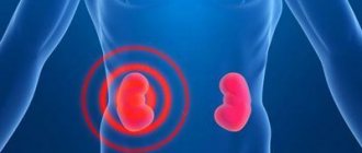
The main symptom is pain in the lumbar region
Ultrasound in adults is performed in the presence of the following symptoms and diseases:
- pain in the lumbar region, radiating to the thigh, groin;
- persistent increase in blood pressure that cannot be treated;
- periodic renal colic;
- pain during urination;
- swelling of the legs;
- deviations in analyzes;
- enuresis (urinary incontinence);
- pyelonephritis;
- after kidney transplantation;
- kidney stone disease;
- cyst;
- pathology of the endocrine system;
- injury to the lumbar and abdominal area;
- Ultrasound of the kidneys in men is prescribed for prostate adenoma, cysts, and adrenal tumors.
The method is actively used in young children to diagnose congenital pathologies. During the examination, the adrenal glands, bladder and ureters are scanned.
Preparing for an ultrasound of the kidneys: main aspects
An ultrasound examination of a paired organ is prescribed if there are patient complaints or severe symptoms, as well as if a preventive examination is necessary.
The study of the kidneys alone is extremely rare; more often they are studied in combination with adjacent structures located in the peritoneum or pelvis - the abdominal cavity organs.
Ultrasound waves perfectly visualize the anatomical components of the kidneys, but excess gases in the retroperitoneal space can become a significant obstacle to the path of the waves. Therefore, preparation for the procedure is very important not only for the doctor, but also for the patient.
What you need to know about the procedure
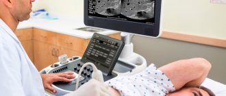
Ultrasound of the kidneys is a highly informative and safe method that allows you to quickly find out information about the state of the urinary structures. It is based on obtaining images of organs and tissues by recording ultrasonic waves, which, when emitted, are reflected from the surface of biological structures and liquids. At the same time, the slightest changes are displayed on the monitor, which facilitates the procedure for making a diagnosis and choosing a treatment regimen.
To perform the procedure, you must have an ultrasonic signal generator (the apparatus itself) and a special sensor-emitter. A monitor is used for visualization, and a gel is used to ensure the tightest contact between the body and the manipulator. It has conductive ability and accelerates the penetration of the wave to the organ under study.
It is important to know! The method is recognized as completely safe, therefore it is prescribed not only to adults, but also to newborns, older children, pregnant women and the elderly.
The advantages of this diagnostic method include:
- high information content;
- harmlessness and absence of injury;
- efficiency;
- real-time visualization;
- comprehensive examination of several organs simultaneously;
- good diagnostic capabilities in the study of soft tissues and internal structures.
But despite all the proven advantages, the method has a number of disadvantages. The most common ones include:
- low image quality compared to x-rays or CT, MRI;
- lack of opportunity to study kidney function;
- difficulties in diagnosing certain disorders;
- insufficient information content of a static image.
However, in recent decades, ultrasound of the kidneys and other organs of the urinary system is among the mandatory procedures that are performed free of charge and prescribed if there are the following indications:
- pain in the lumbar region and lower abdomen;
- urination disorders: cutting, burning and pain during intercourse, frequent urges;
- deviations in the interpretation of the general urine analysis;
- pyelonephritis;
- prostate adenoma;
- pain syndrome like renal colic;
- persistent increase in blood pressure;
- glomerulonephritis;
- thyroid diseases;
- inflammation of the prostate;
- diabetes;
- urolithiasis disease;
- complicated pregnancy.
In some cases, in the presence of diseases of other organs, ultrasound is prescribed to differentiate diagnoses for similar symptoms. In addition, the study is carried out as part of a preventive examination, during which hidden kidney pathologies are often discovered.
General recommendations for preparing for the study
The preparatory stage includes activities designed to ensure high-quality visualization during diagnosis. Despite the fact that modern equipment is characterized by high informative and resolving power, the echogenicity of individual areas and zones can be difficult due to poor nutrition or non-compliance with certain rules. Therefore, doctors advise you to read the memo in advance, explaining how a woman and a man can prepare for an ultrasound of the kidneys.
Attention! To increase the efficiency of diagnosis, two conditions are considered necessary: a full bladder and the absence of gases and feces in the intestines. Liquid media facilitate the free passage of ultrasonic waves, while air and gases interfere with this process.
The following recommendations are considered common for patients of any age and gender, indicating how to prepare for a kidney ultrasound.
- A few days before the procedure, you need to switch to a gentle diet, excluding the consumption of foods that cause bloating.
- On the day of the examination, increase your fluid load to ensure your bladder is full.
- On the eve of the procedure, start taking enterosorbents or laxatives to eliminate signs of flatulence and cleanse the intestines of feces.
- Prepare in advance the necessary minimum of things that will be needed in a medical institution during the manipulations.
As you can see, the preparatory stage begins several days before the scheduled date of the examination, so it is worth considering each point in more detail.
Drinking standards
There are no restrictions on the amount of liquid consumed, but it is worth considering that for the general condition of the body and kidney health, it is better to drink plain water without gas. 3-5 days before the expected date of the ultrasound examination, it is necessary to completely eliminate alcohol (including champagne and beer) and limit the following drinks as much as possible:
- strong tea;
- coffee, cocoa;
- drinks containing dyes and preservatives;
- energy drinks and sweet soda.
In order to conduct an informative diagnosis of the adrenal glands, kidneys, and ureters, it is necessary to fill the bladder with liquid without gas. Pure water, green tea, unsweetened compote, fruit drink or juices are suitable for these purposes. The optimal volume for an adult is 2-3 glasses, that is, 500-700 ml. It takes a certain amount of time for the liquid to get from the stomach to the bladder, so you need to drink the required amount one and a half to two hours before the start of the procedure (if you have the urge to urinate, you should refrain from going to the toilet).
Nutrition rules
Eating habits play a leading role in preparing for an ultrasound. The examination is performed on an empty stomach, that is, on an empty stomach. It is necessary to exclude foods that cause excessive gas formation in the intestines from the diet at least three days before the planned examination. The patient must completely refuse food within 8-10 hours. If the procedure is scheduled for the morning, the last meal should be no later than 19.00 in the evening of the previous day. When the diagnosis is planned to be carried out in the afternoon, eating is allowed until 11.00.
Prohibited Products
The diet involves switching to a lighter diet, eliminating foods that cause bloating and exacerbation of inflammatory processes in this organ. This diet includes fried, hot and spicy foods. In preparation for an ultrasound examination, it is not recommended to consume the following foods:
- vegetables - potatoes, onions, white cabbage, corn;
- zucchini, bell peppers that have not undergone heat treatment;
- legumes – peas, beans;
- milk, cream, kefir, yogurt;
- flour products, black bread;
- fruits – pear, apples, cherries, prunes;
- bran, starch;
- canned food, semi-finished products, fast food.
In addition, you should refrain from a large evening meal, including dishes made from meat, fish, eggs, mushrooms, as well as drinking water during or immediately after eating. Meals during the preparatory period should consist of the following list of products:
- low-fat hard cheeses;
- lean beef, fish, rabbit, chicken;
- vegetables with minimal fiber content;
- berries - strawberries, raspberries, blueberries, watermelon;
- cereals – buckwheat, oatmeal, pearl barley, rice;
- day-old bread;
- walnuts;
- low-fat milk, cottage cheese;
- eggs, but no more than one per day.
All dishes should be boiled or steamed. Doctors advise including in your diet foods that, on the contrary, reduce bloating: yoghurts with bifidobacteria and enzymes, ginger and cinnamon, as well as mint leaves in the form of a decoction or added to tea.
Prescribed medications
For people suffering from flatulence, experts recommend that at the preparation stage they begin taking medications that reduce the amount of gas. This must be done 2-3 days before the procedure. The list of necessary medications includes:
- defoamers – “Meteospasmil”, “Espumizan”;
- enterosorbents – activated carbon, “Sorbex”, “Filtrum”, “Enterosgel”;
- means that normalize peristalsis - “Cerukal”, “Motilium”;
- herbal remedies - fennel or caraway fruits.
For patients suffering from diseases of the pancreas, liver or gall bladder, it is considered advisable to use enzymes with every meal. These are “Festal”, “Pangrol”, “Mezim”, “Pancreatin”.
People suffering from constipation are advised to normalize bowel movements with the help of laxative medications. They should be taken the evening before the test. The following are considered valid:
- "Guttalax";
- "Portalak";
- "Laxomag";
- "Forlax".
If constipation is temporary, you can use herbal preparations: Kafiol, Tisasen, buckthorn bark, senna leaf, as well as suppositories with glycerin or rhubarb. A microenema is considered an effective measure. A medicine (chamomile infusion or starch solution) in a volume of 100-200 ml is injected into the rectum.
Preparation
Preparing the patient for an ultrasound scan of the kidneys and adrenal glands is necessary in order to eliminate possible interference from the intestines and other organs. The existing air in the intestinal loops creates an obstacle to the passage of ultrasound. This distorts the results. Doctors' recommendations usually concern diet before the study. 2-3 days before the procedure, avoid foods that cause flatulence (bloating).
What not to eat before a kidney ultrasound:
- legume products - beans, peas, lentils;
- fruits and vegetables without heat treatment;
- meat broth;
- whole milk;
- carbonated and alcoholic drinks;
- sausage, smoked and fried meat;
- fatty fish;
- Rye bread;
Allowed diet before kidney ultrasound:
- buckwheat, oatmeal, pearl barley porridge, cooked in water without adding milk;
- boiled fish;
- Hard boiled egg;
- boiled lean meat, steam cutlets;
- unsalted hard cheeses;
- crackers or dried bread;
This preparation regimen for kidney ultrasound examination is maintained for 2–3 days. If the patient has increased gas formation, the diet needs to be followed longer (up to a week). To reduce flatulence, it is recommended to take sorbents.
To prepare for a kidney ultrasound, the patient needs to familiarize himself with the main measures aimed at improving the visualization of organs during the examination:
- If you are prone to flatulence for 2-3 days, it is recommended to consult with your doctor and start taking enzymes (Pancreatin, Mezim) with meals. These drugs make digestion easier. In addition, drugs belonging to the group of sorbents (Espumizan, Infacol for infants, Enterosgel, Smecta) which help reduce the formation and removal of gases from the intestines, thereby creating a good ultrasound window for diagnosis.
- The night before or the morning of the procedure, you should try to get stool and empty your bowels.
- If necessary, do a cleansing enema. It is preferable to use laxatives (Guttalax, Picolax). You can make a Microlax microenema or put on a glycerin suppository.
- You must arrive for the procedure on an empty stomach. On the eve of the session, a light dinner is allowed no later than 7 pm. If the study is planned for the daytime, a light breakfast and water are allowed early in the morning. 1.5 hours after eating, take 6-8 tablets of crushed activated carbon mixed with water. You are not allowed to smoke 2 hours before the session.
Do I need to take fluids before the test? Considering the fact that diagnostics of the urinary system, as a rule, also includes an examination of the bladder and ureters, preparation for the procedure requires drinking 500 ml of water 40–60 minutes before the session. It’s even better to come to the procedure with a bottle of water and drink it gradually. Diagnosis is carried out as soon as the desire to urinate appears.
It must be remembered that following the doctor’s recommendations, the patient helps to obtain accurate ultrasound results.
Preparing pregnant women
During gestation, the urinary system experiences a heavy load, and in the presence of toxicosis, these organs are the first to suffer. During this period, you can often see the presence of such a diagnosis as nephropathy in pregnant women. Considering the “interesting” condition of the woman and the many restrictions during this period, diagnosis of diseases is only possible with the help of ultrasound.
Attention! It is not advisable for pregnant women to use sorbents and laxatives. They can harm the baby and affect the tone of the uterus.
Women's preparation consists of following a diet that prevents the formation of gases. If you plan to check only the kidneys, there is no need to follow the diet. During a complete examination of the abdominal cavity, it is recommended to abstain from foods that cause flatulence, and before the session it is recommended to take 500–700 ml of water.
Preparing the child
Screening of newborn children aged 1–1.5 months includes ultrasound diagnostics to exclude congenital pathology. To prepare, it is enough for babies to adhere to the general rules of following a diet that does not allow gas formation (if the child is breastfed, the mother is advised to refrain from foods containing fiber). For flatulence, give Plantex, Espumisan or Bobotik. For newborn children, the study is carried out regardless of bladder filling. Babies under 1 year of age are fed formula or given water 20 minutes before the procedure. Young children are asked to relieve themselves 2 hours before the ultrasound, after which they are given liquid. To fill the bladder, you need to calculate the amount of water you drink depending on your age:
- 100 ml – up to 2 years;
- 200 ml – from 2 to 7 years;
- 300 ml – from 8 to 11 years;
- 400 ml - over 11 years
Before the examination
How to prepare for a woman's kidney ultrasound?
Basic Rules:
| Change food | The accumulation of large amounts of gas in the intestines makes it difficult to obtain a clear image. 2-3 days before the examination it is necessary to exclude from the diet:
Following these simple rules will help eliminate diagnostic errors. Recommended Products:
A routine ultrasound examination is carried out on an empty stomach, in the morning, after a fasting break for 18-19 hours. | Flatulence |
| Fighting increased gas formation | Includes taking the following medications:
| Good drug at an affordable price |
| Empty your bowels | If you are prone to constipation:
| At home |
| Fill your bladder | This measure will help to more accurately determine the condition of the urinary tract. In order to do an ultrasound of the bladder in women, you need to drink 500 ml of still clean water an hour before the study. | Do not urinate before the diagnostic procedure. |
During the examination, a special gel is used. Bring a towel with you to dry yourself after the diagnostic procedure.
Technique of the procedure
Before the session, patients remove metal objects that prevent the passage of waves. How to do a kidney ultrasound:
- The patient lies on his stomach or back, turning to his side at the doctor's request. During the procedure, the doctor asks you to inhale, hold your breath, or inflate your stomach.
- The doctor performs an ultrasound using a probe on which a gel is applied to eliminate the air cushion.
- The study usually begins with an examination of the bladder. Then they move on to other organs of the urinary system. Additionally, the bladder is inspected after emptying.
- At the end of the ultrasound diagnostics, the gel is removed with a paper napkin.
Procedure option
The session lasts no more than 15 minutes. The results of the examination are given to the patient in the form of photographs. They are accompanied by a written ultrasound report of the kidneys. If necessary, the resulting images are transferred to electronic flash drives.
An ultrasound diagnostic specialist examines all organs of the urinary system. What does the doctor pay special attention to during the examination and what does an ultrasound scan of the adrenal glands show:
- location of the urinary organs in the abdominal cavity and retroperitoneal space;
- tissue structure;
- the presence or absence of stones, cysts and neoplasms;
- size, shape and contours of organs;
- blood flow in the renal vessels.
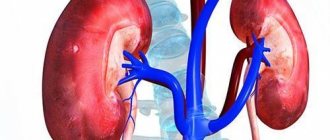
Normal kidney sizes: thickness 4–5 cm; width 5–6 cm; length 10–12 cm. Parenchyma (kidney tissue) to the pelvis is 15–25 mm. This is a paired organ located behind the peritoneum on both sides of the spine at the level of the XII thoracic and I–II lumbar vertebrae. During breathing, they shift by 2–3 cm. The right and left kidneys differ from each other in all respects by 2 cm.
The adrenal glands are located at the upper poles of the kidneys. The left one is shaped like a crescent moon, the right one is a triangle. The average size of the adrenal glands is 5 x 3 x 0.4 cm. The doctor measures the length and thickness of the organs.
Carrying out
To perform ultrasound diagnostics of the kidneys, first of all, it is necessary to expose the abdomen and the entire lumbar region, since to determine the condition of the organ, it is necessary to evaluate it from different angles. This inspection technique is called polypositional. Typically, at the beginning of the procedure, the patient is lying on her back, and the examination is performed through the abdomen. In the second stage, the doctor asks the woman to turn over to the right and then to the left side, performing detailed visualization of all renal structures.
At the third stage, the patient must stand upright, inhale and hold her breath, while the kidney, which has a certain mobility, moves out from under the rib, allowing an examination from one more angle. Determination of the degree of kidney mobility provoked by respiratory activity is carried out during inhalation and exhalation in a lying and standing position.
Diet before ultrasound of the abdominal cavity and kidneys
Ultrasound can also be used to make echographic sections. To do this, an ultrasound sensor is installed in the area of the middle part of the 12th rib, directing the radiation at an angle of 30° to the axis of the body and along the long axis of the kidney.
At the end of the procedure, the patient must remove the gel, which is used to improve contact of the sensor with the body, and get dressed. Such an examination allows you to evaluate the anatomy of the kidney, since the 12th rib overlaps the right kidney in the upper part, and crosses the left one in half.
Important! When an ultrasound of the kidneys is performed, it is not always possible to examine the adrenal glands, since a healthy organ is difficult to visualize. A significant increase in the size of the adrenal glands (up to 2 cm), which makes them easy to detect on an ultrasound image, may indicate the development of a tumor.
Performing an echographic section of the right kidney
Interpretation of results
With pathology of the organs of the urinary system, their shape, parameters and parenchyma density change. Change of size:
- Enlargement of the pelvis indicates possible blockage of the urinary tract at various levels. This part of the kidney is normally visible only during pathological processes.
- When interpreting an ultrasound of the kidneys, a decrease in size from the norm is observed during a dystrophic and destructive process.
- The size of the kidneys according to ultrasound is increased in cases of inflammation and congestion, cancer, diabetes mellitus, amyloidosis, and lymphoma.
Using Doppler scanning, the condition of the vessels and the blood flow in them are assessed. If blood flow changes, renal artery stenosis or venous thrombosis can be diagnosed. In the initial stage of blockage, ultrasound reveals an enlarged kidney with reduced echogenicity along the periphery. At the stage of local hemorrhages, areas with reduced tissue density are identified. Changes in the structure of the kidney are detected in various diseases. Normally, its tissue is homogeneous and does not contain inclusions. Only a doctor can decipher a kidney ultrasound. You should not try to analyze the data yourself.
Only a doctor can interpret the results correctly
Ultrasound diagnostics makes it possible to identify pathologies of the urinary tract of various origins. The results of a kidney ultrasound reflect in the conclusion the changes found and a preliminary diagnosis, which must be compared with the data of the anamnesis and blood tests. Examples of diagnoses:
- nephrosclerosis;
- kidney stone disease;
- chronic pyelonephritis;
- benign and malignant formations;
- kidney abscess;
- congenital pathology of organs and blood vessels;
- nephroptosis – prolapse of the kidney;
- narrowing of the ureter;
- graft rejection.
When monitoring chronic diseases, the procedure allows you to assess the course of the disease, which in turn helps the doctor develop the correct further tactics for managing the patient.
What can a renal ultrasound show?
Let's talk about what ultrasound shows. There are a number of specific parameters and norms, the violation of which should raise concerns.
When performing an ultrasound, the following is considered:
- Quantity, since the kidneys are paired organs. In this case, anomalies cannot be excluded - the presence of an additional kidney, its doubling, or its complete absence may be observed. In some cases, the absence of one of the organs is the result of surgical intervention due to certain reasons.
- The dimensions of the organs, including not only the width and length, but also the thickness of the kidneys. These indicators depend on the age group to which the patient belongs, his body weight and height.
- The location of the organ is also determined using the ultrasound method. Normal indicators imply its retroperitoneal localization, with the right kidney lying slightly lower than the left. If we consider the position of the organs relative to the vertebral region, the right one is located opposite the twelfth thoracic and second lumbar vertebrae. The left kidney is at the level of the first lumbar and eleventh thoracic.
- The shape of an organ is considered normal if it resembles a bean. The contours should be smooth and the tissue structure uniform.
- An important indicator is the structure of the parenchyma, this is the tissue filling the organ. The norm in adult patients is thickness in the range of 14-26 mm. It should be taken into account that over time the parenchyma becomes thinner. Accordingly, for the older generation the norm is 10-11 mm. If the description includes results exceeding the indicated figures, we can talk about inflammatory processes or swelling of the kidneys. In the case when the parameters are less than normal, we are talking about dystrophic phenomena.
- When examining renal blood flow, specialists view the image in color on an ultrasound monitor. In the presence of dark tones, it can be argued that the blood flow is not impaired, amounting to 50-150 cm/sec, but if brightly illuminated areas are observed, it is enhanced.
In addition, ultrasound will show the presence of neoplasms, both benign and malignant, and the presence of stones in the kidney cavities.
Important. Conducting an ultrasound scan in adults requires preliminary preparation and adherence to certain rules; only in this case will it be possible to obtain reliable data.
Ultrasound examination of the adrenal glands
This paired organ is nothing more than the endocrine glands. They produce hormones - cortisol, testosterone, adrenaline, aldosterone, norepinephrine. Their pathology can lead to uncontrolled hypertension and infertility. Main indications for ultrasound of the adrenal glands:
- obesity;
- female infertility;
- changes in skin pigmentation or the appearance of stretch marks;
- decreased or increased blood pressure;
- suspected adrenal tumor;
- identifying the causes of excess weight gain and muscle weakness;
Preparation for an adrenal ultrasound is the same as before a kidney procedure. It is recommended to follow a diet and drink water for several days (exclude tea, coffee, alcohol). The study is carried out in conjunction with a kidney scan.
What does an ultrasound scan of the adrenal glands show? Hematomas, cysts (fluid formations), congenital adrenal hyperplasia, the presence of metastases, benign and malignant tumors. It is not uncommon that if a patient complains of a crisis of arterial hypertension, the presence of pheochromocytoma is detected. Removal of which leads to normalization of blood pressure.
When examining the organs of the urinary system, ultrasound is first of all preferred. This method is safe and informative if the preparation rules are followed. Ultrasound detects many diseases, thanks to which doctors prescribe treatment on time, saving the lives of patients. Patients are only required to follow the doctor's recommendations. In controversial situations or if a serious pathology is detected, the patient is recommended to undergo computed tomography (CT) or magnetic resonance imaging (MRI).
Decoding the results
First of all, an ultrasound of the kidneys during pregnancy determines the size, location and structure of the organ. During the procedure, the presence or absence of various types of neoplasms is noted.
The study compares generally accepted norms of indicators with analysis in pregnant women:
- The length of the internal organ should not exceed 10–12 centimeters. If there is a discrepancy in size, this indicates the formation of tumors.
- Thickening or thinning of organ tissue on the device screen indicates an infectious process.
- Normally, the kidneys are located symmetrically to the spine.
- The organ can move, but only along a limited trajectory.
- Organ tissue in healthy people in a section no more than 2.5 centimeters. The structure is homogeneous.
The normal indicators of the renal pelvis include the absence of foreign impurities. Otherwise, urolithiasis is diagnosed.
If there is a discrepancy with standard indicators during ultrasound of the kidneys during pregnancy, the following diseases are diagnosed:
- Urolithiasis disease. Particles of sand and stones are visible in the kidneys. This indicates a metabolic disorder in a pregnant woman. The echogenic structure and oval-shaped stones are visualized on the device’s screen. With a small size, the acoustic shadow of the stone is not visible.
- Drooping buds. This condition makes it difficult for the body to eliminate urine. As a result, the pregnant woman develops pyelonephritis or hydronephrosis. Ultrasound diagnostics records the degree of prolapse. During pregnancy, the permissible rate of prolapse is 3 centimeters.
- Pyelonephritis of different stages and types. When scanning, the disturbed structure of the organ tissue and dilated pelvis are determined on the screen.
- Neoplasms of various types. When examined, the image shows dense areas in a rounded shape.
- Polycystic. The disease leads to the development of kidney failure. During diagnosis, the doctor determines the number of cysts and their size. Dopplerography is used to accurately determine how the supplement is used.
What is the procedure
The ultrasound diagnostic method is based on the principle of echo - the transformation of sound waves reflected from biological objects into an image that appears on the display screen.
Several ultrasound options are used:
- Transabdominal – ultrasound scans the organ being examined through the abdominal wall.
- Transrectal , when a special sensor is placed in the rectum. It is used when there are difficulties in using the transabdominal method: obesity, ascites, when the integrity of the skin at the site of the bladder projection is compromised, with increased gas formation. This method is very informative, especially for identifying pathological changes in the prostate gland.
- Transurethral , when a probe is inserted directly into the urethra. This method is used in cases where there is a connection between a disease of the bladder and the condition of the urethra. The method is rarely used due to pain and risk of damage to the urethra.
- Ultrasound with Dopplerography , which evaluates urine flow and blood supply to the organs of the urinary system.
Before you begin examining the kidneys, bladder and prostate using ultrasound, special preparation is required.
Preparation for the procedure
The procedure for ultrasound examination of the kidneys itself does not present any difficulties. However, in order for the resulting picture to be most reliable, the patient is required to carefully prepare for the procedure.
Doctors strongly recommend as preparation:
- Adjust your diet in a certain way;
- Maintain a certain drinking regime;
- Carry out intestinal cleansing procedures;
- Take prescribed medications.
Table of normal kidney sizes in adults
Since the size of the kidneys varies depending on age and gender, special tables with normal values in adults and children have been created for ultrasound diagnosticians. They indicate average figures that you need to focus on. But in each specific case, the doctor must consider the data obtained individually.
A doctor with the appropriate qualifications can determine changes in the urinary system. It is necessary to interpret the ultrasound taking into account the patient’s complaints, clinical examination and laboratory tests.
What you need to take with you to the examination
You can take a bottle of water with you to the ultrasound if it takes a long time to get to the clinic or you need to wait for the examination. Before the procedure, to straighten the bladder, you need to drink 0.6-1 liters of clean, still water. Therefore, you need to empty your bladder in advance and drink water an hour before the examination.
Since ultrasound of the kidneys in women is performed in a lying and standing position, you need to take a clean sheet or diaper with you for the examination, which can be laid on the couch. You also need to prepare napkins or a towel so that after the study you can wipe off the gel, which is used to improve contact between the sensor and the skin.
How to prepare for the procedure
To conduct a quality study, the patient must listen to all the doctor’s recommendations and begin to prepare in advance. Usually the specialist issues them in printed form for convenience.
Preparation for an ultrasound examination, in addition to diet, includes:
- drinking regime
- taking enzyme (mezim) and sorbet (activated carbon, enterosgel) drugs;
- abstain from smoking for 3 hours before the procedure;
Ultrasound is performed only on an empty stomach; the last meal should be no later than 7 hours before diagnosis.
Shortly before the examination, the local doctor sends the patient to undergo the necessary tests so that a diagnosis can be made with greater accuracy.
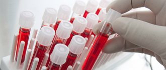
Tests that a doctor may prescribe before performing a kidney ultrasound:
- general urine analysis;
- urine analysis according to Zimnitsky;
- urine analysis according to Nichiporenko;
- general blood analysis;
- blood chemistry.
Diet
The following products should not be consumed:
- unprocessed vegetables;
- dairy and fermented milk products;
- bakery products;
- fat meat;
- any types of beans;
- carbonated drinks;
- alcohol.
Can be used:
- cereal porridge with water;
- meat broth;
- scrambled eggs;
- boiled chicken meat (steamed);
- skim cheese;
- vegetable soups.
Meals should occur every 3-4 hours.
Drinking regime
It is necessary to take 2 liters of water per day throughout the diet. It is advisable to drink water an hour before meals, or 40 minutes after eating.
Doctor Ginzburg L.Z gives recommendations before the procedure for examining the kidneys on the “visus-1” channel.
What does a kidney ultrasound show?
The kidneys are well accessible to ultrasound beams, therefore, in terms of accuracy and specificity, ultrasound of this paired organ is not inferior to such highly informative and expensive diagnostic methods as computed tomography.
Using ultrasound, the doctor can determine the location, shape, size of the kidneys, clarify their relationship with neighboring anatomical structures, assess the clarity of the contours of the capsule, the condition of the parenchyma and pelvic system of the organ. Doppler ultrasound provides very important information about the renal arteries and veins and, in general, about the blood supply to the kidneys.
Having assessed the data obtained during an ultrasound scan of the kidneys, an experienced specialist can identify:
- inflammatory process;
- abnormalities (eg, duplication, absence of kidney);
- pathological changes in the pyelocaliceal system;
- neoplasms and cysts;
- stones;
- pathology of the renal vessels (narrowing, sclerotic changes) and much more.
Note! An ultrasound of the kidneys cannot be considered complete without examining the ureters and bladder (these organs are anatomically and functionally interconnected and, accordingly, must be examined comprehensively), therefore, during such an examination, the doctor can detect pathological changes in these organs.
Features of ultrasound
Ultrasound examination is perhaps the most painless and harmless diagnostic method. To perform an ultrasound, the patient must lie down on a couch. If the doctor is examining the kidneys, the patient should lie on his back, stomach, or side (depending on the situation). In rare cases, kidney diagnostics are performed in a standing position, and the patient must inhale and exhale intensely at this time. As a rule, this position is required to exclude nephroptosis.
How is ultrasound performed?
A special gel is applied to the body, which has conductive properties. The gel improves skin contact with the device used. After this, the doctor passes the transducer over the treated area. The ultrasonic waves emitted during operation of the device are inaudible to humans. They are sent to the kidneys or other organs being diagnosed, and the received signal is displayed on a computer monitor as a picture.
Ultrasound gels
When studying the data obtained, the doctor determines the contours of the internal organs and takes measurements of the kidneys. Based on this, he makes a printout. The procedure lasts about 10 minutes, but during it the patient does not feel any discomfort. The only thing you might not like is the conductive gel. It is quite cool and humid, so particularly sensitive people may experience minor discomfort.
After the examination, the doctor examines the results
Why do you need an ultrasound?
Ultrasound diagnostics are used for the following reasons:
- pain in the lumbar region;
- change in urine color;
- unsatisfactory urine test results, pain when urinating;
- the appearance of edema;
- development of inflammatory processes (cystitis, pyelonephritis, and so on);
Pyelonephritis - what is it, why does it occur?
- mechanical damage to the urinary system or congenital abnormalities;
- development of hypertension. In this case, the doctor may order a kidney ultrasound to examine the renal arteries;
- nephrolithiasis (formation of stones in the patient’s kidneys);
- poor metabolism;
- the patient suffers from gout or diabetes;
- pathologies of the reproductive system.
Kidney ultrasound is required in many cases
On a note! In parallel with ultrasound diagnostics, the doctor may prescribe an examination of other internal organs. These may be organs of the reproductive or digestive system - it all depends on the patient’s condition and the manifestation of symptoms.
How is the research procedure organized?
Ultrasound of the kidneys in men and women is performed mainly by the transabdominal method, through the anterior abdominal wall and a full bladder. The patient lies on the couch on his back, with the abdominal area to the pubic bone and sides open. Organs are examined in different planes and the patient may be asked to turn on their side or even stand up, inhale or exhale.
Ultrasound of the bladder in men and women in some cases may be performed differently. Due to their large weight, women can examine the bladder transvaginally by inserting a sensor in a disposable condom into the vagina. At the same time, the doctor can still look at the condition of the uterus and ovaries.
In men, an ultrasound examination of the urinary system may additionally examine the prostate. The sensor can be inserted transrectally if the patient has obesity or a prostate tumor. In this method, a thin transducer is inserted into the rectum and shows the condition of the bladder very well.
Nutrition and drinking regime before the procedure
To prepare for an ultrasound of the kidneys in women, you need to pay special attention to nutrition, since the degree of gas formation in the intestines depends on its composition. 1-2 days before the ultrasound the following should be excluded from the diet:
- legumes (beans, peas, lentils);
- fresh vegetables, fruits (cabbage, broccoli);
- dairy products, fermented milk are allowed;
- black bread, made from bran;
- baked goods, baked goods;
- carbonated drinks.
Also, in order not to cause irritation to the cells of the renal parenchyma, you should limit spicy, salty, smoked foods and marinades. You can eat your last meal 7–8 hours before the examination.
You can drink plain water if your doctor has not limited its use due to edema or heart problems. Normally, you should drink 30 ml per 1 kg of weight per day.