Synechiae are adhesions in the uterine cavity, fused areas of connective tissue that can form as a result of injuries, as well as inflammation of the mucous membrane. In most cases, injuries to the connective tissue of the uterus can be caused during a diagnostic examination, during curettage, during termination of pregnancy, during the installation of an intrauterine device and during various types of gynecological surgical interventions. These same factors can be causative agents of endometrial inflammation. Experts also call infertility one of the causes of the disease.
- Synechia of the uterine cavity definition
- Asherman's syndrome
Synechia of the uterine cavity definition
After unsuccessful attempts to get pregnant, the doctor diagnosed synechia in the uterine cavity. What is this? How do they affect conception and bearing offspring?
Synechiae or adhesions are fibrous strands that arise in a hollow organ after surgical treatment or as a complication of an infectious process. They can develop in the intestines, fallopian tubes, and uterus.
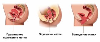
With a small number of fibrous strands, the patient’s quality of life does not suffer. In moderate and severe forms of the disease, adhesions cause complete obstruction of the organ and its dysfunction. Synechiae of the uterine cavity in postmenopause is also found.
Treatment of intrauterine synechiae
The main thing in treating the disease is to eliminate intrauterine contractions in a low-traumatic way, as well as restore menstrual functions and fertility. As a rule, first, for women, adhesions are removed using hysteroscopy. To avoid complications such as uterine perforation, specialists use visual control during surgery through ultrasound or laparoscopy.
The next step is hormonal therapy. It is necessary to restore the endometrium and its cyclic transformation. After surgery, women take estrogen and progestogen in cycles. Taking COC drugs is prohibited, because they cause atrophic changes in the uterine mucosa.
If intrauterine adhesions have developed due to the influence of infection, then after identifying the pathogen and drawing up an antibiogram, the patient is prescribed antibacterial drugs.
Asherman's syndrome
Asherman's syndrome is a set of pathological changes in the reproductive system, which result in the formation of fibrous cords in the uterus. The result of the disease is a decrease in the volume of the cavity or complete fusion of the organ.
There are no cyclic pains. It is possible to assume the presence of synechiae based on the absence of menstruation after invasive procedures on the organs of the reproductive system.
Asherman's syndrome according to ICD 10 is assigned code N85.6 The pathology is classified as non-inflammatory diseases of the uterine cavity.
Symptoms of intrauterine synechia
Patients with intrauterine synechiae usually complain of pain in the lower abdomen, which becomes more frequent during menstruation. When synechiae are dislocated in the cervical canal and the lower third of the uterus, obstacles appear for the rejection of menstrual flow, resulting in increased pain.
Other characteristics of the discharge also change: they become scarcer and shorter lasting. With extensive lesions of the endometrium, menstrual flow occurs in the form of a “spot”, and in the case of complete occlusion of the uterine cavity and cervical canal, it stops altogether.
In patients with occlusion of the cervical canal, but with preserved ovarian and endometrial function, monthly cyclic pain in the lower abdomen occurs on the days of expected menstruation.
Causes and degrees
The cause of the pathological process is surgical intervention performed in a crude manner. These may be the consequences of abortion, polypectomy, endometritis, tuberculosis.
There are 3 degrees of the disease. At the initial stage, synechiae occupy no more than 30% of the volume of the uterus; they are easy to separate during surgery.
The average degree is characterized by the presence of a dense fibrous cord. Separation is difficult. In severe cases, complete fusion of the organ occurs.
Causes of formation of intrauterine synechiae
Factors contributing to the development of the disease are considered to be infectious and traumatic agents, as well as neurovisceral causes. However, mechanical damage to the inner lining of the uterus is considered the most common cause. The endometrium can be injured during an abortion, using intrauterine contraceptives and during diagnostic curettage of the uterine cavity. In addition, uterine bleeding, polyps, myomecomia, metroplasty and conization of the cervix can damage the endometrium.
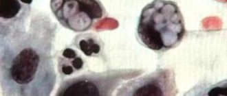
Intrauterine adhesions can develop against the background of genital tuberculosis. This diagnosis is usually confirmed by a biopsy of the uterine endometrium or bacteriological examination of menstrual blood. In addition, the endometrium may be adversely affected by drip injection of solutions into the uterus and radiation therapy aimed at tumors of the reproductive organs. A frozen pregnancy can also cause the development of intrauterine adhesions.
Why do they arise?
Considering that synechiae manifests itself differently in girls and women, the factors that provoke their appearance should be considered by age group.
For girls
During the pre-pubertal period, the female body produces a small amount of estrogens. Namely, they are responsible for the production of a special secretion, which acts as a lubricant in the vagina and vulva area. Its deficiency leads to the labia minora and majora sticking to each other, after which they are “soldered” in this position.
- Disturbances in the gastrointestinal tract. Dysbacteriosis, helminthic infestation and other gastroenterological problems, due to which the supply of nutrients to the mucous membrane is disrupted and additional drying occurs.
- Infections. Tonsillitis, rhinitis, sinusitis (including chronic) are a source of infection in the body. Influenza and adenoviral diseases lead to decreased immunity and increase the likelihood of dysbacteriosis and vulvitis.
- Hygiene. Frequent, intensive washing “to the point of squeaking” using soap often leads to minor injuries to the thin, sensitive mucous membrane of the labia. The healing of these wounds provokes fusion.
- Linen. Synthetic, tight underwear irritates the skin and mucous membranes and contributes to excess moisture in the perineal area. This provokes the appearance of vulvitis.
- Allergy. The reaction to various internal and external irritants can affect the labia: their inflammation occurs and the subsequent formation of synechiae.
Vulvitis in a girl can be the result of infection from her mother when using the same towels and personal hygiene products. Therefore, if a child has synechiae, it is necessary to examine the woman for sexually transmitted infections.
Synechiae in the uterine cavity is called Asherman's syndrome, named after the doctor who first described the disease in detail. Similar adhesions can form in the vagina and cervical canal. The causes of synechiae in women of childbearing age are the following factors.
- Mechanical damage. During abortion, gynecological manipulations, after childbirth, with long-term use of an intrauterine device, after the introduction of drugs into the uterine cavity.
- Inflammation. Colpitis, endometritis.
Curettage of the uterine cavity after a frozen pregnancy often leads to the formation of adhesions. Necrotic areas of the chorion activate fibroblasts (connective tissue cells), which stimulates collagen production and the formation of synechiae.
In menopause
During menopause, under conditions of estrogen deficiency, atrophic processes occur in the genital organs. Synechiae often form in the vagina and cervical canal against the background of chronic colpitis. Moreover, women may not have complaints if they are not sexually active.
What examination do you need to undergo?
Fusion of the labia in girls can be detected during a gynecological examination. Next, the pediatric gynecologist collects secretions from the vaginal vestibule for microscopic examination and bacterial culture. Based on the results, additional treatment is prescribed.
The following methods are used to diagnose intrauterine synechiae.
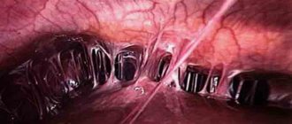
- Hysteroscopy. Synechiae during surgery are visible as thin whitish compounds of varying lengths and densities. They reduce the space of the uterus and most often do not have blood vessels.
- Metrosalpingoscopy. Allows you to determine the degree of adhesions inside the uterine cavity. And also the singleness and multiplicity of pathological fillings of contrast between synechiae, which have different sizes and most often have a lacuna-shaped shape.
Ultrasound examination in the diagnosis of synechiae is in most cases uninformative. Adhesions are not visible, the uterine cavity has a normal structure and shape. In some cases, when performing an ultrasound, synechiae may resemble polyps.
...and adult patients
Synechiae in the uterus are eliminated through surgical manipulation - hysteroscopic dissection. Depending on the length and density of the synechia, the following operations are performed:
- endoscopic scissors;
- tongs;
- hysteroscope body;
- laser;
- hysteroresectoscope.
To monitor the procedure and prevent uterine perforation, a control ultrasound examination or laparoscopy is performed.
The administration of combined hormonal drugs in the postoperative period helps restore the endometrium. In some cases, it is recommended to install an IUD (spiral) to prevent re-fusion of the cavity.
If synechiae is complicated by an infectious-inflammatory process, antibacterial and anti-inflammatory therapy is prescribed.
During menopause, treatment of synechiae can be surgical or conservative, depending on their number and prevalence. Local hormonal drugs are almost always prescribed.
Classification
Adhesions vary in their histological structure. Depending on this, there are three forms of Asherman syndrome:
- light, when the cords consist exclusively of basal endometrial cells, and they are easy to dissect;
- medium, in which fibromuscular adhesions are formed, tightly connected to the mucosa and bleeding when cut;
- heavy, characterized by the formation of dense connective tissue strands that are quite difficult to cut.
Depending on the prevalence of the adhesive process, there are three degrees of pathology:
- I degree - no more than 25% of the surface of the cavity is involved in the process, the cords themselves are thin and do not affect either the fundus of the uterus or the mouth of the tubes;
- II degree - adhesions cover 25-75% of the cavity, the fundus of the uterus and the mouth of the tubes are partially involved in the process of fusion;
- III degree - more than 75% of the cavity is affected, adhesions gradually capture the fallopian tubes and can even close the cervical canal of the cervix, causing the so-called acquired atresia.
The clinical picture of Asherman's syndrome depends on the extent of the adhesions.
The division of synechiae of the genital organs is carried out according to different criteria. The most common classifications are presented in the table.
Table - Classification of synechiae
| Criteria | Division | Characteristics |
| Synechia according to the type of tissue fusion | Children's | - Occurs in girls before puberty; - affects the labia |
| Adults | Formed in the uterine cavity | |
| Synechia according to the severity of fusion of the labia minora | Partial | Up to 2/3 |
| Complete | More than 2/3 | |
| Uterine synechiae by histological structure | Lungs | "Film" that is easily excised |
| Average | — Fibromuscular synechiae; - dense, bleed when cut | |
| Expressed | — Synechiae from connective tissue; - very dense; - extremely difficult to excise without bleeding | |
| Uterine synechiae by prevalence | I degree | - Small adhesions; — affect up to 25% of uterine tissue; - not in the fallopian tubes and the bottom of the organ |
| II degree | — Synechiae cover from 25% to 75% of uterine tissue; — pipe mouths and bottom are covered | |
| III degree | — Synechiae affects more than 75% of uterine tissue; - “sticking” of the walls occurs |
Forecast and prevention of uterine synechiae
Considering the difficulty of removing synechiae, the recurrent nature of the disease and the risks of miscarriage, women should pay special attention to their lifestyle. Let's look at the main rules that help prevent disease.
- Timely identify and treat diseases of the reproductive organs and urinary tract.
- Choose a reliable method of contraception and avoid abortions.
- Observe the rules of intimate hygiene, exclude promiscuity, use condoms to prevent sexually transmitted diseases.
- Regularly undergo preventive examinations with a gynecologist (2 times a year).
Compliance with all the rules of intimate hygiene, a reasonable approach to the choice of contraception, and timely treatment of inflammatory diseases will solve the problem of adhesions at an early stage of their appearance and eliminate all possible negative consequences.
Surgical treatment of adhesions does not exclude the secondary development of synechiae. If a woman becomes pregnant, she has a high risk of spontaneous abortion, miscarriage, delivery at 22-37 weeks of pregnancy, location of the placenta in the lower part of the uterus, covering the area of the internal os, as well as various complications after delivery.
Prevention of Asherman's syndrome consists of excluding abortions, careful and reasonable medical manipulations inside the uterus, treatment of infections affecting the genitals, as well as regular visits to the doctor.
Scar tissue or adhesions are usually removed surgically. Hysteroresectoscopy is often performed. Surgical intervention allows you to remove intrauterine adhesions and restore the patency of the uterus. Treatment of Asherman's syndrome involves two stages:
- Removing adhesions. The first step in treating intrauterine adhesions is to cut through the scar tissue, open the uterine cavity and identify the tubal ostia (the opening of the fallopian tubes). The procedure is performed using a hysteroscope, and doctors use mechanical excision. The adhesions are destroyed after touching the tip of the hysteroscope.
- Preventing the formation of scar tissue after the initial stage of treatment. One of the problems with Asherman's syndrome is that once the scar tissue is removed, it often recurs. There are several different methods used by doctors to prevent its formation. Most often, hormonal treatment is prescribed.
The patient is prescribed immunomodulatory drugs, cyclic or hormone replacement therapy. If the appearance of synechiae is associated with an infectious disease, hormonal therapy will need to be supplemented with the use of antibacterial medications. After surgery to remove adhesions, pregnancy becomes possible; in 32% of cases, after therapy, a woman can give birth to a healthy baby. About 88.2% of women no longer complain of abnormal menstrual periods.
After surgery, doctors recommend undergoing a course of physical therapy. It promotes rapid healing and prevents the formation of new adhesions. This treatment method can be used no earlier than 36 hours after surgery.
Treatment with folk remedies can begin only after a complete examination and confirmation of the diagnosis. Against adhesions, traditional healers recommend using a herbal mixture of coltsfoot, sweet clover and centaury herbs. Well proven:
- flax seeds;
- tincture of marina root;
- St. John's wort;
- aloe leaves;
- Birch buds.
Flax seeds
You should definitely visit your doctor; a full examination cannot be replaced by any information from other sources. Seeking medical help on time will allow you to quickly make a diagnosis and begin immediate treatment.
Even after cutting all adhesions, doctors cannot rule out their reappearance. Such patients still have a risk of miscarriage, premature birth and complications after it. Many such couples are recommended to turn to surrogacy.