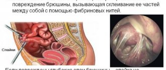
One of the most common malignant skin tumors is basal cell carcinoma or basal cell carcinoma. Oncologists define cancer as a neoplasm made from malignantly altered epithelial cells.
Basal cell skin cancer originates from the basal cells of the epidermis, the stratified squamous epithelium of the skin.
Skin - has a dense connective tissue layer covered with epidermis. Throughout life, the skin epithelium is constantly renewed. Young cells, keratocytes, grow from the basal layer.
During their development, they move to the upper layers, become keratinized and desquamated. In their place, the next protective layer grows. Basalioma is formed from cells of the basal layer or, as it is also called, the germinal layer.
Normally, the germ layer of the skin is clearly demarcated from the underlying tissues. Its cells are located at the same level on the so-called basement membrane. When cells grow malignantly, they penetrate the connective tissue membrane and invade the underlying tissue.
Localization
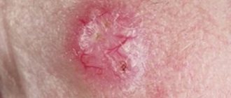
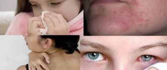
Photo of the initial stage of facial skin basal cell carcinoma
Most often, basalioma is localized on open parts of the body, which accounts for about 80% of all cases. This is usually the skin of the face, less often the scalp. Based on localization, facial basal cell carcinoma affects the following areas:
- frontal region (7%);
- eyelids (14%);
- eyelid angles (9%);
- nasolabial fold (9%);
- area of the wing of the nose (10%);
- parotid region (10%);
- auricle (10%);
- temporal region (11%).
Other tumor locations include the neck, torso, and upper and lower extremities.
Classification of basalioma
There are a number of forms of basal cell carcinoma:
- Cystic - has the appearance of transparent nodes located in the area near the eyes.
- Pigmented - has the form of papillomas or a small tubercle. The color is often dark - black, brown or bluish. Very often, pigmented basal cell carcinoma is confused with melanoma.
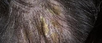
- Superficial - has the appearance of clear edges and is also brown in color. There are no ulcers, but the spots are covered with scabs. The localization of this form of basal cell carcinoma is mainly on the back. Very rarely it can be seen in the parietal scalp. Very often, superficial basal cell carcinoma is confused with psoriasis.
- Sclerodermal - looks like atrophic scars with capillaries. Does not have any scabs or sores. Location: on the face.
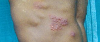
- Mixed is a very aggressive form of basal cell carcinoma that metastasizes. It is quite difficult to treat. It has features of squamous cell carcinoma and basal cell carcinoma.
- Nodular – quite widespread among other forms of the disease. It occurs in the head area (on the forehead, on the wings of the nose), as well as on the back (in its upper part). It looks like a small transparent knot with shiny edges and a depression with a wound in the middle. Sometimes blood vessels are visible on it.
- Ulcerative - forms in the area of the nasolabial fold, near the eyes and ears. It has a painless form, penetrates bone and muscle tissue and destroys them. The ulcer borders are slightly noticeable. Features include penetrating ulcers.
- Solid - has a characteristic development when the affected cells develop not only in depth, but also on the surface. Given its slow growth, the size of the tumor may not change for a long time. Color – light pink.
Nodular basal cell carcinoma
Basalioma in children
In childhood, skin basal cell carcinoma is rare. Factors that promote tumor development are likely to exert their carcinogenic effects on exposed skin over many years. At the same time, according to statistics, of all skin cancers in children, basal cell carcinoma is diagnosed much more often than other forms.
Particular mention should be made of the genetically determined disease Gorlin-Goltz syndrome. This hereditary pathology is accompanied by a combination of anomalies of the skeletal system, reproductive system, and cystic changes in the lower jaw. Multiple basal cell carcinomas are also observed with this syndrome. It is characteristic that skin basalioma in this case is not single, but multiple. Tumor nodes are localized in open areas and number in the tens and hundreds.
Treatment of basal cell carcinoma with folk remedies
Considering that the disease has a low degree of malignancy, you can resort to traditional methods as an additional treatment. However, this does not negate the mandatory contact with a specialist.
There are many herbal remedies that dry out ulcers and start the process of their death.
Among the folk remedies used in the treatment of basal cell carcinoma are:
- Celandine;
- Kalanchoe;
- Tobacco;
- Spurge.
Among the folk recipes you can use the following:
To begin, take 20 cigarettes and remove the tobacco from them, then pour it into a jar and add honey (50 ml) and alcohol (100 ml). The contents need to be sealed and placed on the windowsill for a couple of weeks (don’t forget to shake regularly). Next, you need to strain the liquid and treat the stains with it.
Types of tumor
The following types of basal cell cancer are distinguished:
Nodular-ulcerative
Nodular-ulcerative form
This type of cancer looks like a slightly raised, sometimes ulcerated, node. The size of such a basal cell carcinoma does not exceed 2–3 cm. The tumor is dense in consistency. It is fused to the surrounding tissues.
Dilated blood vessels, or as they are also called “spider veins,” are often visible on the pinkish surface. The node is delimited from the surrounding healthy skin by a pinkish band.
Most often, basal cell carcinoma of this type is found in the area of the nasobuccal fold.
Large nodular (nodular, solid)
Nodular basal cell carcinoma grows outward. This is its difference from nodular-ulcerative. It looks like a yellowish or pinkish hemisphere. This tumor is most often localized in the eyelids and corner of the eye. Localization at the inner edge of the eyelid can lead to severe complications of cerebral circulation.
Perforating
The wing and tip of the nose are completely destroyed.
This is a perforating form of basalioma. Basalioma of this type of facial skin grows with damage to the integrity of the skin.
This type of basal cell carcinoma is localized in areas that are susceptible to injury.
If the initial stage occurs in the form of nodular-ulcerative and nodular forms, then perforating basal cell carcinoma is most likely a consequence of further tumor growth.
Most of the surface of the formation is covered with a crust. Only at the edges there remains a small area of intact pinkish tissue.
Warty (papillary)
This type of basal cell carcinoma resembles a simple viral wart in appearance. The surface of the tumor rises noticeably above the surrounding skin. The node has small protrusions that are similar to warts and are also grayish in color. This facial basal cell carcinoma does not have dilated vessels or ulcerations on its surface.
Clinical signs and stages
Considering the extremely rare metastasis of basal cell carcinoma, its classification into stages is based on the area of spread and the depth of penetration into the underlying tissues with their destruction (destruction) without taking into account the involvement of the lymph nodes. Depending on this, there are 4 stages of development of the neoplasm, which can look like a tumor or ulcer:
- I Art. - size no more than 2 cm, localized within the dermis itself without spreading to surrounding tissues.
- II Art. - dimensions exceed 2 cm, germination of all layers of skin without transition to the subcutaneous fat layer.
- III Art. - significant sizes - 3 cm or more or any size, but spreading to all underlying soft tissues (up to bone).
- IV Art. — tumor growth into bone tissue and/or cartilage.
The initial stage of basalioma (stages I and II) is a small pale pink or flesh-colored tubercle ranging in size from a few millimeters to 1 cm and with gentle edges. It often looks like a bubble or a pearl. There may be several such tubercles, and they gradually merge, resulting in the formation of a plaque with a lobulated surface. Spider veins (telangiectasias) often form on the surface of the tumor.
Subsequently, a ridge of bubbles forms around it, and then a closed dense ridge (a characteristic symptom), which is clearly visible when the skin is stretched, and a reddish “ring,” which is a constant inflammatory process.
Due to the breakdown of tissue, an ulcer or a small erosive surface is formed at the top of the tubercle, which is located at the level of the surface of the surrounding healthy skin and is covered with a flesh-colored crust. When the latter is removed, an uneven bottom of the erosion or crater-shaped edges of the ulcer are revealed, which soon become covered with a crust again.
The ulcerative or erosive surface may undergo partial scarring, but its size gradually increases. In places of ulceration, the color of the neoplasm changes over time. In addition, with any form of tumor, chaotically distributed pigmentation may appear on its surface, which does not indicate anything.
Basalioma slowly increases in size without causing any pain or discomfort. However, over time, severe pain and sensory disturbances may appear due to tissue compression and destruction of nerve branches and trunks.
The larger the affected area, the deeper the tumor spreads. It is its slow growth, on average up to 0.5 cm per year, that makes it possible to detect the disease in 80% within 1-2 years from the appearance of the first symptoms. Timely diagnosis in the early stages makes the prognosis for basal cell carcinoma favorable. In 95-98% of cases, radical excision can be performed with a fairly good cosmetic result and a permanent cure can be achieved.
In the later stages (III and IV), all patients treated are left with significant cosmetic defects that are difficult to correct, and on average, half of the patients (46-50%) experience tumor recurrence after removal. The main reasons for the development of “advanced” stages:
- old age, at which many people become indifferent to their appearance;
- mental, intellectual and personality disorders;
- lack of attention from close relatives;
- living in rural areas far from medical institutions;
- medical diagnostic errors and lack of adequate treatment.
Symptoms of basal cell skin cancer
A characteristic sign of basal cell carcinoma is a reddish, dense ridge surrounding the tumor.
When the skin is stretched, this roller retains its shape. Basal cell carcinoma occurs with minimal symptoms.
Initially, more often on exposed parts of the body, a small nodule appears that resembles a common pimple (see photo above).
Sometimes there are several such nodules located nearby. Later they merge into one large node.
Basal cell carcinoma does not cause pain.
Among the symptoms, patients note only slight itching in the tumor area.
Symptoms of basal cell carcinoma
Types of spots on a child’s skin and the reasons for their appearance
First, a nodule or flat plaque forms on the skin, which turns into an ulcer. It is also possible for a deep formation to appear with a dark red base. Gradually, facial basal cell carcinoma becomes covered with a gray crust. Education gradually increases, but does not cause any unpleasant sensations to a person. At the initial stage of basal cell carcinoma there is no pain or discomfort.
Basalioma can appear at any age; gender also does not affect the development of the disease. However, the disease most often affects people over forty with fair skin. Most often, basal cell carcinoma occurs:
- on the face or nose;
- on hands;
- sometimes - on the chest;
- rarely - on the body.
In rare cases, the nodules in the middle of the plaque disappear or the formation of a thin ridge of cells appears on their contour. Upon closer examination, grains are visible on the surface of the formation, visually reminiscent of pearls.
Gradually, the tumor increases in size, new tumors appear on the skin, and over time the lesions merge with each other. Spider veins form between tumors, as the pathology causes vasodilation. If left untreated, the affected area enlarges and turns into a large ulcer. Deep damage can cause damage to bones and internal organs.
Important! Unfortunately, patients with basilioma of the dorsum of the nose or face usually seek treatment in the later stages of the disease, mistaking the formation for pimples, moles, skin pigmentation or injury. Remember that any formation that does not go away for more than two weeks requires examination by a doctor. The easiest way to cure basilioma is at an early stage.
Stages of basal cell carcinoma
The stages of basal cell carcinoma are determined by the size of the tumor node. Clinicians distinguish five stages of tumor development, including zero.
The initial stage of the disease is characterized by minimal changes. The skin over the tumor that is beginning to grow is not changed. The diagnosis of stage zero is made by chance during a microscopic examination of surgical material taken for other reasons.
1. At the first gradation, basal cell carcinoma is already a clearly visible node with a diameter of up to 2 cm. At this time, the tumor is still located in the upper layers of the skin. It does not grow into the underlying tissues; there are no metastases.
2. The second stage is characterized by the fact that the tumor node reaches a diameter of more than 2 cm, but less than 5 cm. Basal cell skin cancer of this type is already beginning to invade the underlying fibrous layers, but has not yet penetrated into the subcutaneous fatty tissue. There are also no metastases.
3. The third stage is characterized by a formation size of more than 5 cm in diameter. In this case, tumor cords do not penetrate into the subcutaneous tissue. There are no metastases.
Removal or treatment of basal cell carcinoma?
Basalioma, like all malignant neoplasms, requires serious treatment, the approach to which must be individualized.
In addition to surgery, chemotherapy and/or radiation therapy are often used for basal cell skin cancer. In some cases, such methods are the only possible ones. So, if the tumor is localized on the face, it is often not possible to remove it using the traditional surgical method.
In this case, radiation therapy is used to kill the degenerated cells. It is suitable for combating tumors of any location that have not reached a size of 5 cm. For many elderly patients who are unable to undergo traditional surgery, radiation therapy becomes the only salvation. It is often combined with drug treatment.
As part of chemotherapy, local cytostatic drugs are used in the form of applications (lotions) to the tumor area. The most commonly used drugs are fluorouracil and metatrexate.
- A relatively new method in the fight against skin cancer is phototherapy.
Compared to radiation treatment, it has fewer side effects because it does not harm healthy cells. Knowledge of the functioning of malignant cells helps to achieve this effect. They absorb photosensitizing substances more actively than normal ones, and, accordingly, with subsequent exposure to ultraviolet radiation, they die faster.
Diagnostics
A reliable diagnosis of basal cell carcinoma can only be made by microscopic examination.
Basal cell skin cancer can only be reliably diagnosed by microscopic examination of the tumor. The superficial localization of the node makes it accessible to all types of research.
There are 2 main methods of microscopic diagnosis: cytological and histological.
During a cytological examination, tissue is scraped from the surface of a tumor, which may be facial basal cell carcinoma. Alternatively, a small piece of tissue is taken and pressed firmly onto a microscope slide.
In this case, clusters of cells remain on the surface of the glass, which are stained using a special technique.
Under a microscope, cytological examination clearly distinguishes cancerous cells of the basal epithelium.
The histological method is the examination of a piece of tumor tissue in a paraffin block. This tumor fragment is processed in a special way and a block embedded in paraffin is prepared from it. Sections less than a micron thick are made from the block. They are painted with special dyes.
With the histological method of examination, basal cell carcinoma is a part of the altered basal layer of the skin, penetrating deep into the form of strands and individual nests. These are groups of tumor cells that grow into surrounding healthy tissue.
Instrumental diagnostics make it possible to distinguish basal cell carcinoma from lichen planus, lupus erythematosus, scleroderma and many other skin diseases.
Causes of pathology
Basalioma on the face occurs in open skin areas due to the unprotected surface from prolonged ultraviolet exposure or a combination of provoking factors.
The main reasons for the formation and development of neoplasms:
- prolonged exposure to the sun;
- regular stay in the solarium;
- genetic predisposition;
- oncological processes in the body;
- diseases of the hematopoietic system;
- reduced immune defense;
- damage and injury to the skin;
- chronic dermatological diseases;
- exposure to carcinogenic substances;
- bright skin;
- tendency to form moles, freckles, age spots;
- contact and inhalation of combustion products;
- long-term use of medications, especially immunosuppressants;
- the influence of radiation (X-ray or radioactive irradiation, radiation therapy);
- the presence of scars or ulcers on the skin of the face;
- arsenic use or contact with the substance;
- hypothermia or skin burns;
- age-related changes and aging of the body;
- condition after organ transplantation.
Removal of the tumor
Any of the healing methods is aimed at removing or destroying tumor tissue. The following methods of treating basal cell carcinoma are used:
- surgical;
- removal using laser;
- cryodestruction;
- radiation therapy;
- removal by electrocoagulation;
- phototherapy;
- local chemotherapy;
- combined method.
Operating method
This type of treatment for basal cell carcinoma is used most often. The tactics are determined by the fact that the tumor is usually located in areas of the body accessible to intervention (for example, with facial basal cell carcinoma). With the incisional method, the tumor node is excised within healthy tissue.
The localization of the formation is taken into account. Thus, when localized in the area of the inner corner of the eye, the surgeon must be especially careful. It is in this area that the vessels communicating with the cranial cavity pass.
In cases of recurrent scleroderma-like basal cell carcinomas, as well as other tumor relapses, surgical intervention is performed using a special microscopic technique. The method of surgical removal is also indicated when previous radiation therapy is ineffective.
Laser removal
Treatment of basal cell carcinoma with a laser is often carried out in cases where the tumor process is localized on the face. This intervention provides the most beneficial cosmetic effect. Before the operation, local anesthesia of the skin is performed with a special paste or by injecting with local anesthesia.
The tumor tissue is burned out with a laser beam. However, relapses of tumor growth are rare. The rehabilitation period with this intervention is shorter than with the incisional method. The laser method of removal leaves no scars. The defect formed at the site of the tumor is replaced by healthy skin growing from the periphery.
Cryodestruction
This method of removing basal cell carcinoma is based on exposure to extremely low temperatures. It does not matter where the basal cell carcinoma is localized. Two methods of cryodestruction are used: aerosol and application.
In the first method, a cooling substance is applied to the surface of the tumor in the form of an aerosol. When exposed to low temperatures, malignant cells die from the formation of ice crystals in their cytoplasm. This method is more indicated when the stage of the disease is not accompanied by germination.
When applying (overlaying), special probes and tampons are used. The effect of cold in this case is longer. The method is also effective for invasive forms, including when it is impossible to remove basal cell carcinoma surgically. For cooling, liquid nitrogen is used at a temperature of -197°C.
Main forms of basal cell carcinoma
Solid basal cell carcinoma (large nodular, nodular)
The form of skin cancer, which grows primarily not deep into the tissue, but outward, has the appearance of a single hemispherical node measuring from millimeters to 3 cm with an easily bleeding surface. The skin over the formation is light pink or yellowish with telangiectasia. This form accounts for 75% of all basal cell epitheliomas. The most typical location (90%) is the upper areas of the face and neck. Infiltration in depth is insignificant, making surgical removal of basal cell carcinoma effective even if it is 2 cm in size.
Nodular or nodular-ulcerative form
It is considered as a further development of the nodular form. In the center of the tumor, tissue destruction occurs, as a result of which an ulcer is formed with edges in the form of a roller and a bottom covered with purulent necrotic crusts. The most favorite localization of this form of basalioma is the skin of the nasolabial folds, eyelids and inner corners of the eyes. The size of the ulcer can range from millimeters to significant sizes with deep germination and damage to surrounding tissues, which threatens the patient’s life.
Superficial basal cell carcinoma
Accounts for about 70% of all initially diagnosed forms of this disease. It begins with the appearance of a pink spot up to 4 cm in diameter with “pearly” or waxy edges, slightly raised above the surface of healthy skin. It is typically localized on the chest and limbs (60%), less often on the face. The lesions are often multiple. Infiltrative growth is not expressed. The increase occurs mainly due to the superficial expansion of the area of the tumor, which is benign in nature with a long (tens of years) course.
Flat basal cell carcinoma
It occurs in 6% of all cases and is a formation in the form of a flesh-colored plaque. Its edges are raised in the form of a roller with a pearlescent sheen. Localization in 95% is the skin of the head and neck. With this form there is rarely bleeding and ulcers almost never form. Characteristic is a relatively aggressive course with spread deep into the subcutaneous fat and muscle tissue.
Warty form
It is characterized by the external growth of small, numerous hemispherical dense nodules that rise above the surface of the skin and are similar to cauliflower. Their color is lighter than the surrounding skin; spider veins are absent.
The signs of relapse of the disease are the same, but they are significantly more aggressive, more rapid in development, and often the appearance of tumors in other areas of the body. Relapses are most likely in cases where the disease is localized on the face.
Relapses after removal
Recurrence of cancer.
None of the methods for treating basal cell carcinoma provides a complete guarantee of cure. Basal cell carcinoma can recur. The reason for this may be:
- incomplete excision due to inaccessibility;
- tumor growth into deep layers;
- transport of cancer cells with lymph flow;
- the presence of undetected tumor foci;
- treatment errors.
Attention! The most reliable sign of relapse is the appearance of small tumor nodules at the site of the removed basal cell carcinoma, arranged like a necklace.
Other symptoms include a feeling of itching and a slight burning sensation at the site of the removed tumor. In places where small nodules are destroyed, peeling of the skin occurs.
In cases of relapse after surgery, treatment is continued with radiation therapy or chemotherapy. For timely recognition of relapse within several years after surgery, it is necessary to consult an oncologist once every 3 months.
Diagnosis of basal cell carcinoma
First of all, the specialist makes a diagnosis after examination using a dermatoscope, which magnifies the area under study by 20 times.
In most cases, the tumor is first removed, after which it is specially sent for histological examination, and then a diagnosis is made. Sometimes the first thing they do is a biopsy (examination of a specific piece of tissue), then they resort to surgery.
Sometimes (in cases where the disease has affected deep tissues or almost reached the eyeball), an X-ray or ultrasound, MRI and CT scan are done before surgery.
Diagnostics makes it possible to distinguish basal cell carcinoma from diseases with similar symptoms (squamous cell carcinoma, psoriasis, melanoma, lupus, etc.).
Prognosis and prevention
The prognosis for skin cancer such as basal cell carcinoma is favorable. This type of malignant tumor grows very slowly. Some types of basal cell carcinoma do not change their size for years. Metastases to other organs and tissues are extremely rare. To date, such examples of metastasis have been described only a few times in the entire world history of medicine.
To prevent tumor formation, excessive exposure to the sun should be avoided, especially during its active period. Timely treatment of skin damage also plays an important role. It is also necessary to avoid such carcinogenic factors as exposure to ionizing radiation and chemical aggressive substances.
Prognosis and complications of basal cell carcinoma
If you have been diagnosed with basal cell carcinoma, the prognosis for life is mostly positive. Of course, with timely consultation with a specialist and appropriate therapy. But we must not forget that basal cell carcinoma often has recurrent cases in previously discovered places. Therefore, once every few months, a preventive examination by a doctor of scars left after operations is necessary.
The likelihood of complications with basal cell carcinoma in cases of the final stage of the disease, when the tumor enters the bone tissue or cartilage. In this case, it is necessary to remove the bone and perform plastic surgery.
Classification of pathological neoplasms
Based on the location and histological structure of the tumor, neoplasms of basal skin cancer are usually divided into the following types:
- nodular ulcerative tumors;
- sclerodermiformis;
- perforating;
- pigmented;
- pagetoid;
- warty;
- nodular;
- cicatricial-atrophic.
Based on the form of basal cell carcinoma, symptomatic manifestations and patterns of development may differ.
Nodular-ulcerative
A small formation that develops on the upper layers of the dermis as a light pink or red nodule with a dense consistency. The size of such a tumor is up to 5 mm, and thinning of the skin in the affected area with a characteristic greasy sheen can be observed. As the tumor develops, it becomes larger, forming irregularly shaped ulcers. Such ulcers can bleed, and a greasy coating appears at the bottom.
Perforating tumors
The clinical symptoms of perforating neoplasms are similar to nodular ulcerative ones. These tumors most often occur on the skin, which is most exposed to external mechanical influence, while perforating neoplasms are characterized by rapid development.
Sclerodermiform form
At the initial stage of tumor development, a small nodule appears on the skin, having a regular shape with a dense consistency. As it progresses, the neoplasm becomes large in size, transforming into a hard plaque.
Papillary (warty) basalioma
The tumor consists of small nodules with a dense consistency, protruding strongly above the skin. Externally, the tumor has the appearance of a cauliflower and rapidly grows, increasing in size.
Nodular (large nodular) basalioma
The resulting formations do not grow into the deep layers of the dermis, but are characterized by upward growth. These are hemispherical single nodules, which at an advanced stage protrude greatly above the skin.
We recommend reading: Symptoms of sigmoid colon cancer, its stages, prognosis and treatment
Pigment form
Small nodules characterized by rapid progression and ulceration. As a plaque-like nodule develops, ulceration occurs inside the nodule, and a pearlescent ridge appears along the edges of the tumor. Most often, these tumors are located in the facial area on the cheeks and chin.
Pagetoid basal cell carcinoma
A disease characterized by the presence of multiple tumor foci, prone to infiltration into the epidermis, without protruding above its surface. The new growths are light pink, red or brown in color and flat in shape. The size of the tumors does not exceed 5 cm in diameter.
Leading clinics in Israel
Assuta
Israel, Tel Aviv
Ikhilov
Israel, Tel Aviv
Hadassah
Israel, Jerusalem
Important! You need to know what basal cell carcinoma can be confused with. Most often it is confused with lupus erythematosus, lichen planus, psoriasis and epithelioma. You can see cancerous lesions for yourself by carefully examining your facial skin.
Progressive formation provokes the growth of a cancerous tumor. At this moment, new nodules are noticeable on the skin, subsequently merging into one. At this time, spider veins appear between the affected areas, because oncology affects the dilation of blood vessels. After some time, basal cell carcinoma becomes a large ulcer. If treatment procedures are not carried out, the surrounding tissues are affected.
Diagnostic methods
In addition to standard diagnostic methods (history collection, examination), specialists use additional instrumental and laboratory methods to establish a diagnosis of facial skin basal cell carcinoma. These include:
- A smear-imprint (if there is an erosion or ulcer on the surface of the skin) or a scraping from the surface of a tumor formation (in the absence of ulceration) and subsequent cytological examination.
- Biopsy. It is usually carried out to clarify the diagnosis if basal cell carcinoma is suspected based on the results of a cytological analysis.
The dermoscopic method is also actively used. Dermoscopy is especially informative in the differential diagnosis of the pigmented form of basal cell carcinoma of the facial skin and melanoma.
Forecast
Doctors give a favorable prognosis for skin basal cell carcinoma of stages I-III, when it does not grow inside the cartilage, bones and muscles. But even stage IV of the disease is treated surgically, and damaged craniofacial bones are replaced with plates as part of plastic surgery. In all other respects, the consequences of basal cell carcinoma do not in any way affect the health of the patient or his appearance (the skin after recovery does not retain traces of the tumor). So don’t give up and be afraid of surgery - sometimes this is the only opportunity to remove the tumor forever. But if you do not consult a doctor in a timely manner, this is fraught with irreversible changes in the skin:
- first, a harmless (at first glance) painless nodule appears on the face;
- soon it ulcerates (covers with small ulcers), a crust forms on it;
- over the course of 3-4 months (and sometimes years), the nodule gradually grows in width and is constantly accompanied by the formation of bleeding and painful ulcers;
- the tumor becomes clearly lobulated;
- Telangiectasias appear on its surface (vascular networks and asterisks, small capillaries cover it in huge quantities);
- in the absence of proper treatment, basal cell carcinoma turns into a fairly large (more than 10 cm) flat plaque that constantly peels off, or into a mushroom-shaped node protruding above the surface of the skin, or into a deep ulcer that will gradually destroy nearby bones and muscle tissue.
But after removal, basal cell carcinoma will not bother you at all. Modern plastic surgery has reached significant heights, so that after excision or irradiation there are no scars or scars left. It is worth deciding to take such a step in order to forget about this misfortune forever and never return to it again.
You need to understand that basal cell carcinoma on the face is not just a cosmetic problem that can be solved at home. This is a serious skin disease that needs to be treated professionally and as quickly as possible.
Reviews about the treatment of basal cell carcinoma
Basal cell carcinoma is the same as basal cell carcinoma. This term is used by oncologists (including me) when it is necessary to reach common sense or the instinct of self-preservation in careless patients. Also, the term basal cell carcinoma is usually used by histologists, and is written in the histological report after a biopsy of basal cell carcinoma.
From a scientific point of view, it is more correct and better reflects the characteristics of the disease. After all, most tumors ending in oma (except for the concept carcinoma) are benign. Oddly enough, basal cell skin cancer has the same characteristics as basal cell carcinoma: prevalence, causes, external manifestations, varieties, treatment methods, relapse rate, prognosis , risk of metastases and mortality.
Like any cancer, basal cell cancer has many causes, most often a combination of them leads to the disease. Here we distinguish between external and internal. The main cause of basal cell carcinoma is sunlight damaging the DNA of skin cells, leading to mutations in them.
Causes of basal cell carcinoma
| External reasons | Internal reasons |
| Occasional high sun exposure (tanning) during childhood or adolescence | Age |
| Ingestion of arsenic acid through medications or pesticides | Presence of basal cell skin cancer in relatives |
| X-ray or other ionizing radiation | Light skin, light eyes |
| Lack of vitamin intake | Blonde or red hair |
| Excessive nutrition, excessive fat intake | Weak immunity |
| Thermal burns | Rare hereditary diseases: albinism, xeroderma pigmentosum, Rasmussen's syndrome, Rombo syndrome, Gorlin syndrome, etc. |
| Scars, fistulas, ulcers, areas of chronic inflammation | Condition after organ transplantation |
Typically, basal cell carcinoma appears as a pearly pink or light mole with dilated blood vessels. The cancer can also be translucent or slightly reddened with raised, roll-like edges, sometimes barely noticeable. In the area of basal cell carcinoma, the skin may bleed, become scarred, or crust over, but does not hurt.
Photo of basal cell carcinoma with characteristic vasodilation and glassy sheen.
The photo shows a superficial type of basal cell carcinoma. The characteristic raised edges are clearly visible.
The photo shows a superficial type of basal cell carcinoma. Dilated vessels are clearly visible.
The photo shows a superficial basal cell carcinoma with many crusts.
The nodular variety is the most common. Consists of one or more nodules, waxy or red, translucent with dilated vessels. There may be a depression in the center and a crusted ulcer. Superficial basal cell carcinoma, often larger than other varieties and appearing primarily on the torso.
It looks like a red plaque with peeling, slight scarring, and irregular jagged borders. Superficial basal cell skin cancer may resemble psoriasis, fungus, or eczema. Pigmented basal cell carcinoma appears more often in people with dark skin. It looks like a mole or melanoma with its dark coloring.
Other features are approximately the same as those of the nodular variety. Cystic basal cell skin cancer is filled with fluid. The shape is more rounded in appearance than that of nodular cancer. Sclerosing (morpheaform) variety. It resembles a scar, appears as a depressed plaque with poorly defined edges, dense to the touch.
Its treatment is difficult. The basosquamous (metatypical cancer) type resembles a more squamous cell skin cancer in appearance. It is a variant of basal cell carcinoma with faster growth, a greater tendency to recurrent metastases, treatment is difficult. The ulcerative type (infiltrative) is ultimately the result of the further development of any of the other forms of basal cell skin cancer.
Previously, a tumor of this type was called a “gnawing ulcer” abroad. This transition significantly worsens the effectiveness of treatment and prognosis. Pincus fibroepithelioma is also a type of basal cell carcinoma, but is quite rare, resembles a flesh-colored mole, sometimes crusts over, and bleeds. Read more about the types of basal cell carcinoma.
The photo shows pigmented basal cell carcinoma of the temple. The pigment form of growth smoothly turns into an ulcerative form.
In the photo, basal cell cancer of the skin of the neck is of the sclerosing variety, resembling a scar.
Basal cell carcinoma should be treated immediately after it appears. Before treatment, the oncologist assesses the size, determines the type of tumor, and takes a piece of the tumor for a biopsy. Conventional surgical removal, or removal with closure with flaps, is performed in the vast majority of cases, for any type of tumor, if there are no contraindications.
Radiation therapy (irradiation) takes about a month and is acceptable for any type of basal cell carcinoma. It is advisable to perform irradiation only in patients over 65 years of age due to the increased risk of new skin cancer appearing in adjacent areas of healthy skin. Cryodestruction can be performed for any form and size of basal cell carcinoma.
But you will have to take care of the wound in the future. In Russia, the availability of cryodestruction is not high. Laser treatment without a photosensitizer or electrodissection can only be performed for small basal cell carcinomas of a favorable variety, but the relapse rate is still high. Basal cell carcinoma of sclerosing, ulcerative, and metatypic varieties cannot be treated with these methods.
Photodynamic therapy can be carried out for superficial forms, even if the basal cell carcinoma is more than 2 cm in diameter. Its cost is high. Recurrence rate from 12% to 22% If basal cell carcinoma is of a deep ulcerative or sclerosing variety, the laser beams will not penetrate into the depths, you should refrain from this method. Read more about the treatment of basal cell carcinoma.
The reappearance of basal cell skin cancer in the same location after treatment is a relapse. Relapses are less responsive to treatment and have a greater risk of further growth, metastasis and mortality. Basal cell carcinoma gives relapses, the frequency of which depends on the method of treatment, the attending physician, the size and type of basal cell carcinoma.
Recurrence rate of basal cell carcinoma for tumors up to 2 cm in size.
| Method of treatment | Recurrence rate in percentage |
| Conventional surgical removal or flap coverage | 10.1% |
| Curettage and electrodissection. Tumors larger than 2 cm, deep, and sclerosing should not be treated with this method. | From 7.7% to 40% depending on the doctor and the type of basal cell carcinoma |
| Mohs removal | 1% |
| Radiation therapy | 8,7% |
| Cryodestruction | 7,5% |
| Laser treatment without a photosensitizer (not PDT). Tumors larger than 2 cm, deep, sclerosing, basosquamous should not be treated with this method | up to 40% depending on the doctor and type of basal cell carcinoma |
| Laser treatment with a photosensitizer - photodynamic therapy (PDT). In addition to deep ulcerative, sclerosing varieties | from 12 to 22% |
For tumors larger than 2 cm, the relapse rate can double. If basal cell carcinoma is more than 5 cm in diameter, the recurrence rate increases approximately 3 times.
The photo shows a relapse of basal cell carcinoma. The surface of the skin is mostly crusty. The bulk of the skin is in the depths.
Typically, basal cell skin cancer grows slowly over many years and does not bother patients. The development of cancer is unpredictable; it can remain small for many years and then grow very quickly. Or grow in steps, sometimes even significantly decreasing in size, and then increasing even more.
Often, elderly patients expect that they will die from concomitant diseases before basal cell carcinoma causes them any trouble. And, therefore, they refuse treatment, thereby dooming themselves to suffering. Because the tumor grows faster and faster over time. It is necessary to start treatment as early as possible to increase its effectiveness and reduce mortality from basal cell skin cancer.
Such features as size larger than 2 cm, location in the central part of the face, in the ear area, long duration, unclear tumor boundaries have a bad effect on the prognosis. If basal cell carcinoma grows into the underlying tissues and bones and grows to gigantic sizes (more than 10 cm), treatment is often not possible.
Basal cell skin cancer very rarely metastasizes, ranging from 0.0025% to 0.55% of cases. Metastases usually occur against the background of large advanced tumors on the head and neck, mainly in men. The incidence of metastases is most closely related to tumor size and depth. If basal cell carcinoma exceeds 3 cm in diameter, the risk of metastases is 1-2%.
If the tumor is more than 5 cm, metastases appear in 20-25% of people. If the cancer has grown more than 10 cm, the risk of metastasis is already up to 50%. First of all, metastases go to regional lymph nodes, less frequently to the lungs, bones and other organs. The appearance of metastases significantly increases mortality from basal cell carcinoma.
When cellular skin cancer is confirmed, treatment methods are selected depending on the type and how much the tumor has grown and grown into neighboring tissues. Many people want to know how dangerous basal cell carcinoma is and how to treat it so that there are no relapses. The most proven method of treating small tumors is surgical removal of basal cell carcinoma using local anesthesia: lidocaine or ultracaine.
When the tumor grows deep inside and into other tissues, surgical treatment of basal cell carcinoma is used after irradiation, i.e. combined method. In this case, the cancerous tissue is completely removed to the border (edge), but if necessary, they enter the nearest healthy areas of the skin, retreating 1-2 cm from it. With a large incision, a cosmetic suture is carefully applied and removed after 4-6 days. The sooner the formation is removed, the higher the effect and the lower the risk of relapse.
Traditional therapy in the fight against skin basal cell carcinoma
basal cell skin cancer
Basal cell carcinoma of the face and its signs
The pathology develops slowly, but its presence can be detected already at the initial stage. Typical symptoms include:
- The neoplasm has a round shape, but only half of the ball is visible on the surface of the skin.
- The texture of the tumor is tight and uniform.
- Basal cell carcinoma has pronounced borders; along the edges there are compactions and peeling.
- In the middle there is a depression with a thin film and small scales.
- On the surface there are small round nodules - cells of the affected epidermis.
The tumor develops slowly. If no measures are taken, the process takes several years.
Many patients mistake the tumor for an abscess and try to squeeze it out or remove it in another way.
Mechanical damage leads to increased spread of the disease and the appearance of ulcers. The latter affect nearby tissues and are not completely eliminated.
In the photo you can see what basal cell carcinoma looks like. If a tumor appears, you must immediately consult a doctor and under no circumstances take any action at your own discretion.
Introduction
Basal cell carcinoma (synonyms: basal cell carcinoma, basal cell carcinoma) is a malignant skin cancer that is different from melanoma . As the name suggests, this growth originates from the basal cells that are found in the outermost layer of the skin, i.e. in the epidermis.
Like any tumor, basal cell carcinoma is caused by one or more genetic mutations, which are often the result of excessive exposure to the sun's UV rays, but not exclusively. In fact, the risk factors are different.
Basal cell carcinoma appears as a sign on the skin visible to the naked eye and has specific features.
With early diagnosis and early treatment, the disease can be cured. On the contrary, if diagnosis and treatment are delayed, metastases can form and basal cell carcinoma can become fatal.
Treatment of the disease
One of the treatment options for facial basal cell carcinoma is surgery, in which the tumor is removed along with healthy tissue.
The choice of treatment methods depends on the degree of tumor development, symptomatic manifestations, concomitant diseases and general health of the patient, and the depth of the lesion. Treatment of basalioma on the face is carried out in the following ways:
- medicinally;
- surgically;
- combined methods;
- folk remedies.
Drug therapy for basal cell carcinoma uses cytostatics—local drugs that penetrate the cells and destroy the cell cell carcinoma from the inside. Drug treatment methods are used when there are contraindications to surgery or as preparation for surgery. Facial skin basalioma is removed in the following cases:
- increase in tumor size;
- proliferation of the affected area, multiple basal cell carcinoma;
- the addition of discomfort, pain, bleeding;
- constant damage and friction;
- location in places dangerous to life (near the eyes, nose, ears).
Basic removal methods:
- Larez removal. Targeted impact of a laser beam.
- Cryodestruction. Freezing and burning with liquid nitrogen.
- Electrocoagulation. Cauterization with directed electric current.
- Photodynamic therapy. Introduction of light-sensitive drugs that accumulate in the tumor, enhancing the effect of light flashes.
- Radio wave removal. Exposure to high frequency radio waves with further thermal evaporation of the affected area.
- Radiation therapy. X-ray irradiation.
- Surgery. Excision of the tumor with a scalpel.
- Combined. The use of various methods for removing basal cell carcinoma to enhance the therapeutic effect.
Disease prevention
Prevention of the disease consists of protecting the skin from external irritants:
- tanning time should only occur in the morning or evening hours;
- Be sure to use sunscreen, as well as wind and frost creams;
- when under the sun, wear a hat;
- systematically undergo examination by a dermatologist;
- At the first signs of illness, be sure to consult a doctor;
- avoid prolonged contact with harmful chemicals;
- prevent mechanical damage to the skin.
How to treat basalioma
There are many methods for treating basal cell carcinoma of the face. Only an oncologist can determine which therapy will be most effective. The main goal of therapy for basal cell carcinoma is its removal and prevention of relapse of the disease. The treatment method is selected depending on:
- sizes;
- tumor location;
- area of skin damage;
- health conditions;
- patient's age.
Folk remedies
Various folk methods can only slow down the growth of skin tumors, but such remedies cannot completely cure basal cell carcinoma. Alternative medicine in this case can only serve as a complement to the conservative or surgical method of tumor removal. The most effective folk remedies for the treatment of basal cell carcinoma are:
- Celandine. The tumor is smeared with fresh plant juice (to do this, break off a twig from a growing bush) up to 4 times a day for 10 days. The product dries the wound perfectly.
- Golden mustache. The plant is washed with water and twisted using a meat grinder. The pulp is placed in gauze and applied as a compress to the basal cell carcinoma, leaving for a day. Therapy lasts 15 days.
- Tobacco tincture. To make a non-healing sore on the skin disappear, use tobacco infusion. To prepare it, tobacco from one pack of cigarettes is poured with 200 ml of vodka and infused in the refrigerator for 14 days. Afterwards, the solution is shaken and filtered. Cotton wool is moistened with the resulting tincture and applied to the tumor. The course of treatment lasts 10-12 days, after which the scar remaining after the ulcer should disappear.
Ointment
Immunotherapy is often used to treat basal cell carcinoma of the face. It involves the use of a special ointment - imiquod. The drug stimulates the patient’s body to produce interferon, which helps in the fight against atypical cells. As a rule, nasal basal cell carcinoma is treated with cream, since this method of therapy does not leave scars. Imicvod is often used before starting chemotherapy.
Radiation therapy
The method is used if the patient’s condition or the location of the basal cell carcinoma does not allow its surgical removal. Radiation therapy is often used to treat a tumor on the nose. It is carried out using irradiation with short-focus gamma radiation. The results of radiation therapy are aesthetically better than with surgical removal of basal cell carcinoma. The only drawback of the method is the duration of treatment (on average, 20-25 sessions are required).
Photodynamic therapy
Treatment consists of introducing special substances (photosensitizers) under the skin that highlight clear boundaries of the tumor, which is then irradiated with light waves. For facial basal cell carcinoma, the photodynamic method is one of the best treatment options, since it does not lead to cosmetic defects, unlike surgical intervention.
- Papilloma on the face: types of tumors and their removal
- Atheroma - treatment at home with folk remedies and ointments, reviews
- Wen on the back - causes, symptoms, diagnosis, how to treat and removal methods
Classification
There are two types of gradation of this disease.
Based on the cellular composition, basal cell carcinoma in the ears, nose or any other location can be of the following type:
- superficial basal cell carcinoma - changes occur only in the superficial layer of the epidermis;
- nodular or solid – a single node forms in the skin;
- micronodular - the formation consists of several small nodules;
- infiltrative – grows into tissue;
- fibroepithelial - the formation is formed from epithelial and connective elements;
- with adnexal differentiation - cells that are similar in structure to ovarian cells;
- basalioma with keratinization - part of the formation cells is filled with keratin and dies.
TNM classification is also used:
- TIS – not extending beyond the basal layer of the epidermis;
- T1 – extends to the entire thickness of the skin and involves the dermis, but its dimensions do not exceed 2 cm;
- T2 – the formation is more than 2 cm or the patient is diagnosed with more than two risk factors for its growth;
- T3 – formation grows into the skull;
- T4 - grows into the bones of the skull.
Separately, during diagnosis, the following risk factors for degeneration into a malignant formation are identified:
- the size of the formation is more than 2 cm;
- there is germination into the nerve trunks;
- localization of the tumor in the ear;
- low cell differentiation.
Determination of the type of formation is carried out only through diagnostic measures.
Causes of the disease
It is believed that basal carcinoma develops as a result of prolonged exposure to ultraviolet radiation.
In addition, medicine has identified factors that can trigger the development of skin cancer:
- Genetic predisposition.
- Permanent mechanical injuries to a certain area of the skin.
- Prolonged contact with carcinogens and harmful chemicals.
- Viral infections.
- Effect of ionizing radiation.
- Age-related changes in the skin.
- Failures in the functionality of the immune system.
Basal cell carcinoma is characterized by gradual development.
Degenerated cells divide much more slowly than healthy ones, and many of them die. For this reason, the tumor is usually small in size.
People with weakened immune systems and fair skin are at risk.
Manifestation of the disease
One of the sites of localization of malignant manifestations is the nose, and the tumor is pink in color.
Basal cell carcinoma of the skin of the face develops in the form of a small (up to 2 cm) nodule of flesh-colored or red-pink color. Main location of the tumor:
- upper lip area;
- eyelids;
- nose;
- ears, auricle;
- neck;
- cheeks;
- forehead;
- hairy area of the head.
Subsequently, facial basal cell carcinoma is accompanied by the following manifestations:
- gradual tumor growth;
- absence of discomfort and pain at the initial stage;
- the formation of a grayish crust on the surface;
- the formation of a dense ridge of heterogeneous structure around the affected area;
- germination of new nodules and their fusion;
- stretching and dilation of blood vessels on the surface;
- in the center of the neoplasm, an ulcerative process with inflammatory changes begins;
- scar formation;
- tumor growth into surrounding tissues accompanied by pain.
Treatment
Based on the results of statistical studies, approximately 20% of patients who develop basal cell cancer do not seek medical help in a timely manner and carry out self-treatment with folk remedies or with external medications.
Independent attempts to treat basal cell skin cancer at home are strictly prohibited, since such therapy will not only not bring the desired result, but can also aggravate the situation.
This can cause an increase in the depth and area of the lesion, and even increase the risk of metastases.
The main methods of treating basal cell carcinomas include:
- surgery;
- chemotherapy;
- close focus radiation therapy;
- cryodestruction;
- curettage with electrocoagulation;
- photodynamic therapy (PDT).
Treatment methods for basal cell carcinoma are determined only after all diagnostic measures have been carried out, during which the form and degree of development of the neoplasm is established.
Operative method
The operation involves surgical excision of the tumor, capturing up to 5 mm of healthy tissue along the periphery, after which a histological analysis of the edges of the excised area is performed. In case of extensive damage with infiltration into adjacent tissues, it is necessary to perform extensive removal of the tumor with further plastic correction of the wound surface.
The effectiveness of the surgical method of treating such tumors is approximately 95%, with further observation after the operation lasting for 5 years. The risk of recurrence of basal cell carcinoma increases if the tumor is larger than 1 cm in diameter, or if the excised tumor has already recurred. Also, basal cell carcinoma often recurs if the location was the nose, scalp, eyelids and ears.
Typically, a minimally invasive microsurgical operation is performed to minimize the likelihood of damage to unaffected tissue areas. In this case, only the visible part of the tumor is excised, after which horizontal sections of tissue are applied layer by layer for histological examination.
Radiation therapy using x-rays
This method of treatment may be prescribed if the patient has contraindications to surgery to excise the tumor. Irradiation of basal cell carcinomas is also prescribed for elderly patients aged 60 years or more. During the procedure, tumor foci are irradiated with small doses of radiation. The danger of using radiation exposure is that it can provoke the development of radiation dermatitis, alopecia, or even the development of a malignant tumor.
Curettage with electrocoagulation
This method is the most accessible in the treatment of basal cell cancer, and at the same time easy to use. The technique involves removing the bulk of the affected tissue using a metal curette, after which electrocoagulation of the tumor bed is performed. One of the important disadvantages of this method is the lack of possibility of histological control, and the high probability of relapses if the size of the tumors exceeds 1 cm. Also, after tumor removal, patients are left with serious cosmetic defects.
We recommend reading Esophageal cancer - the first symptoms in the early stages, treatment, prognosis
Cryodestruction using liquid nitrogen
The method is characterized by a relatively low cost of the procedure and the ability to carry it out on an outpatient basis, while cosmetic damage is minimal. But cryodestruction is rarely used, since multiple sessions are necessary and there is no possibility of histological control. With this method of treatment, the likelihood of relapse is also high.
Photodynamic treatment
An innovative method of combating basal cell carcinoma, which involves the impact of a laser without a photosensitizer with a low intensity wavelength on the tumor. In this case, as a result of a light-chemical reaction, substances are formed that cause apoptosis of tumor cells. Thus, cancer cells are identified by the body as foreign and it forms an immune response to their development.
Chemotherapy
This method has not proven to be an effective treatment for basal cell skin cancer. Chemotherapy can only be performed for superficial basal cell cancer with a small area of damage. Chemotherapy is usually prescribed as an addition to the main surgical treatment, or if there are contraindications to its use.
Systemic chemotherapy involves the internal drip administration of special medications that negatively affect cancer cells. Creams and emulsions may also be prescribed. The effectiveness of this technique is observed only in the early stages of development, and only in 70% of cases.
Treatment of pathology
Treatment of basal cell carcinoma consists of removing the tumor in one way or another. The advantage of the neoplasm is that it is localized on the surface of the skin - this facilitates access to it by various therapeutic and surgical agents.
Removal of basal cell carcinoma is carried out using the following methods:
- surgical using a scalpel;
- laser destruction;
- cryodestruction;
- radiation therapy;
- chemotherapy (local);
- photodynamic therapy
The choice of method is the prerogative of the oncologist, who will take into account the nature and localization of the tumor, the degree of its spread, the stage of the disease and other important factors. In many cases, combination treatment is used, including a combination of different approaches. In addition, immunotherapy is performed to activate the immune system.
Surgical removal
Removal of basal cell carcinoma can be carried out by excision with removal of surrounding tissue to a depth of 1–2 cm around it. This traditional method is usually used for large tumors and when they grow deep into the skin. The procedure is performed on an outpatient basis and under local anesthesia.
In addition, the removal of basal cell carcinoma can be performed using Mohs micrographic surgery, the essence of which is the layer-by-layer removal of skin segments affected by the malignant process until they are completely cleared of cancer cells, which is ensured by constant microscopic monitoring of the removed material. This operation is also performed on an outpatient basis and under local anesthesia.
Both of these methods have similar disadvantages:
- wound formation followed by scarring;
- possible allergy to pain medication;
- surgical infections;
- possible damage to nerve endings.
Recurrence of basal cell carcinoma of the lower eyelid before surgery (a) and 2 months after total penetrating resection of the lower eyelid with blepharoplasty according to Dieffenbach-Szymanowski (b)
Laser destruction
The method of destroying basal cell carcinoma with a directed laser beam has a number of advantages over surgical removal of the tumor:
- the procedure is practically painless;
- complete sterility of the operation area;
- no subsequent scarring;
- the ability to perform surgery in hard-to-reach places;
- reducing the risk of relapse.
Moreover, the method also has certain disadvantages:
- the use of a laser is possible only for small lesions;
- the need for several sessions (up to three);
- contraindications - diabetes, pregnancy, infections, increased photosensitivity, cardiovascular pathologies, thyroid dysfunction.
Removing basal cell carcinoma using a directed laser beam has many advantages, but the method is contraindicated in some cases
Cryodestruction
The essence of cryodestruction is the destruction of neoplasms at low temperatures. Liquid nitrogen, evaporating, deeply freezes the affected tissue, under the influence of which the cancer cells necrotize. It is possible to freeze small basal cell carcinomas.
The procedure is performed under local anesthesia and leaves virtually no scars.
The procedure for removing basal cell carcinoma with liquid nitrogen leaves virtually no scars behind.
Radiation therapy
Radiation, which was popular in the recent past, is not used as often because it may increase the likelihood of developing squamous cell carcinoma at the site of X-ray exposure. Moreover, the method is quite effective and is successfully used when it is impossible to choose another method of tumor removal. For complete cure, several sessions of radiation therapy are necessary.
The procedure is painless, but leaves behind a radiation burn that requires additional treatment. In addition, irradiation causes general harm to the body - it changes the blood formula, reduces the activity of the immune system, etc.
Local chemotherapy
To combat basal cell carcinoma, local treatment of the affected area with cytostatic chemotherapy drugs in the form of ointments (Colhamin, Methotrexate, etc.) applied to the affected area is used. This method of treatment (unlike general chemotherapy) does not cause injury to other systems and organs, since the drugs act directly on the tumor.
Photodynamic therapy
The essence of the method is to expose the affected area to light pulses after preliminary injection into the tumor of a special drug that makes the tumor sensitive to light.
Phototherapy is used to remove tumors located in hard-to-reach areas.
Folk remedies in the treatment of pathology
It should be understood that folk remedies for the treatment of basal cell carcinoma can, at best, only slow down the development of the tumor, but are not able to completely cure it. It is advisable to use methods of alternative medicine as a complement to the main treatment or to slow down the growth of the tumor until the basal cell carcinoma can be removed.
Burdock and celandine ointment
To prepare the product you need:
- Pour melted lard into a glass of chopped burdock and celandine, taken in equal quantities.
- Then keep the mixture in the oven for a couple of hours, then let it cool and brew for several days.
The prepared ointment should be applied three times a day to the affected area.
Celandine juice
In the spring and summer, it is enough to lubricate the tumor several times a day with fresh celandine juice, which appears if the stem of the plant is broken.
You can prepare the remedy for future use by passing fresh stems and leaves of celandine through a meat grinder and squeezing the juice through several layers of gauze.
Golden mustache juice
You can prepare the juice in the same way as celandine juice. You need to soak a gauze swab in the resulting product and apply it to the tumor for a day.
Basalioma - general characteristics and mechanism of tumor development
Basalioma is also called basal cell carcinoma of the skin, corrosive
or skin carcinoid. All these terms are used as synonyms to refer to the same pathology, namely, skin tumors from atypically changed cells of the basal layer of the epidermis.
Currently, basal cell carcinomas account for 60 to 80% of all types of skin cancer. Tumors develop mainly in people over 50 years of age. At a younger age, basal cell carcinomas practically do not occur. In the population, the tumor affects men somewhat more often. The total lifetime risk of developing this type of skin cancer is 30–35% for men and 20–25% for women. That is, the tumor occurs quite often - in every third man and every fourth woman.
The tumor is specific to the skin and does not affect any other organs, that is, basal cell carcinomas can form exclusively on the skin.
Most often, basal cell carcinoma is localized in the following areas of the skin:
- Upper lip;
- Upper or lower eyelid;
- Nose;
- Nasolabial folds;
- Cheeks;
- Auricle;
- Neck;
- Scalp;
- Forehead.
In 90% of cases, basal cell carcinoma is localized in these areas of the facial skin. In the remaining 10% of cases, the tumor can form on the skin of the torso, arms or legs.
According to the nature of their growth, basal cell carcinomas are classified as malignant tumors, since the neoplasm does not grow in a capsule, but, without any membrane, simply grows into the tissue, destroying their normal structure. Basalioma grows not only in depth, but also in width, which is manifested by a simultaneous expansion of the tumor area and an increase in the volume of damaged underlying tissue.
That is, due to its growth in width, basalioma invades new healthy areas of skin located on the border with the tumor. And due to its growth in depth, the tumor sequentially grows first through all layers of the skin, and then into the subcutaneous fatty tissue. As a rule, the external dimensions of basal cell carcinoma correlate with the depth of its growth in the tissue. That is, the larger the surface area of basal cell carcinoma on the skin, the deeper it has grown into the tissue.
Despite the aggressive nature of growth, which consists in the germination of tissues with disruption of their structure and functions, basal cell carcinoma slowly increases in size - usually no more than 5 mm per year. This makes the tumor slowly progressive and therefore relatively treatable.
However, in addition to aggressive invasive growth, any malignant tumor is characterized by the ability to metastasize, which basal cell carcinoma does not have. That is, basal cell carcinoma does not metastasize to other organs, and this distinguishes it from other malignant tumors of various locations and origins.
Since basal cell carcinoma has only one obligatory property of a malignant neoplasm (an aggressive growth pattern), but does not have the second (the ability to metastasize), it is often classified as a borderline tumor. This means that basal cell carcinoma has the properties of both a benign and a malignant tumor at the same time.
Basalioma develops from degenerated cells of the basal layer of the epidermis. To understand what this means, you need to imagine the structure of the skin and especially its upper layer - the epidermis. Thus, the skin consists of hypodermis, dermis and epidermis. The topmost layer that we see in any person is the epidermis, consisting of five layers.
The lowest layer is called the basal or germinal layer, followed by the spinous layer, followed by the granular and shiny layer, and covering them is the horny layer. It is the stratum corneum that is the outer layer and is in direct contact with the environment. Basalioma is formed from cells of the basal layer of the epidermis that have undergone malignant degeneration.
Since the epidermis and, accordingly, its basal layer are present only on the skin, basal cell carcinoma can form exclusively on the skin. Basalioma can never form in other organs.
Externally, basal cell carcinoma is a spot, mole or elevation on the skin, which gradually increases in size, with a depression and ulcer formed in the central part, covered with a crust. When this crust is torn off, an ulcerated bleeding surface is visible. Basalioma can be mistaken for a wound, but unlike a true wound, it never heals completely.
In accordance with the definition of the World Health Organization Committee, basal cell carcinoma is a locally infiltrating (sprouting) formation that is formed from epidermal cells or hair follicles and is characterized by slow growth, very rare metastasis and non-aggressive properties.
This skin disease occurs mainly in exposed areas of the skin, which are maximally exposed to sunlight. These are, first of all, the skin on the head, mainly in the frontotemporal zone, and neck. Basalioma of the facial skin is especially often localized on the nose, in the area of the nasolabial folds and on the eyelids. On average, in 96% of patients it is single, in 2.6% it is multiple (2-7 or more lesions).
The main risk factors for the development of basal cell epithelioma:
- Chronic and intense nature of the influence of ultraviolet radiation. Moreover, unlike melanoma, chronic exposure is much more important. Therefore, melanoma often occurs on areas of the body protected from the sun by clothing, even several years after a sunburn, and basal cell carcinoma occurs on exposed areas. This fact is confirmed by the different geographical frequency of the disease (in southern climatic zones it occurs much more often than in northern ones) and is the answer to the frequent question “is it possible to sunbathe?”
- Age factor. The incidence rate is significantly higher in older people, and it increases with increasing age. More than 90% of cases of basal cell skin cancer are registered among people over 60 years of age.
- Gender. Men suffer from basal cell carcinoma twice as often as women. However, this indicator is not very convincing, since it may be associated with the different nature of work activity. Due to changes in lifestyle and fashion in recent decades, there has been an increase in incidence among women. Some authors write about the same incidence rate among both sexes.
- Tendency to freckles in childhood and very fair and fair skin types (types I and II). People with darker skin get sick much less often.
- Various genetic diseases associated with increased sensitivity to ultraviolet rays.
- The presence of basal cell carcinoma in other family members.
- Chronic inflammatory skin diseases, burns and the presence of keloid scars, trophic changes.
- Constant chemical exposure to carcinogenic substances (petroleum products, arsenic compounds, resins) and frequent mechanical damage to the same areas of the skin.
- The influence of X-ray and radioactive radiation, previous radiation therapy.
- Reduced immune defense of the body due to: HIV infection, taking cystostatic drugs due to blood diseases or organ transplantation.
Video: Basal cell skin cancer
Despite the fact that basal cell carcinoma of the face is unlike ordinary cancer, since it does not spread metastases to other organs or tissues, it cannot be considered harmless. Find out when, without treatment, the tumor gradually increases in size, destroying nearby skin, muscles and bones. If basalioma affects the nerves, the patient begins to feel severe pain. You can learn more about the disease by watching the video.
“At the doctor’s appointment” Issue 14 - Basal cell carcinoma