The human skull can be divided into a vault and a base, but this distinction is arbitrary - there are no clear limits. The base is the lower part of the skull, which consists of a pair of temporal, occipital, ethmoid, and sphenoid bones. Inside, this part repeats the structure of the brain and has three fossae - posterior, middle, anterior. In the center of the base runs the foramen magnum, which connects the cranial cavity to the spine.
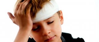
A basal skull fracture is an injury that involves damage to one or more bones. Almost 60% of cases are fatal. The consequences, even with recovery, can be severe and affect subsequent lifestyle. Timely first aid provided by the environment and medical assistance at the scene of the incident preserve the chances that the victim will survive.
Prognosis and survival
Fractures of the vault and base of the skull are often fatal. From 20 to 50% of patients survive the injury. In this case, the prognosis will depend not only on the nature of the injury, but also on the state of the body during the recovery period.
An unfavorable prognosis for the patient is associated with massive hemorrhage, damage to vital structures, infection, as well as combined injury to other bone structures and soft tissues. If a person survives, he may face severe long-term consequences, such as epileptic seizures, loss of hearing, vision, and speech. In addition, loss of motor function often occurs, leading to loss of ability to work and disability.
The development of a favorable outcome is possible if the fracture was not accompanied by hemorrhage, swelling, or the formation of wound defects that act as entry points for infectious factors.
To improve the prognosis for life, it is important to seek medical help early and provide first rescue measures.
Classification of traumatic brain injuries
Types of skull fractures are classified according to location, level of defect, and morphological characteristics. Based on location, injuries to the facial and brain parts are distinguished. Base injuries are divided into fractures of the anterior, middle and posterior cranial fossa. Types of bone defects are classified according to the level of damage as open and closed. Violation of the integrity of the skin and bones indicates the presence of an open skull fracture. Often such situations arise as a result of a strong blow to the head, a fall from a height or into water, or road traffic accidents.
This type of fracture is dangerous due to bleeding and the development of infection in the wound canal. For injuries with intact hairline, not counting scratches and abrasions, a closed type of fracture is diagnosed. The direction of the blow is important. Skull fractures occur at the point of application of force or as a result of transmission of force from other parts of the body (direct or indirect).
Classification according to morphological characteristics includes:
- linear (local and remote);
- comminuted depressed;
- penetrating perforated-depressed;
- multiple linear;
- combined.
According to the severity, contusions are divided into mild, moderate and severe types.
Closed head injuries include:
- brain concussion;
- injury;
- compression;
- base fracture;
- vault cracks.
Types of skull base fractures
In clinical practice, a fracture of the base of the skull has several classifications. Depending on the mechanism of injury, fractures are divided into:
- Linear. A linear fracture of the calvarium is characterized by the appearance of a crack in the thickness of the bone. This option is the most favorable in the clinical course and rarely leads to life-threatening complications.
- Splintered. A fracture of various bones of the cranial vault is accompanied by fragmentation of these elements into several fragments. This option may be accompanied by the development of complications due to traumatization of brain cells by bone fragments. The most common complications include bleeding or innervation disorders with possible death.
- Depressed. Damage to the skull is characterized by compression of bone fragments to the brain structure.
- Perforated. Violation of bone integrity develops as a result of a gunshot wound. The development of a fatal outcome with this type of fracture is possible due to massive damage to soft tissues and vascular bundles.
Structure of the human skull
The cranial bones are divided into two important sections:
- facial;
- cerebral.
Facial department
The facial section consists of the following paired and unpaired bones.
Doubles:
- nasal;
- palatal;
- zygomatic;
- tearful;
- upper jaw;
- inferior nasal concha.
Unpaired:
- lattice;
- sublingual;
- vomer;
- lower jaw.
The facial region plays a very important role in the process of life, since it affects the respiratory, digestive and sensory organs. The unpaired bones have air-filled areas that connect to the nasal cavity. Thanks to the air areas, thermal insulation is provided for the sensory organs, but despite the presence of such areas, the skull is particularly durable and strong.
Air sections include:
- lattice;
- frontal;
- temporal;
- upper jaw;
- wedge-shaped.
An important role is played by the hyoid arcuate bone, which is localized between the jaw and larynx and connects with the help of ligaments and muscles to the bones of the skull.
This element forms paired horns and a body, from which the processes of the temporal bones extend. The upper bones of the skull are flat and consist of special plates with bone substance. These plates are filled with cells containing blood vessels and bone marrow. Some bones of the skull follow the shape of the brain, their irregularities correspond to its convolutions and grooves.
Brain department
The brain section also consists of paired and unpaired bones.
Doubles:
- parietal;
- temporal
Unpaired:
- frontal;
- occipital;
- wedge-shaped.
This section is located above the facial section. It also has an airy frontal bone, which consists of a nasal part and two scales. The frontal bone forms the frontal tubercles and forehead, which form the eye sockets, temporal fossae, and nasal cavities. The parietal bone forms the cranial vaults and the parietal tubercle. The occipital bone forms the cranial vault and hearing organs. All the bones of the skull are connected to each other by special connections - “sutures”.
Symptoms and signs
Nonspecific signs characteristic of a fracture of the base of the skull are expressed in the form of pain in the head (in most cases the patient cannot accurately name its location), nausea and vomiting that does not bring relief.
Specific manifestations characteristic of this type of injury are determined depending on the location of the fracture.
A fracture of the anterior cranial fossa is manifested by profuse bleeding from the nasal cavity, which is difficult to stop using conservative methods. In addition to blood, a small amount of colorless and odorless cerebrospinal fluid may leak. Hematomas around the eyeballs are visible to the naked eye. The symptom of glasses with a fracture of the base of the skull is fully manifested on the second or third day after the incident.
If a section of the ethmoid bone is damaged, subcutaneous emphysema is visualized in its projection.
Traumatic impact affecting the middle cranial fossa is accompanied by massive leakage of blood or cerebrospinal fluid from the ear canal, as well as subsequent impairment of auditory function. Due to damage to the facial nerve, changes in taste sensitivity may occur and facial asymmetry may appear. Upon examination, a hematoma of the postauricular area is revealed. Attempts to stand up end in falls due to disruption of the vestibular apparatus.
Violation of the integrity of the posterior cranial fossa manifests itself:
- Growing hematoma in the area of the ears.
- Change in sensitivity in the affected area.
The development of bulbar or pseudobulbar syndrome is associated with damage to the brain stem. The patient's pulse quickens and blood pressure decreases. Breathing becomes shallow and arrhythmic, which leads to insufficient oxygen supply to the lungs.
The face becomes distorted and asymmetrical, the corner of the mouth drops, and the palpebral fissure widens.
How is the human head structured?
Why did the head appear during evolution?
The head appeared in ancient vertebrates. Before this, the spine had uniform segmentation. From each vertebra, which contained a segment of the spinal cord, a pair of nerves emerged. The pair extending from the very anterior vertebra became olfactory, the second pair received visual function, and the third - auditory. The need to process a large number of signals emanating from the sensory organs led to the thickening of the three anterior segments of the spinal cord and their fusion into the brain. The vertebrae surrounding this most important part of the nervous system also merged, resulting in the so-called brain capsule. It was she who became the prototype of the skull. The modern human head still has the brain and skull divided into segments from which they once developed.
Head structure
The head is rightfully considered the most important part of the body. After all, it contains the brain, organs of vision, hearing, smell, taste, nasopharynx, tongue, and masticatory apparatus.
Brain
The brain is a formation of nerve fibers. The nerve cells of the brain - neurons - form electrical impulses that control the activities of the entire body. 12 pairs of cranial nerves depart from the brain, which innervate the sensory organs, skin, muscles, glands and other organs of the head. Sensory and control signals from the brain to the rest of the body are delivered through the spinal cord.
The brain is covered with connective tissue membranes - hard and soft, between which is the choroid, or arachnoid membrane. Circulating between the membranes and the surface of the brain is cerebrospinal fluid (CSF), produced by a part of the brain called the choroid plexus. Thus, the brain seems to float in a liquid environment, which protects it from contact with the skull when the head moves. The pressure of cerebrospinal fluid on the brain is called intracranial pressure.
The functioning of the brain and other organs of the head involves high energy costs and therefore requires intensive blood circulation. At rest, the brain consumes about 15% of blood volume. The head is supplied by two large arteries - the carotid and vertebral. The outflow of blood occurs through the internal and external jugular veins.
The skeleton of the head - the skull - protects the brain and sensory organs from external influences. The skull is formed by 23 bones, which, with the exception of only the lower jaw bone, are immovably connected to each other. The skull is attached to the cervical spine, which allows the head to turn and maintain a certain position in space.
Head movements are carried out by the neck muscles, and the head muscles mainly perform facial functions. The strongest muscles of the head - chewing - move the lower jaw
The scalp in the upper regions is very rich in sebaceous glands, hair glands and hair follicles from which hair grows. The sebaceous gland secretes a secret that strengthens the hair and protects it from infectious microorganisms.
Head areas
There are 6 unpaired regions and 7 paired regions in the head.
Unpaired areas of the head
1. Frontal region - in the anterior regions it reaches the nasofrontal suture (root of the nose) and the supraorbital margins, in the back - to the parietal region and from the sides - to the temporal regions.
2. Parietal region - corresponds to the contours of the parietal bones.
3. Occipital region - lies posterior to the parietal region and reaches the back of the neck.
4. Nasal area - corresponds to the contours of the nose.
5. Oral region - corresponds to the contours of the mouth.
6. Mental region - separated from the oral region by the chin-labial groove.
Paired head regions
7. Buccal region - limited from the nasal and oral areas by the nasolabial groove
8. Parotid-masticatory region - corresponds to the contours of the parotid gland and masseter muscle. The posterior sections of this area are called the retromaxillary fossa.
9. Temporal region - located on the lateral surface of the head downward from the parietal region and corresponds to the contours of the scales of the temporal bone.
10. Orbital region - corresponds to the boundaries of the orbits.
11. Infraorbital region - lies outward from the nasal region and below the orbital region.
12. Zygomatic region - corresponds to the contours of the body of the zygomatic bone.
13. Mastoid region - located behind the auricle and covered by it. Its boundaries correspond to the outlines of the mastoid process, which can be easily palpated.
Nervous system of the head
The major nerves of the head are the 12 pairs of cranial nerves that arise from the brain. Cranial nerves innervate the scalp, head muscles, glands (lacrimal and salivary) and other organs of the head, as well as a number of organs of the neck, chest and abdominal cavity:
1 - Olfactory nerve - innervates the mucous membrane of the olfactory region of the nasal cavity
2 - Optic nerve - contains about 1 million thin nerve fibers, which are the axons of multipolar neurons of the retina
3 - Oculomotor nerve - muscles of the eyeball.
4 - Trochlear nerve - innervates the superior oblique muscle of the eyeball.
5 - Trigeminal nerve - is the main sensory nerve of the head. The area of innervation of the scalp by the trigeminal nerve is limited by the parietal-ear-mental line. The trigeminal nerve also innervates the eyeball and conjunctiva, dura mater, mucous membrane of the nasal and oral cavity, most of the tongue, teeth and gums. Its motor fibers go to the muscles of mastication and the muscles of the floor of the mouth.
6 - Abducens nerve - innervates the external rectus muscle of the eye.
7 - The facial nerve innervates all facial muscles, as well as the posterior belly of the digastric muscle and the stylohyoid muscle. The cervical branch of the facial nerve branches in the saphenous muscle of the neck.
8 - Vestibulocochlear nerve - conducts irritations from the receptors of the inner ear (and in particular, the vestibular apparatus) to the brain.
9 - Glossopharyngeal nerve - innervates the muscles and mucous membranes of the pharynx, tonsils, tympanic cavity and auditory tube, taste fibers of the tongue and parasympathetic fibers of the parotid gland.
10 - Vagus nerve - has the most extensive area of innervation. It is the main parasympathetic nerve of the internal organs. The vagus nerve provides sensitive and motor innervation to the palate and pharynx (together with the trigeminal and glossopharyngeal nerves), completely innervates the larynx, and participates in the taste innervation of the root of the tongue. The auricular branch of this nerve innervates the skin of the external auditory canal.
11 - The accessory nerve is involved in the motor innervation of the pharynx and larynx, innervates the sternocleidomastoid and trapezius muscles.
12 - Hypoglossal nerve - is the motor nerve of the tongue.
Circulatory system of the head
The functioning of the organs of the head, and especially the brain, involves high energy costs and therefore requires a constant flow of blood. At rest, the brain consumes about 15% of blood volume, and at the same time consumes 20-25% of the oxygen received during breathing.
The main arteries supplying the head and brain are the paired vertebral and carotid arteries.
Venous blood from the bones of the skull, muscles of the head, meninges, brain, eyeball, inner ear and the integument of the face and skull leaves through the (paired) internal and external jugular veins.
Arteries of the head
The main arteries supplying the head and brain are the paired vertebral (2) and carotid (11) arteries.
The carotid arteries are the main channels of supply to the brain. Each carotid artery is divided into two branches - external and internal. The external carotid artery (9) supplies the outside of the head and face (branching into the facial artery 10). The internal carotid artery (1) rises to the base of the skull and enters a special canal of the temporal bone, through which it enters the cranial cavity and gives branches there that supply blood to the eyes and all other parts of the brain.
The blood supply to the head muscles is carried out by arteries both from the external carotid artery system (superficial temporal, occipital) and from the internal carotid artery system (supraorbital, supratrochlear).
The vertebral arteries pass through the openings of the transverse processes of the cervical vertebrae and provide about 15-30% of blood flow to the brain. Having penetrated the cranial cavity, the vertebral arteries merge, forming a larger main (basilar) artery. It supplies blood to the cranial nerves, the inner ear, the medulla oblongata, partly the cervical spinal cord, and the cerebellum.
The blood supply to the brain is maintained at a relatively constant level regardless of the body's activity. Even with significant fluctuations in blood pressure and cardiac output, cerebral blood flow changes very little. This is accomplished by dilating and constricting blood vessels. In the brain, the cross-section of blood vessels can change more than twice. During intense mental or psychophysical activity, blood supply can increase by approximately 50% relative to blood supply at rest. At the same time, in certain areas of the brain, with an increase in their activity, the intensity of blood flow changes quite noticeably, but this has almost no effect on the overall cerebral blood flow.
Veins of the head
Venous blood from the bones of the skull, muscles of the head, meninges, brain, eyeball, inner ear and partially the integument of the skull is collected in the venous sinuses of the brain (14,15) - venous collectors located between the layers of the dura mater. At the exit from the skull, these sinuses form the internal jugular vein (1), which runs parallel to the internal carotid artery.
The external jugular vein (2) is smaller in caliber and is located in the subcutaneous tissue. This vein collects blood from the superficial structures of the head and face (eyes, nose, mouth, chin).
Head muscles
The muscles of the head are divided into masticatory, facial and voluntary muscles of the internal organs of the head (soft palate, tongue, eyes, middle ear).
The muscles of mastication move the lower jaw. The combined and varied movements of these muscles produce complex chewing movements. The main muscles of mastication are the Masseter and Temporalis muscles.
The facial muscles of the head are involved in closing and expanding the openings of the face (eye sockets, mouth, nostrils), provide mobility of the cheeks, lips, nostrils and thus change facial expression. A distinctive feature of facial muscles is that they all begin on the bones of the skull and are attached primarily to the skin of the face. Thanks to this, a certain mobility of individual areas of the skin is achieved.
The upper part of the skull is covered by the supracranial muscle, which consists of the frontalis muscle, the occipital muscle and the tendinous helmet, fused to the scalp. These muscles, in particular, are responsible for the movement of the eyebrows.
In addition, the trapezius muscle is attached to the occipital bone of the skull, which is involved in tilting the head (pulls the head back) and in the formation of posture.
Head bones. Scull
The bones of the head form the Skull, which is attached to the cervical spine. The skull consists of the brain and facial parts.
The brain section of the skull is formed by 8 bones (main: occipital, parietal, frontal, temporal bones). The medullary bones surround and protect the brain and its associated structures. Some bones of the skull have sinuses that open into the nasal cavity. The skull has a number of openings for nerves and blood vessels. At its base is the foramen magnum, which connects the cranial cavity with the spinal canal.
The facial part of the skull is located between the eye sockets and the chin. It forms the skeleton of the initial sections of the digestive and respiratory systems of the body and is the attachment point for the masticatory and facial muscles of the head. The facial part of the skull contains the eye sockets, nasal, oral and tympanic cavities (for the ears). The zygomatic bone, located below the eye socket, next to the tympanic cavity, has many muscles attached to it. The cheekbones protect the eyes and nose from impacts.
The jaws are one of the main bones of the facial skull. The upper jaw is a paired bone, and the lower jaw is unpaired (the only movable bone that is actively involved in the process of chewing food). Strong muscles of mastication are attached to the mandibular bone.
Scalp
The scalp consists of three parts - epidermis, dermis and hypodermis
The epidermis is the outermost layer of the skin, followed by the dermis (the main layer of skin) and the hypodermis (subcutaneous fat). Between these layers there is a basal layer - it is from this layer that hair grows. Hair grows from follicles. Nerve endings, sebaceous glands and scalp muscles extend from the follicle.
The sebaceous gland lubricates the hair, making it elastic, protects the skin from loss of moisture and excessive dryness, and also forms a protective layer against infectious microorganisms.
Blood circulation in the scalp is very intense. Therefore, any (even small) traumatic brain injury is accompanied by severe and prolonged bleeding, which can lead to large blood loss and serious consequences for human health.
Providing first aid to the victim
First aid for a fracture of the base of the skull is provided at the prehospital stage. The victim needs to call an ambulance. In the future, sequentially carry out activities that include:
- Laying the patient on his back. It is necessary to ensure that the head is at the same level as the body. If a person loses consciousness, they must be turned onto either side. This position will avoid possible aspiration of vomit. Clothes in the form of a cushion are placed under the body.
- Fixation of the head and limbs using available material. The patient's lower and upper limbs can be secured using bandages or straps. This is due to the high risk of developing convulsive syndrome.
- Treating the wound with an antiseptic solution, as well as applying an aseptic dressing of sterile bandages.
- Removing dentures, jewelry and glasses that may cause discomfort during resuscitation efforts.
- Ensuring free access of air. To do this, unbutton your clothes and open the windows in the room.
- Pain relief in the absence of signs of respiratory distress. If swallowing function is preserved after injury, oral medications with an analgesic effect are allowed. Injection forms are allowed.
- Applying ice or any source of cold to relieve swelling.
Emergency care for a fracture of the base of the skull with signs of disruption of the cardiovascular system and respiratory function involves the provision of resuscitation measures. They include chest compressions with artificial ventilation.
First aid
If there is a suspicion of a fracture of the skull bones, then the victim should be taken to the hospital. The resuscitation team is immediately called. But before the doctors arrive, it is necessary to provide the person with all possible help, but without much fanaticism: active actions can cause further injury and reduce the likelihood of a positive outcome.
When the patient is conscious, in a more or less normal state (no shock, severe bleeding), all help consists in carefully placing the victim on a hard horizontal surface (a pillow and things that replace it are not needed), fixing the upper body. All! All that remains is to wait for the doctors.
When a person is unconscious, he needs to be placed on his back, and then on his side, securing this position with a roller made from scrap materials (clothing, bedding, etc.) under his back. Turn your head to the side carefully, without sudden movements. This is necessary to avoid suffocation from vomit.
After this, you need to remove constricting clothing if possible, loosen your collar, remove jewelry from your neck, glasses, and dentures if you have them. If there is no breathing, do cardiac massage and artificial respiration.
Medicines you can give are analgin and diphenhydramine tablets. Other drugs with an analgesic, and even more so a narcotic effect, should not be taken by people without medical knowledge. The doctors will do the rest.
When the ambulance is late, you can apply cold to the neck from a towel soaked in water or a compress of dry ice.
Ambulance doctors deliver the patient to the medical facility strictly horizontally, if necessary, slightly to the side, so that he does not choke on vomit. Before being taken to the hospital or on the way to it, the victim’s condition is stabilized. For this, a glucose solution and drugs to normalize the heart rate, diuretics are administered (if the bleeding is severe, replace it with a solution of Polyglucin and Gelatinol).
If the patient has pronounced motor overexcitation, he is given suprastin; if breathing is impaired, he is given oxygen inhalation.
Fracture treatment
A fracture of the vault or base of the skull requires emergency medical care. The patient requires urgent hospitalization in the intensive care unit or intensive care unit. Strict bed rest is prescribed with constant monitoring of vital functions. If the integrity of the skin is damaged, a napkin is applied to the surface of the wound, which is moistened in an antiseptic solution.
In most cases, therapy involves complex treatment with medications and surgery.
Surgical
Indications for surgical treatment include:
- Comminuted and depressed fractures in the parabasal region with damage to the air cavity.
- Epidural hematomas.
- Arteriovenous aneurysm of the cavernous sinus.
- Epidural hemorrhages of the posterior cranial fossa with a fracture of the occipital bone.
- Acute cerebellar edema.
- Long-lasting rhinorrhea.
Surgical intervention consists of decompressing vital parts, removing blood clots that form a hematoma, as well as restoring the integrity of damaged bone tissue and meninges with removal of fragments. If pathological symptoms increase, surgical intervention is performed as an emergency after the incident.
Surgical treatments are also carried out during the rehabilitation phase in order to reduce the severity of long-term consequences caused by decompression or scar changes.
Conservative treatment
The main directions of conservative therapy include:
- Prevention of the development of edema syndrome. To prevent the increase in edema, diuretics and glucocorticoids can be prescribed. Their introduction is carried out as soon as possible after injury. Simultaneously with medications, artificial ventilation of the lungs in the mode of normo- or hyperventilation is prescribed. This will normalize gas exchange and ensure adequate venous outflow. Ice or any cold object is applied to the fracture area, which will reduce blood flow to the damaged area.
- Prevention of infectious complications. At the stage of providing first aid, the patient is administered antibiotics with a wide spectrum of action. They destroy microorganisms that enter wounds. If there are symptoms of an inflammatory reaction, antibiotics are prescribed taking into account sensitivity. The average duration of treatment is 7-10 days.
- Prevention of hemorrhagic syndrome. If there are signs of hemorrhage, therapy is prescribed to stop the bleeding. For this, calcium chloride, vikasol, protease inhibitors, and ascorbic acid can be used. Massive subarachnoid hemorrhage requires the appointment of a lumbar puncture followed by liquor drainage.
Also, drugs aimed at improving metabolic processes in neuronal structures, as well as increasing their functionality, can be prescribed as conservative therapy.
Treatment options
Patient treatment methods are selected based on the nature of the injuries received. Therapy can be carried out through surgery or treated conservatively. Conservative methods are used mainly for linear skull fractures. They are also acceptable for use in mild to moderate injuries, when the leakage of cerebrospinal fluid can be stopped without surgical intervention.
Conservative treatment includes:
- Strict adherence to bed rest.
- Carrying out lumbar punctures 2-3 times a day every day. At the same time, oxygen is introduced into the subarachnoid space of the spinal cord.
- Taking diuretics.
- Daily disinfection of the oral cavity, middle ear and nasopharynx to prevent the development of purulent inflammation.
Surgical intervention is necessary in the following cases:
- Depressed fracture of the skull;
- Linear fracture of the skull bones with the formation of a large number of fragments;
- Damage to bone tissue, resulting in compression of the brain, as well as rupture of blood vessels and nerve endings;
- Recurrent purulent inflammations.
In case of a depressed skull fracture, surgical intervention is necessary.
Treatment of linear, as well as other types of fractures, with the help of surgical intervention is carried out by craniotomy. After removing all fragments and purulent formations, the skull is closed with the previously removed bone or a special titanium plate. The most commonly used prosthetics are plates.
Recovery after injury
Rehabilitation measures are prescribed to all patients after suffering a fracture of the base of the skull. The action plan is selected by the attending physician individually, depending on the severity of the disease.
After a minor uncomplicated fracture you need:
- Follow the recommendations and take medications prescribed by your doctor.
- Limit your level of physical activity.
- Undergo additional examination by specialized specialists in order to exclude long-term consequences.
If the fracture of the base of the skull proceeds with the development of complications, rehabilitation may take a long time. The main measures aimed at restoring the body include:
- Limiting physical activity with the exception of heavy lifting and prolonged overexertion.
- Nutrition correction. The patient needs time to recover; for this purpose, the diet is changed to include sufficient amounts of protein, vitamins and minerals. If swallowing function is impaired, a probe is installed through which nutrition will be delivered. The food is prepared boiled or baked, after which it is thoroughly chopped.
- Physical therapy and massage. After prolonged immobilization, as well as disruption of innervation, gymnastics or massage can reduce the severity of congestion. Improving blood circulation is aimed at preventing trophic disorders.
- Recovery of cognitive impairment. If speech, attention and memory are impaired, the patient is recommended to conduct classes with a speech therapist.
- Consultation with a psychotherapist or psychologist for persons with mental disorders. Severe violations require hospitalization of the victim with subsequent observation.
- Exercise equipment and procedures to restore muscle strength and coordination.
Drug therapy is selected taking into account the remaining pathological symptoms. Most often, the patient is prescribed antihypertensive and anticonvulsant therapy. To restore the functioning of neurons, nootropics or B vitamins are used.
For patients who remain in a supine position, it is necessary to provide care aimed at preventing trophic disorders, contractures and thromboembolic complications. A bed with an anti-decubitus mattress and a nutritious balanced diet are selected.
Consequences and prognosis
Treatment of non-displaced fractures usually does not require surgery. Patients with a basal skull fracture are more likely to experience complications such as meningitis.[6]
Temporal bone fractures can cause damage to the internal carotid artery, which can lead to life-threatening major hemorrhage.[7]
The quality of life of patients is determined by the severity and nature of the traumatic brain injury, the presence of concomitant pathology and possible infection of the pia mater. If non-displaced fractures are observed that do not require surgical intervention, in the absence of purulent complications, the prognosis is usually favorable.
If infectious complications develop, such as encephalitis and meningitis, the future development of encephalopathy, uncontrolled increase in blood pressure of central origin, frequent headaches with periodic epileptic seizures is possible.
Traumatic brain injuries often cause massive bleeding; they can be so voluminous that they lead to the death of patients in the first hours after the injury, or a coma occurs, the prognosis of which is also extremely unfavorable.
With less blood loss, cephalohematomas and intracerebral hematomas can form and encephalopathy can develop in the long-term period of rehabilitation. The favorable outcome of such conditions is noted by the timeliness and adequacy of the treatment.
Fractures of the base of the skull often cause injury to adjacent tissues, which makes medical treatment prognoses not always positive.
Successful treatment and the absence of complications can be predicted in the case of fractures without displacement of fragments or small bone cracks, which are treated without surgery. The chances of successful treatment and returning to a normal lifestyle increase significantly if a purulent complication does not occur.
When a person develops purulent processes (inflammation of the brain or its membranes), this can affect the future condition of the victim:
- Encephalopathy is a local lesion of the brain;
- High blood pressure;
- Migraine;
- Periodic attacks of epilepsy.
If a skull injury causes a discrepancy between the bone attachments of the spine and the skull, this can lead to deformation of the spine and compression of the spinal cord.
Sometimes, the consequences of a fracture manifest themselves in the form of the development of partial or complete paralysis of the body, in case of damage to the spinal cord or brain by fragments of the skull.
The most severe consequence of impaired integrity of the bones of the base of the skull is the death of the victim. With such an injury, it can happen instantly, immediately after injury, or it can result in a long coma.
The chances of negative consequences occurring can be reduced if first aid is provided in a timely and competent manner and the victim is taken to the hospital as soon as possible, where he will receive qualified treatment.
Complications and consequences of a fracture
The consequences of the injury depend on several factors, including the degree of severity, the mechanism of application and the presence of concomitant diseases. The consequences can be direct and long-term.
The direct consequences of a fracture of the base of the skull may appear within the first few hours. Among them are:
- Hemorrhage into the cranial cavity. Damage to blood vessels of various sizes leads to blood saturation of brain cells, as well as the formation of clots. Clinical manifestations will depend on the area of the hematoma. A small hematoma may not be clinically manifest. With a large hematoma, compression of vital structures occurs; the most dangerous is dislocation of the brain stem. In this case, urgent surgical intervention with ligation of the vessel is required.
- Traumatic effects on the pathways of nerve fibers. Skull fragments lead to the development of facial paralysis, impairment of respiratory, visual and auditory function.
- Development of the infectious process. The penetration of microorganisms into a sterile environment leads to their active growth and possible suppuration of brain tissue. The most common forms of infectious diseases are various types of meningitis, encephalitis, and abscesses.
The development of long-term consequences in a patient is possible months or years after the injury. Their appearance is associated with the formation of scar tissue, improper fusion of bone fragments, as well as compression of vascular and nerve bundles. The most common long-term consequences include:
- Epilepsy.
- Psychoneurological disorders.
- Persistent post-traumatic hypertension, which leads to stroke.
- Impaired motor function of the limbs.
- Encephalopathy.
If you suspect a serious injury, it is important to seek medical attention immediately. Providing emergency measures at the prehospital stage will improve the favorable prognosis.
Diagnostics
The first diagnosis is carried out by emergency doctors. They ask the victim or eyewitnesses about what exactly happened, check the general condition, external signs of injury, evaluate reflexes (constriction of the pupils, their uneven diameter, muscle strength), teeth bared, tongue position for deviation.
The next diagnostic stage takes place in a hospital setting. The standard procedure is an x-ray of the skull in several (usually two) projections. Sometimes special projections are made in order to better see the nature of the injury and assess the condition and location of the fragments.
MRI and CT scan of the brain is prescribed. They will help assess the condition of the soft tissues near the fracture, including the brain itself, and calculate the amount of hemorrhage.
The last two methods help the doctor determine whether surgery is necessary.
Sometimes it is difficult to perform instrumental diagnostics if the patient is seriously injured and when he has a special skull structure. Therefore, first the treatment plan is carried out on the basis of symptoms, and once the patient’s condition becomes stable, a fracture of the base of the skull is confirmed by instrumental methods.
Causes
The immediate cause of a fracture of the skull vault is the force applied to it, due to which the integrity of the bones is disrupted.
This damage can be observed:
- at home;
- in production (industrial as well as in agriculture);
- during accidents;
- in case of large-scale disasters (man-made and natural);
- when playing certain sports;
- in situations of a criminal nature.
A fracture of the cranial vault is observed less often in everyday life than under other circumstances. There are two development mechanisms:
- a heavy object falling on the head;
- falling headfirst onto a hard surface.
In the first case, such an injury occurs due to domestic negligence - the following falls on people’s heads:
- mezzanine;
- poorly attached wall shelves;
- flowerpots;
- heavy vases;
- cans of preserves;
- irons;
- books
and so on.
Falling headfirst onto a hard surface followed by a skull fracture can most often occur:
- due to loss of balance when standing on a tree, ladder, etc.;
- when jumping into water without advance determination of the depth of the reservoir and the nature of the bottom.
In everyday conditions, the described type of damage often occurs in a state of severe alcohol intoxication. Funny cases are described when a person, being very drunk, lost his balance while standing, fell - and suffered a fracture of the skull vault. As a rule, such victims were previously diagnosed with bone pathology, which resulted in weakness and decreased strength of bone structures (in this case, the bones of the cranial vault).
The described damage occurs under production conditions due to:
- lack of a clearly established labor protection system;
- ignoring personal protective equipment (helmets) by the workers themselves.
note
Most often, a fracture of the cranial vault in production conditions is observed on construction sites. In agriculture, it can occur due to a strong blow from an animal with a hoof or horn.
This damage often occurs in a person during road traffic accidents - most often when a person loses control of the car. The incidence and severity of a calvarial fracture increases if the victim is not wearing a seat belt. Also, this type of damage is often observed in motorcyclists who ignore the use of a helmet or ride in it, securing it formally and not properly.
A calvarial fracture is one of the ten most common injuries that people receive during large-scale disasters - industrial (man-made) or natural. As a rule, a fracture occurs when heavy objects fall on the head:
- collapsed walls of houses;
- pillars;
- trees
and so on.
Very often, a fracture of the skull vault of varying severity is diagnosed in victims who find themselves under landslides that were formed due to man-made or natural disasters (earthquakes, tsunamis).
This type of traumatic disorder occurs less frequently as a sports injury, since in the modern world of sports the use of personal protective equipment is mandatory.
One of the most common circumstances of a skull fracture is a situation of a criminal nature:
- fights;
- suicide attempts.
In the first case, a fracture of the skull vault occurs due to a strong blow to the head with a heavy and/or massive object - a bat, a piece of reinforcement, a stone, a crowbar, and so on.
In the second case, this type of fracture is diagnosed in people who jumped out of windows, but remained alive due to an unsuccessful suicide attempt.
Often a fracture of the cranial vault occurs in a person as a result of:
- domestic quarrels;
- domestic violence.
The mechanisms of injury in this case can be different - from a blow to the head with a heavy object (a classic example is a frying pan) to a person falling if he was deliberately pushed.
Forecast
The patient’s future life depends on how severe the injury was, what complications there were after it, and how the treatment worked. With the development of infectious complications, for example, when meningitis or encephalitis occurs, encephalopathy, persistent hypertension, chronic migraines and epilepsy can occur.
Almost always, during an injury, severe blood loss occurs, which sometimes causes death, or coma, the outcome of which is also not favorable. If the bleeding is not very large, then intracerebral hematomas may occur.
Skull sutures and their features
The facial part of the skull has flat scars. Basically, all anatomical sutures get their names from the bones that connect in one or another syndesmosis (in Latin). If we examine in detail the connection of the bones of the skull, the seams have names:
- Sagittal suture - it connects the left and right parietal bones of the human skull.
- Coronal suture - it connects the parietal and frontal bones.
- Lambdoid - with the help of such a suture, the occipital and parietal bones are connected.
It is worth noting that the human skull may often contain non-permanent sutures, for example those formed as a result of insufficient ossification of the skeleton.
This is how you can describe the connections of the bones of the skull.
Surgical treatment of skull trauma
If conservative treatment methods do not have a positive effect on the process of cerebral fluid leakage, there is a risk of developing recurrent meningitis. In this case, surgical intervention is prescribed, during which the liquor fistulas are eliminated. To determine the exact location of the defect, an MRI is performed with the injection of a contrast agent into the cerebrospinal fluid.
During trepanation of the frontal region, the lumen is closed by suturing the dura mater; in difficult cases, plastic correction of the aponeurosis or fascia is used. The bone defect is corrected by applying a piece of muscle. When liquorrhea is caused by injury to the wall of the sphenoid sinus, tamponade is performed using a muscle or a hemostatic sponge during transnasal intervention.
Violation of the geometry of the skull bones can lead to damage to the optic canal. The nerve suffers from the pressure of the hematoma. The consequences are blurred vision or total blindness. In such conditions, decompression of the optic nerve is indicated; for this, the canal is opened through transcranial intervention.
Extensive comminuted fractures require surgical treatment with cranioplasty. First, the surgeon removes sharp bone fragments from the wound, and the defect in the calvarium is closed with a plate that is attached to the bone. Special fast-hardening plastic is widely used for prostheses. Tantalum plates are also used.
Urgent surgical intervention is required if an intracranial hematoma forms. The accumulated blood is removed and its source is eliminated.
Antibiotics cannot always stop the development of a purulent infection that has entered the skull after an injury. In this case, surgical treatment is also indicated.
The decision on any surgical intervention is made by a neurosurgeon, based on both the diagnosis and the general condition of the patient’s body, and his age.
Subsequently, the patient requires a long rehabilitation process.
The structure of the frontal bone
The frontal bone has no pair, participates in the creation of the base of the skull and is one of the vault-forming elements. Comprises:
- scales;
- two parts that form the eye sockets;
- bow part.
Frontal bone scales
It has the appearance of an oval. The part in contact with the meninges is concave, and the part opposite it is convex, respectively, has two surfaces:
- brain;
- outer surface.
Ossification of the frontal bone begins with ossification points located on its surface. Usually the process starts on the 60th day of intrauterine development. In a newborn, the scales are divided into two parts using the frontal suture. As you grow older, it disappears and resolves between 7-9 years.
The outer surface is the convex area of the scales. Connecting with the orbital part, it forms two supraorbital edges. In an adult, there is a prominence in the middle of the bone - this is the residual part of the frontal suture. The tubercle of the forehead is located 30 millimeters above the eye socket. In these places, during intrauterine development, the first ossification points appear. Due to the peculiarities of the formation, they can be very prominent in a newborn, which is a variant of the norm. Over time they will become less noticeable. Below are the brow ridges. In women they are weakly expressed, and their volume directly depends on the size of the sinuses.
The brain or inner part of the scale interacts with the brain, which is reflected on its surface and gives it a concave shape. On the upper part of the surface there is a groove that forms the frontal ridge. It is to the ridge that the process of the dura mater is attached. At the end of the ridge, together with the ethmoid bone, a blind hole is formed. Along the entire plane of the bone there are marks left by the convolutions and blood vessels of the brain.
Orbital parts of the bone
Their lower part forms the eye sockets. Connects to the sphenoid bone. The shape resembles a triangle. They have two surfaces:
- orbital;
- cerebral.
The orbital part is involved in the formation of the eye socket; it has a curved shape and is smooth to the touch. On its surface there is a hole intended for the location of the lacrimal gland. The orbital parts are separated from each other using a ethmoidal notch.
The brain part is located above and is in direct contact with the meninges. Because of this, on its plane there are traces of cerebral convolutions and blood vessels.
Nasal area
It is located between the two orbital parts of the frontal bone, and begins on the anterior region of the ethmoidal notch, outwardly resembling an arch. The main function is participation in the formation of the nasal septum.
Rehabilitation period
In addition to the therapy, the injured person requires long-term rehabilitation. While the injury is healing, and thereafter for at least 5-6 months, the patient should not engage in any physical exercise or go to the gym. While rehabilitation is taking place, the patient must periodically wear a Shants collar. Among the physiotherapeutic procedures, the doctor may prescribe acupuncture, magnetic therapy, massage and electrophoresis. In addition, the victim may need to contact such specialists as a psychologist and psychiatrist, and sometimes it is necessary to contact a speech therapist.
Outside surface
What does the base of the skull look like from the outside? Firstly, its anterior section (in which a bony palate is distinguished, bounded by teeth and alveolar maxillary processes) is hidden by the bones of the face. Secondly, the posterior part of the base is formed by the temporal, occipital and sphenoid bones. It contains various openings intended for the passage of blood vessels and nerves. The central part of the base is occupied by the foramen magnum, on the sides of which the condyles of the same name protrude. They are connected to the cervical spine. On the outer surface of the base there are also the styloid and mastoid processes, the pterygoid process of the sphenoid bone and numerous foramina (jugular, stylomastoid) and canals.
Cranial vault fracture: what is it?
A calvarial fracture is a fairly common injury in traumatology in general and traumatic head injury in particular.
As a rule, they suffer in young and middle age (on average from 30 to 45 years), which is associated with high activity. In men, a fracture of the cranial vault is diagnosed, according to various sources, 2.5-4 times more often than in women - this is due to the fact that males more often perform dangerous work associated with the risk of traumatizing the skull, and also sort things out by force. , which also risks breaking his bones.
note
Slightly more often, a skull fracture occurs in patients with systemic pathologies of bone tissue, against the background of which it becomes weaker.
Consequences
Any fracture entails certain consequences; in case of injury to the base of the skull, they can be direct (occurring immediately at the fracture or shortly after it) and remote (appearing several months and even years after the injury).
Direct consequences:
- Intracranial hematoma. If it is minor, it can resolve on its own; if it covers large areas, it can be treated surgically.
- Rupture or damage to brain tissue. This consequence may result in disruption of various body functions (vision, hearing, facial expressions, etc.)
- Purulent formation. As mentioned above, the development of a purulent infection can lead to the occurrence of complex diseases, including a blood abscess.
Long-term consequences
The cause of such consequences may be incomplete restoration of the structure of brain tissue or the formation of scars on the bone, as well as:
- Epilepsy.
- Paralysis.
- Encephalopathy.
- Hypertension.
- Mental disorders
Bessonova Elina Sergeevna, doctor, medical observer
8, total, today
( 173 votes, average: 4.68 out of 5)
Contracture of the shoulder joint: why it occurs, what to do
Fracture of the thoracic spine: symptoms and treatment
Related Posts
The consequences of such injuries
If the skull fracture is linear, without displacement of bones or large hematomas, and also if purulent infection has been avoided, then the prognosis for recovery is usually favorable. But a skull fracture does not always go away without complications. The consequences of such an injury can be very serious:
- meningitis, encephalitis;
- intracerebral hematomas can lead to encephalopathy;
- excessive bleeding most often ends in death;
- after a comminuted fracture of the base of the skull, paralysis of the entire body may develop;
- Patients often suffer from psychological and emotional disorders and decreased mental abilities.