The human liver is a large unpaired organ of the abdominal cavity. In an adult conditionally healthy person, its weight on average is 1.5 kg, length - about 28 cm, width - about 16 cm, height - about 12 cm. Size and shape depend on the physique, age, and ongoing pathological processes. The mass can change - decrease with atrophy and increase with parasitic infections, fibrosis and tumor processes.
The human liver comes into contact with the following organs:
- diaphragm - a muscle that separates the chest and abdominal cavity;
- stomach;
- gallbladder;
- duodenum;
- right kidney and right adrenal gland;
- transverse colon.
The liver is located on the right under the ribs and has a wedge-shaped shape.
The organ has two surfaces:
- Diaphragmatic (upper) – convex, dome-shaped, corresponding to the concavity of the diaphragm.
- Visceral (lower) - uneven, with impressions of adjacent organs, with three grooves (one transverse and two longitudinal), forming the letter H. In the transverse groove is the portal of the liver, through which nerves and vessels enter and lymph vessels and bile ducts exit. In the middle of the right longitudinal groove is the gallbladder, in the posterior part is the IVC (inferior vena cava). The umbilical vein passes through the anterior part of the left longitudinal groove; the remainder of the ductus venosus of Aranti is located in the posterior part.
The liver has two edges - a sharp lower edge and a blunt upper-posterior edge. The upper and lower surfaces are separated by a lower sharp edge. The superior posterior margin looks almost like the posterior surface.
The structure of the human liver
It consists of very soft fabric, its structure is granular. It is located in a Glissonian capsule made of connective tissue. In the area of the hilum of the liver, the Glissonian capsule is thicker and is called the hilar plate. On top, the liver is covered with a layer of peritoneum, which tightly fuses with the connective tissue capsule. There is no visceral layer of peritoneum at the site of attachment of the organ to the diaphragm, at the site of entry of blood vessels and exit of the biliary tract. The peritoneal layer is absent in the posterior area adjacent to the retroperitoneal tissue. At this point, access to the posterior parts of the liver is possible, for example, to open abscesses.
In the center of the lower part of the organ there is Glisson's gate - the exit of the biliary tract and the entrance of large vessels. Blood enters the liver through the portal vein (75%) and the hepatic artery (25%). The portal vein and hepatic artery in approximately 60% of cases are divided into right and left branches.
The falciform and transverse ligaments divide the organ into two unequal-sized lobes - right and left. These are the main lobes of the liver; in addition to them, there is also the caudal and quadrate lobes.
The liver consists of parenchyma and stroma
Parenchyma is formed from lobules, which are its structural units. In their structure, the lobules resemble prisms inserted into each other.
The stroma is a fibrous membrane, or Glissonian capsule, of dense connective tissue with septa of loose connective tissue that penetrate the parenchyma and divide it into lobules. It is pierced by nerves and blood vessels.
The liver is usually divided into tubular systems, segments and sectors (zones). Segments and sectors are separated by depressions - grooves. Division is determined by the branching of the portal vein.
Tubular systems include:
- Arteries.
- Portal system (portal vein branches).
- Caval system (hepatic veins).
- Biliary tract.
- Lymphatic system.
Tubular systems, except for the portal and caval veins, run next to the branches of the portal vein parallel to each other and form bundles. Nerves join them.
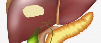
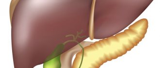
The photo shows tubular systems according to Sinelnikov: 1 – inferior vena cava; 2 – right lobe of the liver; 3 – common hepatic duct; 4 – portal vein; 5 – lymphatic vessels; 6 – common hepatic artery; 7 – left lobe of the liver; 8 – hepatic veins
Segments are sections of the parenchyma that are pyramid-shaped and adjacent to the branches of the second-order portal vein, branches of the hepatic duct, and branches of the hepatic artery. They are located around the gates in radii.
There are eight segments (from right to left counterclockwise from I to VIII):
- Left lobe: caudate – I, posterior – II, anterior – III, quadrate – IV.
- Right lobe: middle upper anterior – V, lateral inferoanterior – VI and lateral inferoposterior – VII, middle upper posterior – VIII.
Segments form larger areas - sectors (zones). There are five of them. They are formed by certain segments:
What are liver diseases?
- Left lateral (segment II).
- Left paramedian (III and IV).
- Right paramedian (V and VIII).
- Right lateral (VI and VII).
- Left dorsal (I).
The outflow of blood occurs through three hepatic veins, which come together on the posterior surface of the liver and flow into the inferior cava, which lies on the border of the right part of the organ and the left.
The bile ducts (right and left), which remove bile, merge into the hepatic duct at the Glissonian hilum.
The outflow of lymph from the liver occurs through the lymph nodes of the Glissonian hilum, the retroperitoneal space and the hepatoduodenal ligament. There are no lymphatic capillaries inside the hepatic lobes; they are located in the connective tissue and flow into the lymphatic vascular plexuses that accompany the portal vein, hepatic arteries, biliary tract and hepatic veins.
The liver is supplied with nerves from the vagus nerve (its main trunk is the Lattarget nerve).
The ligamentous apparatus, consisting of the semilunar, falciform and triangular ligaments, attaches the liver to the posterior wall of the peritoneum and the diaphragm.
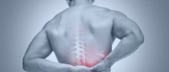
Reasons why pain in the right hypochondrium may occur
Even if you know in which side the liver is located, there is still a risk of missing the first symptoms of its disease. The fact is that the liver parenchyma does not have nerve receptors. Even significant damage will go unnoticed if the outer layer is not damaged. The capsule contains nerve cells, so it is able to capture and transmit pain impulses. All pathologies of this organ, which are accompanied by pain, appear only when the capsule is damaged or stretched under pressure from the parenchyma.
Before the full picture of the disease appears, complaints of general weakness, digestive disorders and headaches appear. When the liver increases in size due to the inflammatory process, pain appears in the right hypochondrium. The next stage, which will indicate congestion in the liver and biliary tract, will be jaundice, often together with itching.
Pain in the liver area may indicate various liver diseases:
- hepatitis (liver inflammation) of infectious, non-contagious, toxic, including medicinal and alcoholic origin;
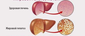
- hepatosis - the appearance of fat inclusions in normal liver tissue;
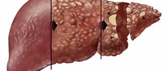
- cirrhosis - destruction and necrosis of areas of the liver parenchyma that are replaced by connective tissue;
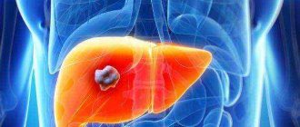
- oncological processes;
- formation of cysts, including parasitic ones;
- the appearance of abscesses.
Human anatomy is a compulsory subject in the school curriculum. Every person should know whether his liver is on the right or on the left. It is also advisable to know at least the approximate location of other vital internal organs. However, it is worth understanding that painful sensations can radiate. If the primary source of pain is located on the left or right, after a while it will respond with pain throughout the body.
Liver topography
The liver is located on the right side under the diaphragm. It occupies most of the upper abdominal cavity. A small part of the organ extends beyond the midline into the left part of the subphrenic region and reaches the left hypochondrium. From above it is adjacent to the lower surface of the diaphragm, a small part of the anterior surface of the liver is adjacent to the anterior wall of the peritoneum.
Most of the organ is located under the right ribs, a small part in the epigastric zone and under the left ribs. The midline coincides with the border between the lobes of the liver.
The liver has four boundaries: right, left, upper, lower. The organ is projected onto the anterior wall of the peritoneum. The upper and lower boundaries are projected onto the anterolateral surface of the body and converge at two points - on the right and left sides.
The location of the upper border of the liver is the right nipple line, the level of the fourth intercostal space.
The apex of the left lobe is the left parasterial line, the level of the fifth intercostal space.
The anterior lower edge is the level of the tenth intercostal space.
The anterior edge is the right nipple line, the costal edge, then it departs from the ribs and stretches obliquely upward to the left.
The anterior contour of the organ has a triangular shape.
The lower edge is not covered with ribs only in the epigastric zone.
In diseases, the anterior edge of the liver protrudes beyond the edge of the ribs and is easily palpable.
Palpation of the pancreas
Palpation of the pancreas is carried out;
- with the patient lying on his back
- in a position on the right side
- in a standing position
First point. The slightly bent fingers of the right hand are placed horizontally 2-3 cm above the found lower border of the stomach to the left of the midline of the abdomen.
https://www.youtube.com/watch?v=yTkeM0R3AZg
Second point. With a superficial movement of the fingers, the skin is shifted upward so that a skin fold is formed in front of the nail surface of the fingers.
Third point. Taking advantage of the relaxation of the abdominal muscles when exhaling, gradually plunge the palpating fingers deep into the abdomen until they reach the back wall.
Fourth point. Without removing your fingers from the posterior abdominal wall, make sliding movements with your hand from top to bottom. If the test result is positive, a formation is detected in the form of a transversely located cylinder about 1.5-2 cm in diameter.
Functions of the liver in the human body
The role of the liver in the human body is great; gland is one of the vital organs. This gland performs many different functions. The main role in their implementation is given to structural elements - hepatocytes.
How does the liver work and what processes occur in it? It takes part in digestion, in all types of metabolic processes, performs a barrier and hormonal function, as well as hematopoietic during embryonic development.
Basic liver functions
What does the liver do as a filter?
It neutralizes toxic products of protein metabolism that come with the blood, that is, it disinfects toxic substances, turning them into less harmless ones that are easily removed from the body. Thanks to the phagocytic properties of the endothelium of the liver capillaries, substances absorbed in the intestinal tract are neutralized.
It is responsible for removing excess vitamins, hormones, mediators, and other toxic intermediate and end products of metabolism from the body.
What is the role of the liver in digestion?
It produces bile, which then enters the duodenum. Bile is a yellow, greenish or brown jelly-like substance with a specific odor and a bitter taste. Its color depends on the content of bile pigments, which are formed during the breakdown of red blood cells. It contains bilirubin, cholesterol, lecithin, bile acids, mucus. Thanks to bile acids, fats are emulsified and absorbed in the gastrointestinal tract. Half of all the bile produced by liver cells goes into the gallbladder.
What is the role of the liver in metabolic processes?
It is called the glycogen depot. Carbohydrates that are absorbed by the small intestine are converted into glycogen in the liver cells. It is deposited in hepatocytes and muscle cells and, when there is a deficiency of glucose, begins to be consumed by the body. Glucose is synthesized in the liver from fructose, galactose and other organic compounds. When accumulated in excess in the body, it turns into fat and settles throughout the body in fat cells. The deposition of glycogen and its breakdown to release glucose is regulated by insulin and glucagon, the hormones of the pancreas.
The liver breaks down amino acids and synthesizes proteins.
It neutralizes ammonia released during the breakdown of proteins (it turns into urea and leaves the body with urine) and other toxic substances.
Phospholipids and other fats needed by the body are synthesized from fatty acids obtained from food.
What function does the fetal liver perform?
During embryonic development, it produces red blood cells - erythrocytes. The placenta plays a neutralizing role during this period.
Due to the variety of functions, it is not entirely clear which organ system the liver belongs to. This is an exocrine gland and is considered an auxiliary organ of the digestive tract.
Innervation
The process of innervation of the liver occurs due to the action mainly of the vagus nerves, as well as those localized in the celiac plexus and the right phrenic nerve.
The splanchnic nerves are responsible for the innervation of the sympathetic type, and the vagus nerves in this case are responsible for the parasympathetic system.
The vagus nerves, the plexuses of which are localized at the hepatic gate, are divided into anterior and posterior hepatic plexuses. The branches that arise from the phrenic nerve are localized along the inferior vena cava. The fibers of this nerve are in the structure of the hepatic plexuses and therefore these fibers become effective sources in the process of innervation of both the liver and the gallbladder.
Pathologies
Liver diseases are caused by its functions. Since one of its main tasks is the neutralization of foreign agents, the most common diseases of the organ are infectious and toxic lesions. Despite the fact that liver cells are able to quickly recover, these capabilities are not unlimited and can quickly be lost during infectious lesions. With prolonged exposure to pathogens, fibrosis can develop, which is very difficult to treat.
Pathologies can have a biological, physical and chemical nature of development. Biological factors include viruses, bacteria, and parasites. Streptococci, Koch's bacillus, staphylococci, viruses containing DNA and RNA, amoebas, lamblia, echinococci and others have a negative effect on the organ. Physical factors include mechanical injuries, and chemical factors include medications with long-term use (antibiotics, antitumor drugs, barbiturates, vaccines, anti-tuberculosis drugs, sulfonamides).
Diseases can appear not only as a result of direct exposure to harmful factors on hepatocytes, but as a result of poor nutrition, circulatory disorders, and other things.
Pathologies usually develop in the form of dystrophy, bile stagnation, inflammation, and liver failure. Further disturbances in metabolic processes: protein, carbohydrate, fat, hormonal, enzymatic depend on the degree of damage to the liver tissue.
Diseases can occur in a chronic or acute form, changes in the organ can be reversible or irreversible.
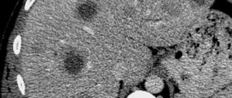
In the course of research, it was discovered that tubular systems undergo significant changes during pathological processes such as cirrhosis, parasitic diseases, and cancer.
Liver failure
Characterized by disruption of the organ. One function, several or all at once may decrease. There are acute and chronic insufficiency, based on the outcome of the disease - non-fatal and fatal.
The most severe form is acute. In acute renal failure, the production of blood coagulation factors and albumin synthesis are disrupted.
If one liver function is impaired, there is partial failure, if several - subtotal, if all - total.
If carbohydrate metabolism is disturbed, hypo- and hyperglycemia may develop.
If fat is disturbed, cholesterol plaques are deposited in blood vessels and atherosclerosis develops.
If protein metabolism is disrupted, bleeding, swelling, delayed absorption of vitamin K in the intestine.
Possible assumptions when moving the border
The displacement of the lower line is an important diagnostic criterion that can be interpreted variably, depending on other indicators. Percussion and palpation during a physical examination, even with the presence of numerous hardware diagnostic methods, have not lost their importance during a physical examination. These manipulations, which have been in practice for several decades, are constantly used and are an important component for the initial diagnosis, assumptions about the nature of the pathology or decisions about relative health:
- In 5 out of 6 subjects, they are quite easy to carry out, both along two lines (the midclavicular muscle on the right and the rectus abdominis muscle of the same dislocation), and along all 5, used if a more detailed examination is necessary. This is difficult to do with ascites: the presence of fluid makes it difficult to palpate the edges and distorts the sound during percussion.
- The combination of simultaneous movement of the upper and lower lines indicates that the organ is pubescent. But if only that edge, which normally protrudes slightly from under the costal arch, is shifted down, we can talk about an increase in the exocrine gland, in relation to the conditional size norm. In this case, the range of possible assumptions is wide - this phenomenon is observed during inflammation and stagnant processes, during acute infectious diseases, at the initial stage of progression of cirrhotic cell changes and in many other cases.
- Ascites, pregnancy, severe flatulence can lead to movement of the upper border higher than normal, but sometimes this is a sign of parasitic infestation (echinococcosis) or liver cancer. It happens that this symptom is not caused by damage to the organ itself - the detected decrease occurs with lung disease - emphysema or pneumothorax, accumulation of gases due to intestinal pathologies.
- If the lower border, on the contrary, has risen in relation to the normal location, it can be assumed that the vital organ has decreased in size, and this is a sign of dystrophy, the so-called wrinkling, a decrease in size (this occurs in the last stage of portal cirrhosis).
Dimensions and location are not the only data obtained using traditional methods. An experienced hepatologist, proficient in both techniques, draws conclusions based on pain, surface irregularities, compactions - consistency features and defects felt by the fingers, which should not normally exist. If the liver is detected to deviate from its usual boundaries, in most cases we can speak of an existing pathology, but further research is necessary for a final diagnosis.
an important organ has decreased in size, and this is a sign of dystrophy, the so-called wrinkling, a decrease in size (this occurs in the last stage of portal cirrhosis).
Cirrhosis. First signs
It’s a paradox, but even a dying liver practically doesn’t hurt. It begins to tingle only when the organ has only 30% of its working area left. But, at the same time, the liver can recover if it has at least a little working surface left.
These symptoms can be called minor. They do not directly talk about the development of cirrhosis, they only hint at problems with the organ that purifies the blood. But if you notice one or a combination of signs, this is a reason to think about it and get checked!
Advice. Almost every woman has spider veins. Meanwhile, this is a clear sign of impending cirrhosis. The brighter the stars, the more actively the tissues die.
"Minor" symptoms:
- hormonal changes in men. Enlarged mammary glands, hair loss, plump buttocks and thighs. The cause of such symptoms is reduced liver function, which cannot cope with the neutralization of female sex hormones;
- changes in finger joints. Nails may become more convex, and joints may become spherical;
- red palms and lips. A shiny tongue of rich crimson color is a clear sign of illness.
One of these signs does not mean anything, but their totality already inspires serious concern and gives reason to consult a doctor and undergo an examination.
What needs to be done?
If at least one of the listed symptoms is familiar, it is important to visit a doctor as soon as possible. Timely and adequate treatment of the liver is a guarantee of further fulfilling life.
First aid for pain
Before going to the doctor, when it is necessary to relieve a painful condition, you can use special medications that relieve spasm and severe pain. Such remedies may be No-Shpa or Papaverine.
No-Shpa
Intramuscular administration of the drug is preferable; injections quickly relieve spasms and alleviate the condition. However, it is necessary to take into account that No-Shpa is not used for liver failure.
The adult dosage ranges from 40 to 240 mg per day, with injections given from one to three times a day. An effective one-time dosage for colic is from 40 to 80 mg.
Papaverine
Papaverine injections are also prescribed to suppress spastic pain. The direct purpose of the drug is to relax the smooth muscles of the digestive and genitourinary systems. Just like in the situation with No-shpa, the use of Papaverine in case of liver failure is contraindicated.
Papaverine can be administered subcutaneously, intramuscularly and intravenously.
- For subcutaneous administration, as a rule, 0.5-2, maximum 5 ml of the drug is recommended, not exceeding the daily dosage of 15 ml;
- The intramuscular dosage for an adult is also from 0.5 to 2 ml, the maximum is 5 ml, the maximum daily dosage is 15 ml;
- Papaverine is administered intravenously dissolved in saline solution, the highest daily volume of the administered drug is 6 ml.
Analgin
It is not recommended to take Analgin on your own, because this can dull sensations, change the signs of the disease, which, in fact, misleads the doctor, and also creates additional stress on the liver.
Analgin by injection relieves pain better; if the doctor deems it necessary, he can prescribe 1 or 2 ml of the drug.
If the pain is intense and difficult to endure, you must call an ambulance and do not refuse transportation to the hospital.