When is it prescribed?
If the doctor suspects the development of a pathological process in the pelvic organs, he will most likely prescribe a transvaginal ultrasound. The reasons for prescribing such an examination may be:
- the need to learn about the fixation of the fertilized egg in the fallopian tube;
- control of the treatment;
- prevention of a whole range of diseases and pathological conditions.
Also, direct indications for prescribing a pelvic ultrasound are:
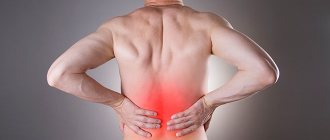
- pain in the abdomen;
- searching for the causes of infertility;
- control of follicle maturation;
- hormonal disorders, including cycle disorders;
- inflammatory processes in the appendages;
- problems with the patency of the fallopian tubes;
- bleeding;
- fibroids and cysts;
- endometriosis;
- monitoring the presence of pregnancy after artificial procedures such as ICSI, IVF;
- suspicion of pathological attachment of the fetus (outside the uterus);
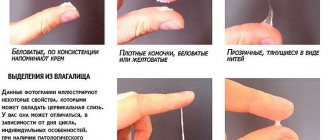
- abnormal discharge;
- oncological neoplasms;
- the need to clarify or confirm a previously made diagnosis.
There are certain restrictions on performing a transvaginal procedure. So, this type of examination is not suitable for girls who are not sexually active, pregnant women whose term exceeds 12 weeks, and women with pelvic inflammation . Most likely, the doctor will recommend a transrectal ultrasound.
Our doctors
General Director Margarita Beniaminovna Anshina Make an appointment
Obstetrician-gynecologist-reproductologist Anna Anatolyevna Smirnova Make an appointment
Director of the Rizhinashvili clinic Semyon Iosifovich Make an appointment
Obstetrician-gynecologist-reproductologist Zhordanidze Diana Omarovna Make an appointment
Obstetrician-gynecologist-reproductologist Torchinov Aslanbek Ruslanovich Make an appointment
Obstetrician-gynecologist, oncologist Kira Evgeniy Fedorovich Make an appointment
Obstetrician-gynecologist, reproductive specialist. Efimova Maria Sergeevna Make an appointment
Obstetrician-gynecologist-endocrinologist Sharyafetdinova Failya Abdulkhaevna Make an appointment
Oncologist-mammologist Victoria Borisovna Akimova Make an appointment
Obstetrician-gynecologist Pitskhelauri Elena Germanovna Make an appointment
Obstetrician-gynecologist-reproductologist Olga Aleksandrovna Iskorostinskaya Make an appointment
Urologist-andrologist Iskorostinsky Evgeniy Vladimirovich Make an appointment
Embryologist Vladimir Viktorovich Ovsyankin Make an appointment
Perinatologist Nelly Idrisovna Kokhno Make an appointment
Therapist Alexandra Vladimirovna Kabakova Make an appointment
Ultrasound diagnostics doctor Olga Ivanovna Babaeva Make an appointment
Urologist-andrologist of the highest qualification category Gablia Mikhail Yurievich Make an appointment
Urologist Kondratenko Vladimir Ermolaevich Make an appointment
Psychologist Barskaya Ekaterina Sergeevna Make an appointment
Obstetrician-gynecologist Margarita Yuryevna Skvortsova Make an appointment
Endocrinologist Anna Zakarevna Sarkisyan Make an appointment
Obstetrician-gynecologist Ignatenko Olga Viktorovna Make an appointment
Obstetrician-gynecologist Pushkina Valeria Vadimovna Make an appointment
Obstetrician-gynecologist Olga Borisovna Kuharkina Make an appointment
Endocrinologist Anzhelika Vladimirovna Manovitskaya Make an appointment
Anesthesiologist-reanimatologist Isaev Oleg Vladimirovich Make an appointment
Hemostasiologist Lopukhin Vadim Olegovich Make an appointment
Head of the Embryology Laboratory. Ph.D. Sergeev Sergey Alexandrovich Make an appointment
Embryologist Kalinina Irina Ivanovna Make an appointment
Embryologist Matveeva Elona Olegovna Make an appointment
Embryologist Kalyuzhny Sergey Andreevich Make an appointment
Preparation
Before visiting the ultrasound room, you need to prepare a diaper or towel to lay on the trestle bed (if the procedure is carried out for a fee, the provision of a disposable sheet is usually included in the cost of the ultrasound).
Preparation for an ultrasound examination includes taking medications that reduce gas formation (smecta, filtrum, activated carbon and others). On the eve of the ultrasound, it is advisable to limit the consumption of foods that can cause flatulence.
Reference! It is not necessary to fast before a pelvic examination.
If you need to be examined urgently, you can do without preparation, but the content and accuracy of the procedure will suffer.
Transvaginal ultrasound: indications
Ultrasound through the vagina is performed on all women who have become sexually active. For virgins, transabdominal (through the anterior wall of the abdomen) or transrectal (through the rectum) types of ultrasound examination of the reproductive organs are used.
Indications for transvaginal ultrasound examination:
- Abdominal pain.
- Bloody vaginal discharge between periods.
- Menstrual irregularities (too infrequent or frequent, too long or short, painful, scanty, heavy menstruation, etc.).
- Diagnosis of pregnancy (uterine and ectopic).
- Infertility.
- During an examination of a patient, a gynecologist discovered an enlarged uterus, appendages, and pathological mass formations in the genital area.
- Preventive examination (ultrasound examination is not harmful to health, so it can be done regularly, like a regular gynecological examination).
How do they do it?
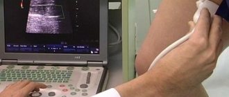
Scanning of the reproductive system organs occurs through a sensor that is inserted into the woman’s vagina.
Before the examination, you need to be naked from the waist down and take a comfortable position on the couch. The doctor puts a medical condom on the sensor, then lubricates it with gel and, after insertion, begins examining the internal organs. The sensor is a plastic rod with a diameter of 3 cm and a length of about 12 cm with a beveled handle. At its end there is a channel with a needle for collecting material during a biopsy.
Internal examination of the uterus and adjacent pelvic organs greatly simplifies the diagnosis, which is why the procedure has received such wide recognition in medical circles.
Important! If necessary, the study can be done many times without harming the patient's health.
Ultrasound using a vaginal probe is considered more informative and meaningful than traditional examination through the abdominal wall. During the manipulations, the patient should feel quite comfortable, but if unpleasant sensations arise, they should be reported to the doctor immediately.
Video. Transvaginal pelvic ultrasound: order of execution.
How is a vaginal ultrasound performed?
You come to the diagnostic room. While the specialist gets acquainted with your chart, you take off your clothes as during a gynecological examination. Depending on the arrangement of the ultrasound room, transvaginal ultrasound is performed either on a gynecological chair or on a regular couch, but you will only need to lie down and bend your knees.
The transducer (ultrasound device sensor) is not very large, its length is 12 cm, and its diameter is about 3 cm. A condom is put on it, and then a special gel is applied to conduct ultrasonic waves. Also, with this gel it will be easier to insert the sensor; it performs the task of the lubricant. Then the doctor will move the sensor, then up, then down, then to the sides. This is done for a more accurate examination of the internal organs. If you experience severe pain during a transvaginal ultrasound, immediately inform your doctor. Typically, an ultrasound does not take much time, about 20-30 minutes.
Norms and decoding
Transvaginal ultrasound provides a unique opportunity to timely detect the development of a number of diseases of the female genital organs, both inflammatory and oncological etiologies. And when used together with Doppler ultrasound (Dopplerography) is an excellent opportunity to timely identify the potential risk of thrombosis , find out about the presence of atherosclerosis and examine blood flow in the pelvis.
The ability of ultrasound waves to detect pregnancy at its very beginning is of great importance for women awaiting IVF results.
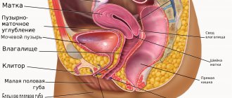
Normal ultrasound results of the reproductive area look like this:
- Uterus. Normally, it is deviated anteriorly, outlined evenly and clearly. Parameters: 71 mm (length) x 62 mm (width) x ~40 mm (diameter). The tissue is homogeneous in density, the thickness of the inner layer of the mucous membrane ranges from a few millimeters to 1-2 cm.
- Cervix. The normal length is about 4 cm, the anteroposterior size is about 2.5 mm, the structure is homogeneous. On the eve of menstruation, the cervical canal fills with fluid (mucus).
- Ovaries. The length of each organ is 30 mm, width – 25 mm, thickness – 15 mm. Lumpy contours can be visualized, tissue density is mostly homogeneous, fibrous areas are acceptable. Several follicles are observed, among which the dominant one stands out.
- Fallopian tubes. Normally, they do not differ on ultrasound or are barely noticeable.
- Free fluid – on days 13-15 of the cycle can be observed in small quantities, this is not a pathology.
Preparing for a transvaginal ultrasound
First of all, it is necessary to decide on the date of the study, since during the menstrual cycle the functional state of the reproductive organs and, accordingly, the ultrasound picture change.
The most optimal time for a scheduled ultrasound is 8-14 days of the cycle. However, in each clinical situation it may be different - for example, if severe pain or bleeding occurs, no one will postpone the study, especially since bleeding from the genital tract is not a contraindication for transvaginal ultrasound. When studying the functional capacity of the gonads or determining the type of cyst found in the ovary, it may be necessary to repeat ultrasound scans over several menstrual cycles.
Therefore, in order to get the most accurate results from the diagnosis, questions related to the dates of the ultrasound are best discussed with your gynecologist.
As for the immediate preparation for transvaginal ultrasound, it is as follows: on the eve of the procedure, it is advisable not to pass and empty the intestines, and if there is a tendency to bloating, drink espumizan (in the doses indicated in the instructions for the medicine) to reduce discomfort. And, of course, a woman should take care of genital hygiene.
To summarize, I would like to note that transvaginal ultrasound is the optimal way to effectively examine the female reproductive system without resorting to expensive and unsafe research methods.
Zubkova Olga Sergeevna, medical observer, epidemiologist
14, total, today
( 66 votes, average: 4.55 out of 5)
Torsion of the pedicle of an ovarian cyst: symptoms and treatment
Cervical cancer
Related Posts
Which days of the menstrual cycle are suitable for transvaginal ultrasound?
Transvaginal ultrasound in the area of the uterus and pelvis is mainly performed in the first days of the cycle, immediately after the end of menstruation. That is, it is recommended to choose days 5 to 8 of the cycle if we are talking about a routine scan. If the doctor suspects that a woman has uterine endometriosis, the procedure is postponed to the second part of the cycle. If there are inflammatory diseases of the pelvic organs, or you need to monitor the dynamics of follicle development, then the procedure is carried out several times in one cycle. If a girl starts bleeding, which is definitely not menstruation, then an ultrasound is done urgently any day.
How to prepare for a pelvic ultrasound
A few days before the procedure, it is necessary to exclude gas-forming foods from the diet - brown bread, legumes, milk, fresh vegetables, so that the accumulation of gases in the intestines does not interfere with the diagnosis.
On the day of the transabdominal ultrasound, you should drink about a liter of fluid and try not to urinate before the examination. Transvaginal ultrasound usually does not require preparation or is performed with an empty bladder.
Your doctor will tell you more about preparing for ultrasound diagnostics.
ON CLINIC is currently one of the best clinics in Russia in terms of medical equipment, team of professionals and working methods used. We invite you to see this for yourself!
Happy stories
Maria
Dec 10 2019
Two years of unsuccessfully visiting doctors with my husband. We were advised to contact Zhordanidze Diana Omarovna in Fertimed. Wonderful doctor! Rezul
Read the story
Alipat
Oct 21 2019
Thanks to you, your clinic and Anna Anatolyevna Smirnova, I came to my cherished dream after 7 years of wandering from one doctor to another. Thankfully, I found out about
Read the story
Natalia
09 Oct 2019
I'll write a story too. 7 years of infertility, 3 IVF attempts. Out of desperation, I go and ask for a referral for laparoscopy. And in the intensive care unit I got into a conversation with a woman. TO
Read the story
Natalia
20 Feb 2019
Hello. I want to express my deep gratitude to your center. I got the impression of a close-knit team focused on results. Were treated by
Read the story
Andrey and Alexandra
14 Feb 2019
Many thanks from our entire large family to all the employees of the center for your work, for the smile on your face and faith in a positive result! Mihai
Read the story
Valentina
15 Jan 2019
My husband and I’s path to the happiness of being parents began in 2012 after the sounds of Mendelssohn’s wedding march died down. Like everyone else, we thought
Read the story
Catherine
May 16, 2019
After marriage, I couldn’t get pregnant at the age of 22; it turned out to be problems with the fallopian tubes. When I found out that I could never have anything on my own
Read the story
Olga
02 Mar 2019
I would like to express my deep gratitude to the FertiMed clinic, and especially to doctor Aslanbek Ruslanovich, for his good, caring attitude, and also for his
Read the story
Irina
23 Jun 2018
I would like to express my deep gratitude to the wonderful doctor of the FertiMed clinic, Aslanbek Ruslanovich Torchinov, for our long-awaited son. On hold
Read the story
Yuri
May 15, 2018
It will probably be an unusual review, since this is a review from a new father, and in general, I don’t really understand how I can express ours to you,
Read the story
Catherine
15 Dec. 2017
We first turned to FertiMed back in 2010, after 3 years of visiting doctors and undergoing all sorts of exotic tests. Unfortunately, the first
Read the story
Catherine
25 Sep. 2017
I got married at 22, my husband was 23, I never thought that there might be problems with conception, it turned out after I had appendicitis in childhood.
Read the story
Elena
May 23, 2017
My husband and I dreamed of having a child for 6 years. I have an adult daughter (20 years old) from my first marriage; my husband had no children. Passed several attempts at Eco in p.
Read the story
Elena
27 Feb 2017
We are happy parents! Thank you very much from the bottom of my heart to the FertiMed clinic and personally to our doctor Diana Omarovna for what you do, for helping
Read the story
Nina and Peter
27 Sep. 2017
My husband looked at his spermogram as if it were a death sentence - not a single sperm count. And the reason is unclear. There was only one hope - for TEZU. It was explained to us that
Read the story
Faith
18 Jul 2016
I was ready to do anything to have a baby. By the age of 32, I had already undergone five operations on my ovaries and only one tiny piece remained from them. Nasty
Read the story
Olga
29 Jul. 2016
I never thought there would be problems with the children, but it turned out that both pipes were impassable. The first IVF attempt at FertiMed ended in birth
Read the story
Maria
27 Jun 2016
I had two ectopics and no chance of having my baby. The irony was that my closest
Read the story
Valentina and Mikhail
Oct 14 2016
My husband and I flew from Kamchatka. They really wanted a second child. FertiMed was recommended by friends. They say that small and thin women like me have
Read the story
Sophia
27 Jul 2016
At FertiMed, at first they didn’t believe that I had 43 IVF attempts. There was clearly distrust and sympathy in the eyes of the staff - they say the woman went crazy.
Read the story
Alya
30 Sep. 2016
When I left my husband, with whom we had three children, no one, of course, could understand this, and no one wanted to. And even more no one could understand what m
Read the story
Valentina and Victor
01 Nov 2016
Our Dasha is growing up, at 1 year and 9 months she is a very smart girl, learning the alphabet on her own tablet (a real one, not a toy one)
Read the story
Christina
05 Apr 2016
I was 45 when I was ready for IVF. At the first meeting, the doctor at FertiMed said: there is no chance of having a child with my own eggs. At first there were sh
Read the story
Yana
13 2016
I am 29 years younger than my husband. He has three adult children, but I really wanted my child and ours together. Over the past few years, my husband has been very
Read the story
Anna
01 Mar 2016
I'm a happy mom! 5 years of standing with a birch tree, measuring basal temperature, tracking ovulation, hysteroscopy, laparoscopy (for my husband too), clostil
Read the story
Cost of services
| Name of service | price, rub. |
| Ultrasound of the pelvic organs transabdominal | 1800 |
| Ultrasound of the pelvic organs transvaginal | 2500 |
| Two-stage ultrasound of the pelvic organs | 3300 |
| Recording an ultrasound examination on DVD | 350 |
Dear patients!
You can view the full list of services and price list at the reception or ask a question by phone.
The administration tries to promptly update the price list posted on the website, but in order to avoid possible misunderstandings, we advise you to clarify the cost of services on the day of contact at the reception or call center by calling 8(495)223-22-22
. The posted price list does not constitute an offer.
Transvaginal scan during pregnancy
TVUS is also used in obstetrics and gynecology to visualize the uterus and embryo in the early stages of pregnancy.
This research method allows you to see the presence of pregnancy and the characteristics of the patient’s condition.
Typically, the intravaginal method is prescribed when determining a woman’s health before undergoing IVF or ICSI.
In the first trimester, it is included in mandatory screening to check for the absence of fetal developmental anomalies, diagnose the health of the uterus and adjacent anatomical structures, and determine various risks for the pregnant woman.
Diagnosis of gynecological diseases
Transvaginal ultrasound is performed to detect a wide range of gynecological diseases in women:
- intrauterine synechiae;
- polycystic ovary syndrome;
- polyps of the uterine body;
- hyperplasia of the inner muscular layer of the uterus (endometrium);
- uterine fibroids;
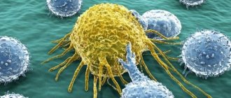
- ovarian tumors;
- fallopian tube cancer;
- torsion of ovarian cyst;
- uterine perforation and other diseases.
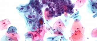
When scanning the endometrium, it is necessary to take into account the phase of the menstrual cycle. In this case, it is recommended to do an ultrasound on days 5-7 of the monthly cycle. In the absence of deviations from the norm, the endometrium is visualized on photographs as a homogeneous structure with smooth contours. Signs of endometrial pathology on an echogram are the following:
- heterogeneity of structure;
- thickening or thinning of the muscle layer;
- the presence of formations that poorly reflect ultrasonic waves.
For postmenopausal women, with an endometrial thickness of more than 5-8 mm (normally it should be 4-5 mm), a biopsy is indicated. If the patient takes hormonal drugs, the thickness of the endometrium is assessed individually. Curettage with histological examination of the biopsy specimen is also carried out in case of uneven contours of the uterus and the presence of inclusions in its cavity. For a more informative diagnosis of uterine polyps and fibroids, transvaginal echography with the introduction of a contrast agent (hydrosonography) is used, but it does not replace hysteroscopic examination. A reliable assessment of the presence of fibroids using hydrosonography is 100%.
The study of small tumor-like formations in the ovaries is carried out using the transvaginal method, and large ones - by the transabdominal method. When scanning, the size of the formation is measured, its structure, the presence of septa, changes in blood supply, thickness and structure of the tumor capsule are assessed. The ovaries are visualized in the image as a darkened structure.
One of the emergency conditions that require a transvaginal ultrasound is bleeding from the female genital organs that is not associated with the menstrual cycle. The examination allows you to determine the source of bleeding - uterine or ectopic (changes in the vagina or cervix) and its cause.
To diagnose fallopian tube cancer, a transvaginal ultrasound and abdominal scan are performed. On echography, the affected fallopian tube is visualized as a sausage-shaped tube with thickened walls and prominent growths. The more complex the structure of the fallopian tube, the more likely the formation of a malignant tumor.
Transvaginal ultrasound should be performed in women with diagnosed uterine fibroids twice a year. If an operation was performed to remove a pedunculated myomatous polyp arising through the cervix, then the examination is carried out after surgery 4 times during the first year, and then 2 times a year.
Which examination method is best for men?
A pelvic examination is usually prescribed for females, but men also periodically face the need to examine organs located in the pelvis. To examine men, a specialist individually selects a method and determines which method is best to use in a particular case. For men, two research methods are also considered acceptable:
- Transabdominal. The rules for its implementation are standard for all cases: it is carried out through the outer surface of the abdomen at the moment the bladder is completely filled. However, this method is not very suitable for studying the prostate gland. This examination is also less effective for men who are overweight or have problems with urinary incontinence.
- Transrectal (TRUS). Ideal for examining the prostate gland because the prostate is small in size and is in close contact with the inner wall of the rectum. Therefore, the transrectal technique makes it possible to assess the condition of the prostate with high accuracy.
Loading...
Share with friends!
When is it necessary to perform an ultrasound and in what cases is the study prescribed?
A vaginal ultrasound may be prescribed by a gynecologist as a preventive examination. Also if you suspect the development of diseases of the internal genital organs in women. If some alarming symptoms appear, the gynecologist prescribes diagnostics to identify the causes of discomfort.
It can be:
- Drawing and cutting pain in the lower abdomen;
- Uterine bleeding during menstruation and in the intermediate cycle;
- Delayed menstruation (determines the presence of uterine or ectopic pregnancy).
Also, such diagnostics determine the functionality of the ovaries, their size, and the presence of formations and cysts.
Interpretation of examination results
During the examination, the specialist takes parameters of the uterus, assesses the echogenicity and condition of the internal organs. This way he is able to determine the presence of pus, cysts, neoplasms (fibroids, cancer), polyps, cystic hickeys, ectopic pregnancy and other pathological processes.
You can get more information about the study or sign up for it by contacting the consultant of the FertiMed Center for Reproduction and Genetics at the indicated numbers.
Don't waste time! Make an appointment now!
By clicking the “Submit Application” button, you consent to the processing of your personal data in accordance with the terms
Privacy Policy and User Agreement
* - required fields
How is the procedure done?
The methodology also depends on the type of study. Before visiting a doctor, you should find out how a pelvic ultrasound is performed:
- Abdominal. The patient frees his stomach from clothes and sits on the couch. Acoustic gel is applied to the lower abdomen. Using a sensor, the uterus, appendages, fallopian tubes, and bladder are examined.
- Vaginal. The patient lies down on a couch or gynecological chair and bends her legs. A medical condom is placed on the sensor, it is inserted to a shallow depth, and an examination is carried out.
- Rectal method. The patient frees the lower part of the body from clothing. Lies on the left side, bends the legs, pulls them towards the stomach. A medical condom is put on the sensor and inserted into the anus. During the procedure you need to relax as much as possible, then discomfort will be minimized.