Author of the article
Anatoly Shishigin
Reading time: 3 minutes
AA
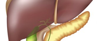
The urinary system in the human body consists of a pair of ureters, two kidneys, a bladder and a urinary canal. The structure of the ureter in the female and male body is different, but in both it is a hollow tube up to 30 cm long. The organ is designed to transport urine from the pelvis of the kidneys to the bladder. Its movement occurs with the help of muscles in the walls of the bladder.
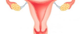
In women, the ureter is located in the outer part on the side where the uterus is located. At the same time, it crosses the uterine artery and enters the bladder near the lateral section of the vagina, near its top. Such anatomical features in the structure of the genitourinary system are taken into account by operating surgeons during gynecological and urological manipulations.
Characteristics of the pathology
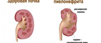
Urolithiasis is a fairly common disease. Its appearance is provoked by many different factors. Most often, pathology occurs against the background of poor nutrition and unsatisfactory quality of drinking water. Initially, stones form in the kidneys.
Most patients do not even suspect the presence of stones for a long period of time. After all, signs of the disease do not appear immediately. Meanwhile, the stones “grow” in the kidneys. And as a result of certain factors, stones may appear in the ureter.
What are the symptoms in women? This is, first of all, severe pain. It indicates renal colic (indicates the sinking of a stone into the ureter). In such situations, you should immediately consult a doctor.
Reasons for development
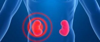
The kidneys play an important role in the human body; they serve as a filter. Blood is passed through them, and unnecessary, harmful toxins or waste are cleansed. Then urine begins to form. The secretions move through the ureters, filling the bladder. The urethra serves as the urethra.
Men and women are susceptible to inflammatory processes of this kind. Most often this occurs due to damage to the walls of the ureter. These can be kidney stones that are large in diameter.
In females, this kind of inflammation provokes the development of cystitis, urethritis, and pyelonephritis. At a time when the disease is just beginning to develop, symptomatic manifestations of the disease that provoked the process are observed.
A very important point, early diagnosis will help avoid further complications without affecting other important organs. If the channels become clogged, this threatens stagnation of urine, the development of infectious diseases, and sepsis. There are cases of untimely treatment, when severe cases and complications can lead to death.
For preventive purposes, it will be enough to undergo annual examinations to monitor your health. People who suffer from diseases of the urinary system are at risk.
Among the most common causes of inflammation of the ureter - urethritis, there are 9 main ones:
- Inflammatory processes in the kidneys, for example, pyelonephritis, pyelitis;
- colds that contribute to the development of cystitis;
- defective innervation processes;
- kidney stones, their difficult passage;
- development of inflammation in the urethra;
- pain can move from the groin, abdominal area to the external part of the genital organs;
- frequent urination;
- the patient feels nausea and vomiting;
- a person experiences increased blood pressure.
Penetration of stone into the ureter
Stones usually form in the renal pelvis. However, there are cases where stones have formed in the ureter. Symptoms in women and treatment are points that must be discussed with a doctor. Fighting pathology on your own is completely unacceptable.
So, if a stone forms in the kidney, why does it end up in the ureter? Many different factors can provoke such a movement. Most often this occurs as a result of the following reasons:
- carrying heavy objects;
- long bumpy ride;
- drinking plenty of fluids and food;
- horseback riding.
It is very important to remember what signs appear if stones are localized in the ureter. Symptoms in women indicating the advancement of a stone manifest themselves in the form of severe pain. Acute discomfort appears in the abdomen and back. This condition is called renal colic.
Developmental anomalies
The most common anomaly occurs in which two tubes emerge from the left kidney to drain urine. Also, there may be two holes for exiting the bubble, or one. Treatment of such an anomaly can only be done surgically, and not in all cases successfully. Duplication of the left kidney also occurs, which also requires surgical intervention.
A pregnant woman often experiences renal colic, and the ureteral valve or its middle part is affected. To determine pain, the specialist uses two points by palpation. One of them is located in the rectus abdominis muscles, and on the outside they are located near the navel. The second is located on the same muscles below, and their doctors find them, focusing on the protrusions of the iliac bones in the pelvic area.
In a pregnant woman, such landmarks may not be sufficiently informative, since the urinary tubes are pushed aside by the uterus and the developing fetus in the body. If there is a stone in the ureter, especially in the distal section, it becomes greatly enlarged and becomes overstretched, accompanied by pain.
More on the topic: What causes ureteral stricture?
https://youtu.be/Dd8ttQb6F4g
Irradiation of paroxysmal pain occurs in the external genitalia and groin. At the same time, the patient cannot find a position for herself in which the pain subsides. Non-narcotic analgesics are not able to relieve pain, so you need to contact a gynecologist or therapist who can prescribe an effective drug regimen to relieve symptoms and treat the cause.
Causes of the disease
Ureteral calculi are formed from various substances:
- uric acid;
- cystine;
- calcium phosphate;
- struvite.
Most often, the process of stone formation is influenced by the following factors:
- Genetic predisposition. Doctors say that the disease is more often diagnosed in patients who have a family history of urolithiasis.
- Impaired outflow, stagnation of urine. The development of the disease may be based on congenital pathologies. Most often, the disease is provoked by narrowed ureters in women, their underdevelopment, kinks or abnormalities of the bladder.
- Chronic urinary tract diseases. Infectious diseases can lead to the development of pathology. For example, pyelonephritis.
- Disturbed exchange. Acquired or congenital ailments may be accompanied by the penetration of lithogenic substances into the urine - calcium (if hyperparathyroidism is diagnosed), urate (in the case of gout).
- Diseases of the digestive system. If the absorption function is impaired, stones may form.
- Use of medications. Some medications can lead to the development of the disease. For example, such consequences are provoked by uroseptics from the nitrofuran category.
Doctors say that uroliths often form in women living in hot and dry climates. High-calorie foods rich in animal proteins can trigger the development of the disease.
Dimensions
In an adult, the length of the ureter is 28-34 cm. It depends on growth and is determined by the height of the kidneys when they are laid in the embryo. In women, the length of the organ is 2-2.5 cm shorter than in men. The right ureter is one centimeter shorter than the left one, since the location of the right kidney is slightly lower.
The lumen of the tube is not the same: narrowings alternate with areas of expansion. The narrowest parts are:
- next to the pelvis;
- at the border of the abdominal and pelvic sections;
- when entering the bladder.
Here the diameter of the ureter is 2-4 mm and 4-6 mm, respectively.
In diagnosis, pathological changes are determined segment by segment
Segments are distinguished between the narrowed areas:
- above – pyelourethral segment;
- area of intersection with iliac vessels;
- lower – vesicoureteral segment.
The abdominal and pelvic sections of the ureter differ in lumen:
- in the area of the abdominal wall it is 8-15 mm;
- in the pelvis – uniform expansion of no more than 6 mm.
However, it should be noted that due to the good elasticity of the wall, the ureter is capable of expanding up to 8 cm in diameter. This ability helps to withstand urinary retention and congestion.
In cross section, the lumen of the organ has a star-shaped shape.
Symptoms of the disease
There are cases when severe pain is not caused by stones in the ureter. Symptoms in women that characterize the movement of a stone completely depend on its size and shape. Stones not exceeding 2 mm in diameter can move painlessly along the ureter. In this case, there may be no symptoms. The woman will not even know about the unpleasant pathology in the body.
But most often large stones are observed in the ureter in women. Signs of pathology are provoked by stone getting stuck.
In this case, the symptoms are pronounced and are called renal colic:
- Sharp, severe pain localized in the lumbar region. In women it spreads to the perineum and labia.
- Urination may be impaired. But such a sign is observed extremely rarely and characterizes the simultaneous passage of stones from both ureters. Most often, women experience a frequent urge to urinate.
- The urine contains blood and the inner epithelium of the kidney. Such symptoms appear as a result of damage to the ureter by the sharp edges of a calculus. If the stone completely blocks the path, then there will be no such sign, since urine flows only through a normal ureter not affected by the disease.
- Sweating, chills. There is an increase in temperature to 37–37.5 degrees. The pathology may be accompanied by nausea, flatulence, and often vomiting.
The calculus usually advances periodically. This leads to the fact that painful symptoms in a woman appear and then disappear. Such colic can be annoying for several hours or days.
Dimensions and blood supply
The anatomical average norm for an adult is considered to be a size from 28 to 34 cm. The length of this organ is determined at the stage of embryonic development and largely depends on the height of the site of kidney formation in the embryo.
The ureter in men is always longer than in women, by 2–3 cm, and the right tube of the organ in all people is shorter than the left by 1–1.5 cm, since the development and activity of the left kidney in the body is always higher.
The lumen of the tube cavity is also different; in cross-section, the organ resembles an accordion. The most significant narrowing of the internal lumen is located:
- at the end of the abdominal part and the beginning of the pelvic part;
- behind the pelvis;
- when passing into the bladder.
Structure of the bladder
It is these parts of the ureter that are most often susceptible to various pathologies, congestion, and infections. The diameter of the narrowest parts of the organ varies from 2 to 4 mm, and is capable of expanding to 6–8 mm.
The abdominal and pelvic parts of the organ differ in the diameter of the lumen in the internal cavity:
- behind the abdominal wall the largest diameter of the lumen is from 6 to 8 mm, and this part can expand up to 12–14.5 mm;
- The ureters passing through the pelvis are no wider than 4 mm, with an expansion of up to 6–8 mm.
All parts of the organ are nourished and filled with arterial blood. The vessels are located in the adventitia, that is, the outer part of the membrane, and from them capillaries pass into the organ.
In their upper part, the arterial branches come from the renal artery. The middle section is connected by the abdominal aorta and the common iliac internal artery. The lower section is fed by branches of the iliac artery, such as:
- uterine;
- cystic;
- rectal.
In the abdominal part, the plexus of vessels is located in front of the organ, and in the pelvic area - behind the organ.
As for venous blood flow, it is provided by the veins of the same name, located near the arteries. The lower section of the organ “drains” blood into the internal iliac veins, and the upper section into the testicular veins.
The flow of lymph is provided by the lumbar and internal iliac lymph nodes.
Symptoms of the pathology, depending on the location of the stone
Most often, the stone is found at the site of narrowing of the ureter. This is the area where the renal pelvis connects to the canal. This area is called the pyeloureteral segment. The next area in which a stuck stone is often diagnosed is the area where the ureter passes from the large to the small pelvis. Another “dangerous” area is the connection of the canal with the bladder.
If a stone blocks the ureter in the upper zone in women, the following symptoms are observed:
- severe pain appears in the lower back;
- acute discomfort is wave-like, sometimes subsiding, sometimes intensifying;
- changing body position does not reduce the intensity of pain;
- discomfort covers the lateral areas of the abdomen.
The localization of the stone in the middle zone of the canal is indicated by the following signs:
- pain is acutely felt in the lateral region of the abdomen (below, along the edge of the ribs);
- discomfort extends to the groin and ileum.
If the stone has descended into the lower part of the ureter, then the woman’s symptoms appear as follows:
- the pain is localized in the lower abdomen and groin area;
- severe discomfort covers the outer labia;
- urination becomes more frequent;
- there is a feeling of fullness of the bubble;
- the process of urination does not bring relief (there is no feeling of emptying).
Structure and development of the ureter
The mucous membrane has a special opening in the ureter called the orifice. A mucous fold has formed near the bladder. Peristalsis of the ureteral region is determined by the presence of muscle fibers, which are muscle bundles located lengthwise, obliquely and transversely. The mucous membrane of the organ consists of elastic fibers that form folds.
The outer part consists of the adventitia and fascia. The anatomy of the ureter has three layers: muscular, external and mucosal, which consists of connective tissue with lymphatic and blood vessels. There is also an epithelial layer called the urothelium that can stretch and surround the organ. Directly below the urothelium is the submucosa.
X-ray examination reveals the existence of 2 types of curvatures, differing in planes. Normally, in the pelvic region the curvature goes to the lateral side, and in the lumbar region - to the medial side. There are rare cases of anatomical structure in which there is no curvature at all.
When the glomerular vessels are dissected by the organ, some narrowing is formed, at the same time there are also gaps characterized by expansions. This point is very important for diagnostic examinations, since it is in narrow places that congestion can form due to stones and other concretions getting into them. There is also a narrowing at the junction with the bladder. Near its entrance there are special ureterovescial valves that prevent reflux.
Cases of congenital anomalies are very rare, but do occur. The most common are atresia, when there is no ureteral tube at all or there are no holes in the ureteral canal, megaloureter, characterized by strong expansion without narrowing along the entire length, or ectopia, when the ureter is connected to the intestine without the participation of the bladder. It is also possible that it is abnormally attached to the genital organs, internal or external.
More on the topic: What complications occur after installing a stent in the ureter?
Diagnostic methods
To make sure that severe discomfort is caused by the movement of a stone along the ureter, the doctor will conduct an initial examination. It involves palpation.
Then the patient will be prescribed more accurate studies:
- urine analysis, determining protein, salts, pus, blood cells;
- bacterial sowing;
- urine analysis to study its acidity;
- X-ray examination;
- blood analysis;
- urography;
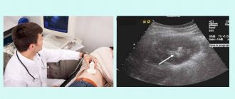
- Ultrasound of the urinary tract;
- Kidney CT;
- radioisotope diagnostics.
A complex of such examinations makes it possible to determine the location of the stone, identify the sources of the disease and select adequate therapy.
Characteristic symptoms
Bacteria and viruses develop and contribute to the occurrence of urethritis. The chronic manifestations of this disease are vivid and difficult to miss. In cases of the acute phase, renal colic is observed. They are accompanied by pain of an acute, excruciating nature. Its location is the lower back, as well as the abdominal part. Painful and uncomfortable sensations appear when the stones begin to move. There may be discomfort in the pubic area. When the disease spreads to the bladder, cystitis develops.
To summarize, the following are the causes of the inflammatory process in women and men:
- A sharp, unpleasant odor of urine, its color becomes cloudy, with bloody particles;
- pain sensations are predominantly in the abdominal area from top to bottom, the location of the pain depends on which ureter has inflammation;
- the patient has an elevated temperature that lasts for a long time;
- the patient feels constant weakness, uncontrolled loss of strength, rapid, unreasonable fatigue.
Treatment options
If during diagnosis stones are identified in the ureter in women, only a competent specialist can decide how to remove them.
Treatment methods depend on the complexity of the situation and the size of the stone. Depending on these factors, they can develop in 2 directions:
- Conservative expectant therapy. It is undertaken in cases where the stone in diameter does not exceed 2-3 mm and does not clog the duct. In this case, there is a high possibility of the stone coming out on its own.
- Active treatment. Used if conservative therapy is not possible or has not yielded positive results.
Anatomical characteristics
In the body of an adult, the orifice is anatomically located in the central part of the bladder, forming minor folds in the tissue walls of the organ, consisting mainly of muscle tissue. In the middle between the ureters there is also a fold formed by smooth muscle, which is the so-called triangle of the bladder or a small area of tissue consisting exclusively of mucous membranes.
The orifice of the ureter is the narrowest section, which predisposes to blockage of the lumen during the formation and subsequent release of stones, that is, sand and stones. This pathological process is accompanied by painful sensations and can lead to the development of a number of complications.
The length of the ureter can vary somewhat and is usually from twenty-eight to thirty-two centimeters. At the same time, the sizes of the right and left elements of the urinary system also differ, and this is due to the fact that the kidneys are located at different levels.
The diameter of the ureter also has different numerical values. The orifice, for example, is one of three anatomical narrowings, each of which is characterized as a segment at risk of blockage by stones. It should also be noted that at rest the diameter of the mouths is no more than a millimeter, but against the background of active activity the value increases slightly, as a rule, up to three millimeters.
There are three conventional sections of the ureters, the anatomical features of which may differ slightly depending on gender and location:
- Abdominal. Normally, this section of the ureter is located at the very beginning, in close proximity to the outer walls of the muscle tissue of the lumbar region.
- Left. The location of this section is as follows: the posterior surface of the bend located between the duodenum and jejunum.
- Pelvic. The location of the given segment of the ureter in women is characterized by the following: on the front side of the ovaries, passing behind the wall of the uterus and located between the tissues of the bladder and the vagina. The ureters in men pass in close proximity to the seminal ducts.
Due to the peculiarities of the anatomical structure of the ureters in men, with pathologies of this organ, it is quite possible to disrupt the activity of the organs of the reproductive system.
Functional tasks
The ureters flowing into the bladder, as well as the orifices, perform identical functional tasks - due to the predominance of muscle tissue, these elements of the excretory system push urine and prevent its return to the kidneys. This activity is possible due to the structure of the ureters and the elastic muscle fibers located in the structure of their tissues.
Depending on the negative influence of various factors, disruption or complete loss of this function is possible. Against the background of such pathologies, the manifestation of such variants of dysfunction of the ureters and orifices is possible, such as the reflux of urine into the kidneys, blockage of the lumens with stones, stagnation of urine, as well as a number of others.
Drug treatment
How to remove a stone from the ureter?
Conservative expectant therapy includes:
- Prescription of urolitic drugs. Medicines "Nifedipine" or "Tamsulosin" speed up the passage of stones.
- The use of painkillers and antispasmodics. Often the patient is recommended NSAIDs, such as Ibuprofen, Naproxen.
- The woman is prescribed physiotherapy and special physical therapy.
In addition, the doctor recommends that the patient reconsider her diet.
Structural features in childhood
In a newborn baby, the length of the ureter is 5-7 cm. It has a convoluted shape in the form of “knees”. Only at the age of four does the length grow to 15 cm. The intravesical part also gradually grows from 4-6 mm in infants to 10-13 mm by the age of 12.
In the pelvic part, the ureter extends at an angle of 90 degrees, which is associated with the formation of the renal pelvis during the first year of the baby’s life.
The muscle layer in the wall is poorly developed. Elasticity is reduced due to thin collagen fibers. However, the contraction mechanism provides a fairly large evacuation of urine, the rhythm of contractions is constantly frequent.
Congenital malformations are considered:
- atresia - complete absence of the ureteric tube or outlet;
- megaloureter – pronounced expansion of the diameter along the entire length;
- ectopia - disturbed location or connection of the ureter, includes communication with the intestines, entry into the urethra, bypassing the bladder, connection with the internal and external genitalia.
Diet
Diet therapy will be of particular benefit. It is based on excluding from the diet foods that contribute to the formation of stones in the body, and recommends increasing the consumption of food that accelerates the removal and dissolution of stones.
To provide such recommendations it is necessary:
- Avoid foods containing oxalic acid (cabbage, spinach, nuts, currants, legumes).
- The above foods should not be combined with dairy products rich in calcium.
- Include foods containing vitamin A in your diet (broccoli, carrots, pumpkin).
- Every week, have a fasting day (watermelon or cucumber).
- Adjust your drinking regime. You should drink about 2 liters of water daily.
Possible diseases, their causes and symptoms
The ureters, like other parts of the urinary system, are susceptible to disease. Most often these are inflammatory processes. Often there is a blockage of the lumen with a stone that has passed from the kidney.
Inflammation of the ureter
Ureteritis is a secondary urological disease caused by infection, abnormal structure of the organs of the urinary system, and tumor lesions. In urology, types of inflammation are distinguished:
- septic – infectious inflammation of the mucous membrane provoked by pathogenic microorganisms;
- aseptic - inflammation of the urinary ducts caused by mechanical damage, disruption of innervation, trauma or tumors.
Septic (microbial) ureteritis occurs 3 times more often than aseptic.
The infection penetrates through an ascending or descending route. In the first case, pathogenic microorganisms enter the ureters from the urethra and bladder, and in the second, from the kidney. Ureteritis is manifested by urination disorder (dysuria), pain in the affected area, and fever. Inflammation of the ureters in women and men is provoked by the same diseases. The most likely causes of ureteritis include:
- cystitis;
- pyelitis;
- pyelonephritis;
- urolithiasis;
- urethritis;
- renal failure.
Damage to the ureters in men is sometimes provoked by male diseases - prostate hyperplasia and adenoma, sluggish prostatitis. When the prostate gland enlarges, the urinary ducts are compressed, which increases the risk of stagnation and reverse flow of urine.
Symptoms of inflammation of the ureter:
- temperature increase;
- pain along the ureters;
- cloudy urine;
- burning when urinating;
- nausea or vomiting;
- lack of appetite;
- feverish condition;
- urodynamic disturbance – sluggish or intermittent stream.
If ureteritis is provoked by cystitis, a false or urgent (imperative) urge to go to the toilet occurs.
Congenital anomalies
In 15% of cases, ureteral diseases arise due to the abnormal structure of the organs of the urinary system. Narrowing and scarring of the ducts leads to stagnation of urine, proliferation of bacteria and inflammation of the mucous membranes. Factors that provoke ureteritis include:
- underdevelopment (hypoplasia) of the muscle layer;
- congenital deformity;
- erosion of the mouth of the duct;
- duplication of the ureter.
The abnormal structure of the organs of the urinary system leads to disruption of urodynamics, which increases the likelihood of septic inflammation. If the ureter on the right hurts, you need to consult a urologist. Congenital diseases in most cases require surgical treatment.
Attempts at self-medication are fraught with obstruction of the urinary tract, impaired outflow of urine and inflammation of the kidney.
Dilatation
To determine why ureteritis occurred, you need to undergo an instrumental examination - ultrasound, CT diagnostics. If pathological enlargement of the ureters is detected without changing their structure, dilatation is diagnosed. Depending on the cause and manifestations, there are 3 forms of the disease:
- vesicoureteral - caused by obstruction at the level of the mouth of the bladder, due to which urine begins to circulate between it and the ureter;
- reflux - provoked by the reflux of urine back into the ducts due to dysfunction of the urinary stop valve;
- obstructive - occurs due to a narrowing of the mouth of the urinary duct, preventing the outflow of urine.
Dilatation is manifested by lower back pain, urinary dysfunction, and cloudy urine.
Ormond's disease
Retroperitoneal fibrosis, or Ormond's disease, is a rare pathology associated with inflammation of the retroperitoneal tissue. It surrounds the vessels at the intersection with the ureters. Therefore, the urinary tract is involved in inflammation, which leads to their obstruction.
Signs of Ormond's disease:
- urinary obstruction;
- pain in the affected area;
- fever;
- painful urge to go to the toilet;
- stabbing pain in back.
The disease is detected 2 times more often in men. Without proper treatment, it is complicated by hydronephrosis and kidney failure.
Diverticulum
Ureteral diverticulum is a rare disease caused by a pouch-like protrusion of the wall. Congenital diverticula are single, they are located mainly in the lower part of the organ. Acquired protrusions are multiple in nature.
Disease provocateurs include:
- urethral obstruction;
- neurogenic bladder;
- blockage of the urethra by polyps or tumors.
The disease is manifested by impaired urination and pain when emptying the bladder.
If left untreated, diverticula enlarge, leading to thinning of their walls. This is dangerous due to perforation (perforation) and leakage of urine into the retroperitoneal space.
Leukoplakia
It is considered a precancerous condition. This is the replacement of the urothelium (normal epithelium of the ureter) with keratinizing epithelium. Leads to a decrease in the elasticity and motility of the urinary tract, which causes stagnation of urine. Lesions form in one or several parts of the organ at once.
The main manifestations of ureteral disease:
- decreased amount of urine;
- pain along the ureter;
- sluggish stream;
- acute urinary retention.
Leukoplakia is dangerous due to severe complications - obstruction of the urinary ducts, hydronephrosis, chronic kidney failure.
Cutting pain in the ureter is a typical symptom of urolithiasis. With urolithiasis, stones form in different parts of the urinary system. The movement of stones can cause blockage or damage to the wall of the urinary tract. As a result, aseptic inflammation occurs, which manifests itself:
- pain in the lumbar back;
- feverish condition;
- slight increase in temperature;
- blood in the urine;
- sluggish stream.
Urolithiasis leads to obstruction of the ureters, decreased contractility of the muscle layer and stagnation of urine.
Neoplasms
In clinical urology, primary urinary tract tumors occur in only 1% of cases. Most often they are secondary in nature, that is, they arise against the background of kidney or bladder cancer. Up to 80% of tumors are detected in patients 45-60 years old.
Conventionally, all neoplasms of the ureter are divided into 2 groups:
- epithelial – papilloma, papillary and squamous adenocarcinoma;
- connective tissue – neurofibromas, lipomas, fibromas, rhabdomyosarcomas.
Neoplasms in the ureter in men are provoked by diverticulosis, tobacco smoking, alcohol abuse, chronic prostatitis and pyelonephritis. Characteristic manifestations of tumor disease include:
- pain in the lower back;
- blood in the urine (hematuria);
- lack of appetite;
- weight loss;
- urinary disorders.
If the tumor blocks the ureter, pressure in the urinary system increases. Because of this, the disease is complicated by hydronephrosis.
Ureteral prolapse
The disease belongs to a group of congenital anomalies caused by malformations of the urinary system. It is characterized by prolapse of individual areas of the urinary duct into the lumen of the urinary tract. It is more often diagnosed in women of reproductive age.
Causes of ureteral prolapse:
- uterus removal;
- overweight;
- deficiency of sex hormones;
- physical inactivity;
- connective tissue dysplasia;
- inflammation in the ureter in women.
Pelvic organ prolapse occurs against the background of diseases that provoke weakening of supporting muscles and ligaments, as well as multiple/multiple pregnancies.
Endometriosis
The disease occurs exclusively in women. It occurs due to the growth of the endometrium - the inner layer of the uterus - into the organs of the urinary system. Extragenital endometriosis is caused by gene mutations and hormonal imbalances.
Symptoms of ureteral disease in women:
- renal colic;
- pelvic pain;
- blood in urine;
- urinary retention.
The pain intensifies during bowel movements and before menstrual periods, which is associated with an increase in the level of estrogen in the blood.
Schistosomiasis
The parasitic disease is provoked by trematodes - fluke helminths of the genus Schistosoma. Invading the ureters, they secrete enzymes that provoke allergic reactions. Characteristic signs of genitourinary schistosomiasis include:
- skin itching and rashes;
- blood in urine;
- pain during sexual intercourse;
- vaginal bleeding;
- infertility.
Urogenital schistosomiasis is a rare disease that occurs primarily in the Middle East and Africa.
If left untreated, helminths penetrate the bladder and kidneys, causing them to scar. Possible complications include bladder cancer.
Tuberculosis
The infectious disease is provoked by Koch's bacillus and occurs against the background of kidney tuberculosis. The lesions are localized mainly in the lower part of the ureter, closer to the ureter. Infectious ureteritis is manifested by the following symptoms:
- dull pain in the lower back;
- low temperature;
- hematuria;
- drowsiness;
- rapid fatigue;
- weight loss.
The disease is complicated by renal failure and testicular tuberculosis in men. Women complain of menstrual irregularities and heavy vaginal bleeding.
Other diseases
The ureters may suffer due to other reasons:
- achalasia is a congenital pathology caused by improper development of the neuromuscular layer of the ureter;
- ureterocele - a decrease in the orifice of the ureter, resulting in a hernia-like protrusion inside the ureter;
- malakoplakia - inflammatory damage to the urinary ducts, accompanied by the formation of soft plaques on their surface;
- neurogenic bladder - urinary dysfunction caused by acquired or congenital diseases of the nervous system;
- phimosis is a pathological narrowing of the foreskin in men, leading to urination disorders.
Symptoms of urinary tract diseases are similar to those of other urological problems. To determine the cause and further treatment tactics, contact a urologist or therapist.
Why do you need specialist help?
Sometimes the above-described conservative therapy is ineffective, and stones in the ureter are still diagnosed. It is important to discuss symptoms in women and stone removal with a professional urologist. It is strictly forbidden to fight the disease on your own.
Self-treatment can lead to quite sad consequences. Among these complications, urinary tract infection often occurs. And this is a direct path to the development of sepsis. Unfortunately, in advanced situations, the patient may even be prescribed removal of the ureter, and sometimes the kidney.
Blood supply
The tissues of the ureter receive nutrition from arterial blood. The vessels lie in the adventitial (outer) membrane and accompany it along its entire length, penetrating deep into the wall with small capillaries. Arterial branches arise in the upper part from the ovarian artery in women and the testicular artery in men, as well as from the renal artery.
The middle third receives blood from the abdominal aorta, internal and common iliac arteries. In the lower section - from the branches of the internal iliac artery (uterine, vesical, umbilical, rectal branches). The vascular bundle in the abdominal part passes in front of the ureter, and in the small pelvis - behind it.
Venous blood flow is formed by veins of the same name, located parallel to the arteries. From the lower section, blood flows through them into the branches of the internal iliac vein, and from the upper section into the ovarian (testicular) vein.
Lymph drainage goes through its own vessels to the internal iliac and lumbar lymph nodes.
Surgery
Most often, the following methods are used to remove a stone stuck in the ureter:
- Lithotripsy. The most effective way to crush stones. At the same time, it is low-traumatic. Litrotripsy involves the remote crushing of stones using waves. The event lasts on average about 1 hour. In most cases it is performed without anesthesia.
- Urethroscopy. This removal of the stone is carried out using a special device inserted into the canal through the genitourinary system. Sometimes, before inserting the urethroscope, the stones are pre-crushed with a laser. The intervention is performed under general or partial anesthesia.
- Ureterolithotomy. This is a surgical intervention that is justified for fairly large stones. During this operation, the calculus is removed through dissection of the walls of the ureter. Of course, the procedure involves general anesthesia.
Stones in the ureter are a serious pathology, in which it is extremely dangerous to delay a visit to the hospital. The disease is a serious illness that can lead to disastrous results. Therefore, do not practice getting rid of stones on your own. Seek help from competent specialists.
Functions of the ureter
The urinary system synthesizes hormones, removes waste products from the body, and also maintains water-salt balance. The ureter moves urine through its autonomic and motor functions, as well as through a system of rhythmic contractions. The generation of rhythm is carried out by a pacemaker or pacemaker located at the very top of the anastomosis of the pelvis.
When changing body position, under stress, or when changing the rate of urine production, contractions can change their rhythm. Muscles contract due to an increase or decrease in the number of calcium ions. This difference in pressure causes urine to move around. Valves in the bladder prevent reflux, in which fluid begins to flow in the opposite direction.
Diagnostic features
As soon as this delicate problem is discovered in a child, it is necessary to contact a specialist and undergo examination. They will help rule out congenital malformations, inflammation or neurogenic bladder dysfunction. The urologist will assess the patient’s somatic status and conduct the necessary nephrological and neuropsychiatric studies. These include:
- assessment of the daily rhythm of voluntary urination;
- urine culture and general clinical tests;
- Ultrasound of the kidneys and bladder;
- if necessary, retrograde cystometry, cystoscopy with urethral calibration, urethral profilometry and other specific studies are prescribed.
Sometimes at the diagnostic stage a neurologist is involved to assess the child’s neurological status
How to prepare for an ultrasound of the kidneys and bladder? Recommendations from a reputable doctor:
ul
Tumor processes
Neoplasms in the ureter can be of malignant or benign origin, and the further prognosis of the disease directly depends on this. The most commonly diagnosed is transitional cell carcinoma, which rapidly grows into surrounding tissues and organs.
The most common signs of the disease are:
- the appearance of blood in the urine (hematuria is caused by damage to the walls of blood vessels by a growing tumor, their ulceration and destruction);
- pain in the lumbar region (on the side of the affected ureter), which has varying degrees of severity (from a feeling of discomfort to a sharp attack of colic);
- dysuric disorders (blockage of the lumen of the ureter occurs with necrotic masses, blood clots or pieces of tumor, which leads to stagnation of urine and disruption of the urination process);
- general weakness, drowsiness, apathy, decreased ability to work, causeless loss of body weight, increased sweating during sleep and other signs indicating the onset of the disease.
General information
With ectopic ureter, its mouth may open distal to the angle of Lieto's triangle or flow into adjacent organs (urethra, vagina, uterus, intestines, seminal vesicles). The pathology in most cases is combined with complete doubling of the ureter and pelvis, while the ectopic, as a rule, is the ureter connecting to the upper pelvis. Less common is the incorrect location of the base of the solitary or main ureter. Ectopic ostium is a congenital anomaly of the ureter, which manifests itself already in childhood. In practical urology, this malformation is detected 4 times more often in girls than in boys.
Prognosis and prevention
With timely initiation of treatment and the absence of complications, the prognosis is usually favorable. In the early postoperative period, urinary tract infections and long-term hematuria may occur. In men after heminephrectomy, in rare cases, disturbances in the blood supply to the kidney are observed, requiring complete removal of the organ. Patients of both sexes are advised to observe a urologist, monitor blood pressure, and undergo periodic tests to exclude proteinuria. Preventive measures include the exclusion of teratogenic effects.
The degree of abnormality is determined on a scale from moderate to severe. In male dogs, the disease can be latent and the disease is discovered in adulthood. Veterinarians believe that males hide urine leakage at a young age due to the physiological characteristics and structure of the genitourinary organs. Dogs with one ectopic ureter can urinate normally. Most often, the disease can be detected in a pet by six months of age.
If an animal has such a pathology, the following symptoms are observed:
- urinary incontinence (constantly or periodically; most often this indicates a bilateral pathology);
- cloudy urine (pyuria) - different in color and consistency from the urine of a healthy dog;
- constant leakage of urine (especially in young dogs);
- the external genitalia are almost always wet;
- urine acquires an unpleasant odor (with a urinary tract infection);
- The external genitalia of dogs may become inflamed and red.
How are stones removed from the ureter? Methods and preparations
Treatment methods for urethral stone disease are selected depending on the complexity of the process, determined by the size and location of the formations. Only a doctor can determine how it is possible to remove stones from the ureter. Treatment methods are based on two directions - conservative expectant therapy and surgical intervention.
Expectant therapy is justified for small stone sizes (up to 3 mm), when under the influence of medications it is possible to provoke spontaneous passage of stones. For this purpose, drugs are prescribed, the basis of which is natural raw materials - terpenes. Their properties are due to different actions:
- enhance blood circulation processes, causing hyperemia;
- increase the volume of urine excreted;
- eliminate spasm of the pelvic and urethral muscles;
- promote increased peristalsis, thereby increasing the likelihood of tumors emerging;
- have a bactericidal effect on infectious flora.
Preference is given to the drugs “Enatina”, “Olymetina”, “Spasmacistenal”, “Rovatinex”, “Canephron”, “Cyston”, “Fitolysin” or “Palina”.
The final choice of drug is based on the indications of the chemical type of stones, their size and location. These indicators determine how long it takes for the stone to leave the ureter. Typically, this happens after a two-week course of full therapy, including:
- Painkillers – “Ketanov”, “Baralgin” and antispasmodics – “Papaverina”, “Drotaverina”, which promote painless passage of stones through the urethral canal, which is relaxed by antispasmodics and has an increased diameter.
- NSAIDs - Ibuprofen or Naproxen.
- When an infection occurs, antibiotics are prescribed.
- As emergency measures to relieve renal colic, narcotic analgesics, antispasmodics and blockade drugs are prescribed.
- Long-term course use of herbal preparations that can dissolve stones of any composition, alleviate symptoms and restore impaired urination - “Curly silkworm”, a collection of 17 medicinal herbs - “Litolysin”.
- Exercise therapy techniques.
- Physiotherapy in the form of diathermy, diadynamic currents and subaqueous baths.
Diet and drinking regimen. In addition to drug treatment, patients are advised to balance their drinking and nutritional diet. Patients need to drink at least 2 liters of fluid per day.
When choosing a diet, the doctor is guided by the chemical nature of the stones. For example, when diagnosing oxalate stones, dishes from cabbage, spinach, sorrel, parsley, currants, legumes, nuts, etc. should be excluded from the patient’s diet. The menu must include products that help dissolve stones and dilute urine. A watermelon diet, foods, drinks and decoctions that have diuretic properties are useful.
The same criteria are used to select a diet for phosphate, urate, struvite, carbonate, cystine and coral stones.
As endovesical techniques , to enhance peristalsis and facilitate the passage of stones, solutions of Papaverine, glycerin or Novocaine are injected into the lumen of the urethra. Electrical stimulation of the canal with urethral catheters is possible. Sometimes, to remove stones from the ureter, an endourological method is used - ureterolithoextraction.
- Removal of stones is carried out using special loop devices (traps) inserted into the urethra using a urethroscope.
When removing strangulated stone formations at the mouth of the urethra, or large stones, a procedure is performed for non-invasive laser crushing of stones in the ureter, using an ultrasound or electrohydraulic method (lithotripsia), followed by stenting of the urethra, which ensures the complete passage of urine, sand and broken fragments.
In the active treatment of stone disease in the urethra, methods are used - remote, contact and endoscopic crushing. Laparoscopic or open ureterolithotomy is indicated in exceptional conditions:
- the presence in the ureter of formations larger than 1 cm.
- in case of failure of treatment of infectious processes;
- in severe cases of intractable pain;
- immovable stone;
- in cases of obstructive processes, especially if there is only one kidney;
- failure of other methods.
Removing stones from the ureter at home
Patients often ask questions about how to remove stones from the ureter at home. Despite the many supposedly proven methods advertised on the Internet, you should not self-medicate. Since with stones in the ureter, delay in drug treatment is very often complicated by an infection of the urethra.
The ascending spread of infection very quickly invades the kidney tissue. As a rule, lesions in the kidneys are noted within a day, in the form of varying severity of inflammatory processes, and after two weeks, you can lose your kidneys due to the development of an abscess and sepsis. Therefore, in principle, home treatment methods cannot be an alternative to adequate drug treatment.
What's the prognosis?
The prognosis is not alarming if stones are removed from the urethra in a timely manner and subsequent treatment is adequate. Multiple neoplasms complicated by chronic renal pathologies may cause concern.
To avoid relapses of the disease, patients need periodic follow-up examinations to identify the possible activity of inflammatory processes and the degree of urodynamic disorders.
Diseases
Based on the symptoms, certain categories of ureteral disease can be distinguished, including:
- Congenital type (dilation, ureteral hypoplasia, ureterocele).
- Inflammatory (ureteritis).
- Obstructive (obstruction).
- Tumor nature (fibroepithelial polyp, various tumors).
- Traumatic (rupture of the ureter).
The size of the ureter is an imprecise value, since not only the difference in the structure of the body of each individual individual plays a role here, but also differences in diameter as the organ moves forward.
You can also learn about the ureter from this video.
https://youtu.be/F2LD0ax36oI
What are the different types of ureteral diseases?
In view of the provoking factors, all diseases of the ureters can be divided into:
- congenital;
- inflammatory;
- obstructive;
- tumor;
- traumatic.
In medical practice, several types of ureteral diseases are defined; let’s look at some of them.
Dilatation
The pathology is caused by a change in the stricture of the ureter. The lumen of the canals expands, which leads to serious disruptions in their operation.
To eliminate the pathological problem, in frequent cases, doctors resort to surgical interventions. The consequence of neglect of the process is the formation of stones and obstruction of the ureter.
Expansion of the lumen of canals in medicine is divided into the following forms:
- vesicoureteral (urine circulates from the bladder to the ureters and back);
- refluxing (reverse outflow of urine occurs);
- obstructive (urine output is prevented by various anatomical obstacles).
Ureteral dilatation is an expansion of the walls of the organ.
Ormond's disease
In scientific terminology, this pathology is called fibrous stenotic periureteritis. It is characterized by compaction in the area of the retroperitoneal cells, which provokes the development of stenosis.
Medical experts are confident that the precursor to the disease is a failure in the development of collagen in organ tissues. As a result of this process, fibrous growth occurs, spreading along the entire length of the ureter. The progression of the pathology leads to complete obstruction of the canals.
The disease is classified into two types:
- Segmental periureteritis.
- Diffuse periureteritis.
Achalasia
This is the most dangerous pathological condition. Ureteral achalasia is the last stage of neuromuscular dysplasia.
In the lower parts of the ureter, expansion occurs due to the underdevelopment of the muscular structure of the organ. The disease can affect one organ or both, with the latter case occurring much more often.
The disease is caused by the reverse flow of urine up the channels and only a small part ends up in the bladder.
Achalasia is considered the most dangerous pathology that threatens human life.
Hypoplasia
Rarely, but it can still form as an independent pathology.
The provoking factor is congenital anomalies of the ureters, due to:
- underdevelopment of one of the layers of the canal wall (usually muscle);
- narrowing of the lumen of the urinary ducts;
- complete blockage of the canals.
Often accompanied by further spread of inflammation to the kidneys and other genitourinary organs.
Leukoplakia
It is a rare pathological condition in which the epithelium of the ureters is replaced by a keratinized layer. Can form in any area of the canals. In medical practice, pathology is compared to a precancerous condition.
Without timely treatment, severe complications may develop:
- blockage of the lumen of the ureter;
- dramatic changes in the layers of the epithelium;
- decreased function of muscle tissue contraction.
Leukoplakia leads to serious pathological consequences.
Malakoplakia
It is also considered a fairly rare phenomenon in which ulcers form on the mucous layer of the ureters. Unfortunately, the origin of the disease is unknown to medicine. At the beginning of the development of pathology, nodules appear on the mucous membrane of the organ, which, without appropriate medical control, turn into ulcers.
The appearance of the formed nodules is characterized by a soft structure with a yellowish tint; neoplasms are located inside hyperemic rings.
Diverticulum
Like leukoplakia, it is a rare pathology. Diverticulum is a disease caused by the attachment of a hollow formation to the lumen of the ureteric canals, that is, protrusion of the tubular cavity of the ureters. In almost all cases recorded in medical practice, this developmental anomaly occurs precisely in the lower part of the organ.
The walls of the diverticulum have the same structure as the ureters themselves. The results of a urogram can determine their presence. In the image, diverticula form a spherical darkening localized in the pelvic region. The sizes of the formations vary, in some cases they reach the size of a bladder.
A diverticulum is the attachment of a hollow formation to the ureteric tube.
Cystic pyeloureteritis
With this disease, vesicles filled with liquid transparent contents form on the walls of the ureters. Newly formed cysts can be located on the mucous or submucosal layer of the ureters.
The bubbles are localized in maximum proximity to each other and represent rows of irregular shape. Neoplasms subsequently lead to the development of inflammation and cause swelling of the canals. As a result, dilation of the ureters occurs, which leads to urinary obstruction.
Ureterocele
The development of the disease often occurs during the period of intrauterine development. The disease is caused by the formation of a sac-like protrusion of a separate part of the ureter, which passes through the wall of the ureter.
Anomalies in the intrauterine development of the organ lead to obstruction, which subsequently makes it difficult for urine to pass into the bladder. As a rule, treatment is carried out using surgical techniques.
Ureterocele is a pathology that often develops in utero.
Schistosomiasis
In medicine, the pathology in question is also called bilharzia, which often affects the lower parts of the ureter. Pathological changes inevitably affect the bladder and lead to the formation of stones. The lower part of the canal gradually expands, which subsequently leads to blockage of the lumen and disruption of the normal outflow of urine.
Urolithiasis disease
The formation of stones in the ureters in medicine is called the disease ureterolithiasis. The presence of stones in the ducts makes it difficult to remove urine and contributes to stagnation. The constant movement of stones scratches the mucous epithelium of the organ, irritates the walls and relaxes the muscle tissue.
As a result, serious complications may occur. After some time, atrophy of the nerve and muscle fibers of the organ is noted, which significantly reduces the tone of the ureters. Prolonged presence of stones violates the integrity of the walls, and when infection occurs, secondary diseases develop.
This:
- cystitis;
- pyelonephritis;
- urethritis;
- perforation of the walls of the ureters.
Prolapse of the ureteral canal
This pathology, like many others, refers to congenital anomalies of the development of internal organs of the MVS. Caused by intussusception (invasion of a section of the ureter through the mouth into the lumen of the ureter). The fallen part has the shape of a tube.
Diagnosis of the pathological condition is carried out using a cystogram, which clearly reveals a violation of the longitudinal filling of the canal cavity.
Attention. Prolapse of both ureteral canals in women is extremely rarely diagnosed. If such a phenomenon occurs, then this is necessarily facilitated by the formation of large stones in the ureters.
Ureteral tuberculosis
Such a disease is always a consequence of kidney tuberculosis and occurs as a secondary pathology.
Pathogenic bacteria spread throughout the intercellular space and lymph. At the initial stage, the lower part of the canals, closer to the bladder, is affected. In severe cases of the disease, the bladder and urethra are affected.
Fact. The bacteria that cause tuberculosis always move from the kidneys to the bladder and never in the opposite direction.
Endometriosis
The danger of the disease lies in the fact that it causes stenosis of the ureters. As a rule, it is a secondary pathology provoked by endometriosis of the ovaries or uterus. A characteristic feature of the disease is the active proliferation of endometrial cells outside the walls of the canals.
The pathological phenomenon leads to:
- urinary retention;
- pyelonephritis;
- structural shrinkage of the kidneys;
- kidney dysfunction.
The disease can be recognized by its characteristic symptoms:
- renal colic;
- presence of blood impurities in urine;
- pain in the lower abdomen;
- severe itching in the urethral area.
Tumor formations in the ureters
It is a secondary pathology.
In medicine, tumors are divided into two types:
- Connective tissue.
- Epithelial.
Epithelial tumors manifest themselves as papillomas, adenocarcinomas and squamous cell carcinomas. Tumors begin to form in the lower region of the canals, but at the same time they quickly grow and metastasize.
Attention. The most basic sign of the presence of a tumor in the organs of the urinary system is blood in the urine.
With the development of the pathological phenomenon, the outflow of urine is disrupted and due to the accumulation of fluid, a significant expansion of the renal pelvis is noted. At the location of the neoplasms, the organ expands, which leads to absolute blockage of the lumen of the ureter.
Pathologies of the ureters
Developmental defects
Anomalies in the development of the ureters account for up to 25% of all defects of the urinary system. Changes in structure lead to improper outflow of urine from the body and kidney dysfunction. The presence of a defect in a person means that the treatment of any disease will be complicated. There are such vices:
- quantitative anomalies - doubling, tripling;
- structural - dysplasia, hypoplasia;
- change in geometry, when the shape of the organ looks like a ring or a corkscrew;
- wrong location.
Damage
Damage occurs under the influence of traumatized mucosa or when a suture is roughly applied due to surgical operations. Unsafe for organ integrity:
- electrocoagulation;
- radiation exposure;
- dissection;
- gunshot wounds.
Endoscopic manipulations, that is, examination of ureteral stones, cause iatrogenic damage. Trauma can be diagnosed using the following methods:
- clinical;
- instrumental;
- operational.
Diseases
Diseases may be associated with impaired urine flow, injuries and intoxication. Symptoms:
- renal colic;
- lumbar pain;
- blood when urinating;
- frequent urge to go to the toilet;
- high blood pressure.
If a person has two or more symptoms, urgent consultation with a doctor is necessary. There are such common diseases of the ureter:
- hypoplasia, dilatation;
- achalasia;
- endometriosis.
Neoplasms
Neoplasms can be primary and secondary. Primary tumors are of epithelial origin. They are formed in the lower or middle part of the organ. Secondary tumors are metastases of cancer that are more common than primary tumors. The accumulation of carcinogens in the body and smoking are considered to be the causes of tumors. Symptoms are the presence of traces of blood in the urine, pain when going to the toilet, colic in the kidneys, increased body temperature. Treatment of tumors is possible only by surgical removal of the tumor. The appearance of tumors can be avoided by avoiding smoking, timely examination and therapy as prescribed by a doctor.
Treatment
Treatment and its success for inflammatory disease of the ureter depend on whether all important factors have been taken into account when choosing tactics. Things to keep in mind:
- etiological factor;
- pathogenetic mechanism.
If the ureter hurts in the presence of a primary focus of infection, the primary task is to sanitize that focus to eliminate the causes that support inflammation.
Etiotropic treatment for bacterial inflammation is the use of antibacterial therapy, taking into account antibiotic sensitivity/sensitivity to chemotherapy.
In the presence of urolithiasis, drug therapy, lithotripsy and surgical treatment are used as indicated. If there is an obstruction, its elimination is a priority task.
https://youtu.be/jV_1k_Ni2l8
Diagnostics
Diagnostic measures include a number of standard procedures: collection of anamnesis, complaints, objective examination, laboratory urine testing. Typical complaints were described in the previous chapter.
During the examination, the doctor pays attention to the presence or absence of swelling in the upper body and on the face. In the lumbar region, redness of the skin and pain when palpated may be detected.
A mandatory step is palpation of the kidneys, and it is also necessary to check for pain points:
- superior ureteric point;
- mid ureteric point;
- costovertebral;
- costo-lumbar.
The first two points are checked on the front surface of the abdomen. Detection of pain in the ureter at projection points indicates their damage.
If an infection of the lower urinary tract is suspected, the urethra is palpated, which, if pathology is present, reveals pain and, in some cases, infiltration. When performing this manipulation, a portion of pus is sometimes released from the external opening of the urethra.
The next stage of diagnosis is laboratory and instrumental methods. Laboratory tests include urine analysis. It is recommended to examine fresh urine to determine the presence and type of pathogen and antibiotic sensitivity/sensitivity to chemotherapy. For this purpose, bacterioscopic (microscopic) and bacteriological (cultural) studies are carried out. Leukocyturia and possibly hematuria are also detected in the urine. With the cultural method, routine urine culture is performed, which is a decisive and diagnostically significant method, as well as special cultures to identify a specific pathogen.
In instrumental diagnostic methods for ureteral disease, radiological methods are widely used, which include: plain radiography, excretory urography, retrograde ureteropyelography, antegrade pyeloureterogram, as well as computed tomography, which also visualizes other diseases of the ureter well. Survey radiography is performed at the beginning of all other radiological methods or as an independent method for detecting radiopaque stones in the urinary tract.
Excretory urography is a common study that involves contrasting the urinary system using intravenous administration of water-soluble contrast agents. Retrograde ureteropyelography is an invasive method and is more often used when excretory urography is not informative, since it is labor-intensive and carries a risk of complications. In addition, ultrasound methods are used in diagnosis, including retrograde ultrasound ureteroscopy and magnetic resonance imaging.
Blood supply and innervation
The upper segments, such as the proximal ureter, receive their blood supply from the renal arteries. The central parts are supplied with blood through the iliac arteries, the vesical and rectal, if these are male organs. And if this is a female body, then the juxtavesical section of the ureter is supplied with blood with the participation of the uterine and vaginal arteries. Vessels in the form of a loop are located in the adventitia. Blood enters the intramural section of the ureter thanks to its own small arteries. Parallel to the arterial blood supply, venous and lymphatic drainage occur. Anastomization of the arteries allows surgical interventions in the urinary system without disrupting the blood supply.
Innervation
Innervation is the connection between an organ and the central nervous system. Following neuromorphology, two sources of innervation pass through the ureter:
- Nervous connection between the renal pelvis and calyces. Provided by resources located higher than the branches of the renal plexus.
- Communication of the upper sections is carried out by intramural nerve ganglia. They pass through the intravesical and juxicavesial parts. The blood supply to the ureter passes near the nerve supply system.
The upper section is innervated by nerve fibers from the renal plexus, as well as by parasympathetic branches originating from the vagus nerve. The middle parts are supplied by branches of the lumbar region. There are α- and β-receptors in the walls of the organ, as in other organs. Therefore, spasms in renal colic can only be blocked by drugs that stop alpha receptors. And drugs that block beta receptors relieve spasms only in 3% of cases.
Diet for urolithiasis
When urolithiasis is diagnosed, treatment begins with diet correction. As noted above, some foods promote the formation of new stones and their further growth. Products containing oxalic acid must be excluded from the diet:
At the same time, increase the amount of foods containing vitamin A (carrots, pumpkin). It is important to monitor the amount of liquid you drink, consuming at least 2 liters of liquid per day. This helps reduce urine concentration and eliminates the accumulation of salts in the kidneys. People try to completely eliminate table salt from their diet in order to make them feel better and speed up the treatment process.
https://youtu.be/0AycQEZXPxA
Quick page navigation
Stone disease of the urethral canal in medicine is called ureterolithiasis. Compared to other localizations, calculi (stones) in the ureter are dangerous for the development of more serious complications. Their presence in the urethral canal leads to disruption of the passage of urine, loosening of its internal coating (mucous), hemorrhages in the tissues of the submucosal layer and hypertrophic processes in the muscle tissue of the walls of the urethra.
Long-term stone disease provokes atrophic processes in the nerve and muscle fibers of the urethra, fatigue of tone, dilatation (ureterectasia) and urethral dilatation (hydroureteronephrosis).
The addition of an infection leads to the rapid development in the affected canal of ascending renal infections (pyelonephritis) and descending infections (cystitis), inflammatory processes in the surrounding structures (periureteritis) and tissue (peripyelitis).
The prolonged presence of tumors in the ureter provokes stricture (narrowing of the canal), the formation of bedsores and rupture of all layers of the ureteral wall (perforation).
Primary stone self-formations in the ureter are formed in rare cases; the lion's share of formations in the urethra are stones that have entered the canal from the renal pelvis. They appear in various sizes and shapes. Single pebbles usually get into the ureter, but there are also multiple formations.
Genesis of stone disease
The basis for stone formation in the ureter can be of various origins - this pathology can be caused by genetic predisposition, the influence of diet, or external conditions. Primary, initial formation can be promoted by defects in the structure of the urethra (ureterocele - a hernia-like protrusion), its abnormal location (ectopia) or narrowing of the lumen (stricture), tumor formations and ligatures.
But the main causative factor is due to metabolic dysfunctions that change the chemical composition of urine and reduce its dissolving properties.
Often, inflammatory reactions developing in any organs lead to pathological processes in the renal or vesico-urinary structures, which provokes the development of ureteral stone disease. Contribute to this:
- Various destructive processes in the gastrointestinal tract;
- Infectious damage to the kidneys and structural abnormalities in its pyelocaliceal structure;
- Chronic retention of urine in the bladder organ;
- Pathologies of bone tissue and endocrine diseases;
- Traumatic effects on the urethra;
- Limited fluid intake, as well as a provocative diet.
Diagnosis and treatment
To make a diagnosis, at the Ros-Vet VC, a complete examination of the animal is carried out, tests are prescribed: blood, urine, ultrasound and checking the patency of the urethra. Contrast radiography is indicated; cystometry and urodynamic studies will help to objectively assess the pet’s condition.
Ectopia can only be eliminated surgically. The purpose of the operation is to restore the normal course of the ureter, returning it to the correct anatomical position. Competent actions will help the animal return to a full life, although veterinary surgeons do not recommend breeding such pets and producing offspring.
On average, rehabilitation and restoration of normal urination takes up to 4 weeks, in complex cases it will take several months. Antibiotic therapy is mandatory to prevent the development of genitourinary infections.
The goals of any surgical operation performed at the Ros-Vet EC for ectopia are to return the animal to normal life. Therefore, if you notice symptoms indicating urinary incontinence, be sure to take the animal to the Ros-Vet EC or call +7 (495) 256-11-11.
Characteristics and causes of the disease
This disease has an official medical name - ureterolithiasis.
With it, calculi (stones) appear in the human kidneys, which mainly consist of cholesterol, sodium and potassium salts, which are part of uric acid. Stones may contain sulfur and even soap. The stones actively settle in the lower part of the kidneys, increasing in size. A disease that has similar manifestations is called ureterolithiasis. Both adults and children suffer from the pathology. Males more often consult a doctor with complaints, but women are characterized by more severe forms of the disease.