18 Feb 2019 at 01:32 pm MRI of the head in Tushino 27857
The formation of cells in physiological tissues is subject to various disturbances, resulting in the formation of tumors. They can be benign or malignant. The first type usually does not affect the work of other systems and organs. One of these neoplasms is osteoma of the frontal bone.
Leading clinics in Israel
Assuta
Israel, Tel Aviv
Ikhilov
Israel, Tel Aviv
Hadassah
Israel, Jerusalem
Choroidal osteomas are very rare tumors that are formed by mature bone tissue in the choroid of the eye. Most cases are unilateral, but sometimes both eyes are affected. One type of osteoma is osteoblastoma, which, unlike osteoid osteoma, develops quickly and ranges in size from 3 to 10 cm. Enostoma is a type of osteoma. They are determined in the metaphyseal zone and are located on the surface of the bone, in very rare cases inside the bone.
Bone growths of traumatic origin are not osteomas. An example is exostosis, a pathological bone formation of various origins.
Osteomas can be solitary (single) or multiple. Multiple neoplasms are systemic diseases and are classified as ecchondromas. In exceptional cases, osteoblastoma develops from osteoblasts, which is considered a transitional type between benign tumors and malignant sarcomas. Based on the type of structure, such neoplasms are divided into solid, medullary and spongy.
Solid (compact) osteomas consist of a dense substance, similar in structure to ivory and not containing bone marrow. Often appear in the facial sinuses and bones of the cranial vault. Spongy tumors are formed by a porous, spongy substance. In most cases they are found in tubular bones. Medullary osteomas contain large cavities filled with bone marrow. They usually form in the maxillary and basal sinuses of the facial bones.
By origin, osteomas can be hyperplastic (formed from bone tissue) and heteroplastic (formed from connective tissue).
Hyperplastic osteomas are those that have the structure of normal bone tissue. They are localized on the bones of the skull (on the forehead, back of the head, behind the ear), in the walls of the paranasal sinuses (right and left). Organs close to them are often compressed and are often complicated by visual impairment and epileptic seizures. Osteoid osteomas are bone neoplasms that differ from the structure of healthy bone tissue. Osteoids consist of vascular-rich osteogenic (Osteogenus) tissue, randomly arranged bone beams and zones of osteolysis (areas of destroyed bone tissue). In most cases, they do not exceed 10 mm in diameter. Such tumors do not develop on the bones of the head and sternum. Osteoid osteomas are localized mainly on the long tubular bones of the lower extremities. Most often the tibia and femur are affected (in more than 60% of cases).
Osteophytes are classified as heteroplastic osteomas. Osteophytes are external (exostoses) and internal (enostoses).
Exostoses develop on the surfaces of bones, affecting the bones of the face, skull, pelvis, thigh, lower leg, knee joint, shoulder and forearm. They also form on the ilium, hand, scapula, collarbone, vertebrae and ribs. The number of exostoses ranges from one to hundreds. Bone formations range in size from a small pea to a child’s head. They arise for no apparent reason or from the influence of any pathological processes in the body. Such tumors can develop without obvious symptoms, or they can manifest themselves as cosmetic defects, putting pressure on adjacent organs, and in some cases, bone deformation is observed. Exostoses occur due to disruption of the formation of epiphyseal growth cartilage and the proliferation of osteochondral formations.
Enostoses (also called a compact bone island) grow into the bone marrow canal and, as a rule, are single (the exception is osteopoikilosis, a hereditary disease in which multiple enostoses are noted). The focus of enostosis in some cases leads to overgrowth of the bone marrow canal. These tumors develop without symptoms and are discovered incidentally on X-rays.
Heteroplastic osteomas are localized not only on bones, but also in the pleura, cardiac membrane, brain tissue, diaphragm, and tendon attachments.
Prevention
The prognosis of an osteoid tumor is always favorable, since the tumor does not become malignant and does not grow into deep or surrounding tissues. Surgical removal of the tumor leads to complete recovery and prevents recurrence of the disease.
As a preventative measure, it is necessary to adhere to a healthy diet, lead an active lifestyle, avoid injuries, and also undergo an annual examination by an osteopath, rheumatologist, orthopedist, and otolaryngologist.
Timely detection of a tumor gives a better chance of removing it without any consequences.
Causes of osteomas
The exact reasons contributing to the onset of osteoma formation are currently unknown. However, it is believed that the development of the pathological process is caused by several factors:
- Hereditary (genetic) predisposition;
- Metaplasia (degeneration of tissue from one type to another);
- Abnormal development of embryonic cells;
- Lack of vitamin D in the body;
- Disturbance of calcium metabolism in the body (gout);
- Chronic inflammatory foci and infectious diseases (severe injuries, rheumatism, syphilis, osteophages, apostematosis with cutaneostomy);
- Complications of long-term purulent processes after inflammation (in the nasal and frontal sinuses);
- Injuries (especially repetitive ones).
When several factors are combined, the risk of developing tumors increases. Prolonged stress, the use of low-quality foods in the diet, and unfavorable ecology also predispose to the formation of osteomas.
Drug therapy
Osteoid osteoma does not respond to conservative treatment. But with its help you can relieve the symptoms of the disease. First of all, of course, to relieve the patient of pain. For this purpose, non-steroidal anti-inflammatory drugs are used in the form of ointments, creams or gels locally, as well as in the form of tablets to achieve a complex effect.
For small tumors, surgery is not indicated, since it is accompanied by discomfort for the patient and can be more traumatic than the tumor. Therefore, clinical monitoring is established for the patient, so that if the size of the pathological focus increases, appropriate measures are taken in a timely manner. There is no specific drug therapy.
Symptoms
The symptoms of osteomas depend on their location. The diagnosis is usually made using clinical and radiological examinations. Often these tumors develop without obvious symptoms and are discovered when pain appears, the tumor presses on a nerve or impedes motor functions. Small tumors may not manifest themselves for a long time. And the symptoms of large growths depend on their location. A clear sign of osteomas in the later stages of development is the formation of a hard lump. Common manifestations of osteomas, which do not depend on the location, are a feeling of pain in the soft tissues, which intensifies at night and a feeling of tightness.
Symptoms of osteoma of the face, upper and lower jaw, maxillary sinus: very severe headaches, shortness of breath, nosebleeds, painful sensations in the throat (if the osteoma is on the cheekbone), difficulty opening the mouth, breathing through the nose is difficult due to the narrowing of the lumen of the nasal cavity. Symptoms of osteoma of the paranasal sinuses: decreased sense of smell and hearing, blurred vision, eye diseases.
Bone tumors can be localized in the mouth; those located on the jaw lead to bone deformation. When osteoma is located near the hard palate, difficulties arise in prosthetics. Osteoma of the middle ear of a large size leads to decreased hearing, tinnitus is felt, and the ears protrude. Osteoma of the frontal sinus (left and right lobes) is characterized by a tubercle-shaped bone growth that does not cause pain.
Symptoms of osteoma formed in the eye orbit:
- A sharp deterioration in vision;
- Pupils of different sizes (anisocoria);
- drooping eyelid (ptosis);
- Inflammation of the lacrimal sac;
- Limited mobility of the eyeball;
- Exophthalmos (displacement of the eyeball forward, the so-called bulging eyes, often with a displacement to the side);
- Double image (diplopia).
Osteomas, located on the outside of the skull, are extremely dense, immobile tumors with a smooth surface, most often not causing pain.
Symptoms of osteoma located inside the skull: sudden memory loss, increased intracranial pressure, epileptic seizures. Tumors at the base of the skull (in the “sella turcica”), in the frontal sinus and in the occipital protuberance provoke neurological pain. And due to the close location of the pituitary gland, hormonal disruptions may occur. Osteomas of the parietal and occipital bones create only a cosmetic defect; there are no other signs of a pathological process.
Important! You need to know that osteomas formed on the head can provoke the development of a brain abscess, which has serious consequences. Symptoms of osteomas located on the vertebrae (near the nerve root): intense pain, deformity of the spinal column and spinal cord compression syndrome.
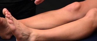
Tumors that have formed on the navicular bone of the leg are manifested by very severe pain, worse at night. Osteoma of the calcaneus is localized on the plantar part of the heel or behind. Such a tumor, even a small one, can interfere with walking and cause severe pain. It often compresses the nerve plexuses, resulting in loss of skin sensitivity and numbness of the foot.
With rib osteoma, there is a lesion in the form of a dense formation up to 3 cm in diameter. Typically, such a pathology does not cause severe pain. No changes (redness) in skin color or swelling are observed. Symptoms of osteomas located on the long tubular bones of the leg and pelvis: (iliac, ischial and pubic bones, head and neck of the femur) pain that increases with movement, swelling at the site of the lesion, lameness. Neoplasms formed on the upper extremities have the same signs.
Would you like to receive an estimate for treatment?
*Only upon receipt of data on the patient’s disease, a representative of the clinic will be able to calculate an accurate estimate for treatment.
How dangerous is the disease?
Growths in the cervical spine provoke insufficient blood supply to the brain.
It is dangerous when the formation grows in the vertebrae of the thoracic and occipital areas of the spine. The cervical vertebrae are penetrated by a large number of vessels and nerve fibers. If ventral exostosis is formed, the intervertebral disc begins to deform, as a result of which the arteries and nerves are compressed, and the brain begins to experience hypoxia.
Ventral hernias prevent the affected vertebra from functioning normally, and if such disorders are not removed in a timely manner, the situation can be fatal. And also, if a combination of unfavorable factors occurs, bone exostosis on the leg, right or left arm can degenerate into a malignant form.
Diagnostics
Diagnosis of osteoma begins with clarifying the patient’s general well-being, carefully studying his medical history, determining the type and size of the tumor, and identifying associated complications.
The main task of diagnosis is differentiation from other bone formations, often malignant, such as fibroma, osteogenic sarcoma, osteochondroma, osteochondrosarcoma, myoblastoma (myoblastus), fibrous dysplasia, osteomyelitis, etc. First of all, the patient is sent for an X-ray examination. The photographs, usually taken in two projections, reveal the type of formation outside the bone (solid or spongy) and the presence of destruction in the adjacent bone tissue. If the tumor is small, then x-rays are ineffective. In such cases, a CT scan is prescribed, which provides more accurate information about the degree of homogeneity of the tumor and its location. MRI more accurately determines the type of osteoma. For example, the bones of the ankle (talus and calcaneus) or the bones of the foot are very well visualized using MRI.
Examination of a tissue sample for histology determines the structure of the tumor, the existing areas of sclerosis and the types of bone tissue canals. Rhinoscopy of the nose is also performed (examination using a special mirror).
Osteogammascintigraphy of bones (examination using preparations containing radioisotope particles) is prescribed. With static scintigraphy, a small number of images are taken to study the morphology of the tumor. With dynamic, a whole series of pictures is recorded, conveying information about the condition of the skeletal bones and existing foci of inflammation. Laboratory tests of blood tests are carried out.
Possible complications
Often, exostosis of the bones of the foot proceeds without a trace, but there are negative manifestations, especially when the growth has formed in the spine area. Its growth can provoke compression of discs and areas of the spinal cord, which leads to serious troubles.
Rapid growth of tumors may indicate the onset of a malignant course. Often this process is observed in the area of the hip, shoulder blades, vertebrae, and pelvic bones. Rarely affects the knees, metatarsals and feet.
Treatment
How to treat osteoma? Depending on the location of formation, osteoma can be treated by maxillofacial surgeons, traumatologists and neurosurgeons. In cases where osteoma is asymptomatic, surgery is avoided. Monitoring the development of the tumor is sufficient.
If there are significant cosmetic defects or signs of compression of organs adjacent to the tumor appear, surgical treatment is used. Surgical intervention consists of radical removal of the tumor. During the operation, the tumor is completely excised, then part of the periosteum and healthy bone tissue located under the tumor are resected. This is done in order to prevent re-development of the tumor.
Another surgical method for treating osteomas is evaporation (vaporization), that is, burning the surface of the tumor with a laser. Using the endoscopic method, it is possible to evaporate osteomas of any location. This method is not as traumatic as surgery and promotes rapid rehabilitation of patients.
Drug treatment is used to relieve pain. Painkillers and anti-inflammatory drugs are prescribed. You can use folk remedies to treat osteoma only on the recommendation of a doctor. Elderberry tincture and hawthorn flower decoction reduce pain. As a distraction technique, it is recommended to apply a mixture of red hot pepper with 6% apple cider vinegar and honey to the affected area (for 10-15 minutes). It has been established that regular herbal treatment over a long period of time reduces the size of osteomas detected in the early stages of development.
Note! Osteomas do not degenerate into malignant neoplasms and, as a rule, do not prevent a sick person from living a full life. But with pronounced manifestations of the disease and complications that arise, surgery is necessary.
When is resection necessary?
Removal surgery is necessary when the osteoma has grown to a large size and can cause negative consequences. Exostosectomy is mandatory if occipital exostosis is diagnosed, in which there is a high probability of destruction of intervertebral structures and compression of nerve fibers and blood vessels.
During surgery, the growth is removed and the periosteum that is located next to it is scraped off. The manipulations are carried out through a small puncture; due to the low level of trauma, the patient can leave the hospital on the same day; rehabilitation in this case takes no more than 2 weeks. Often, postoperative complications do not arise, the person recovers completely, and the disease is not prone to relapse.
Lipoma
This tumor is essentially benign and consists of fatty tissue. Most often it appears in areas covered with hair, in some cases on the forehead. The cause of the appearance may be certain pathologies, hereditary predisposition or a metabolic problem.
In this case, the lump in the back of the head is dense in structure, has a spherical shape and can move under the skin. If you do not take action, it sometimes reaches large sizes, thereby complicating the functioning of blood vessels and causing headaches. Self-medication will not help here, so you need to contact a surgeon to remove the lump.
Which doctor should I contact?
First of all, you should contact a therapist, who, if necessary, will prescribe an examination and refer you to another specialist. This could be, for example, a surgeon, dermatologist or oncologist.
X-rays can be used as a diagnostic method, with the help of which osteoma (tumor of the skull bones) can be easily determined.
Sometimes it is necessary to undergo an ultrasound to obtain information about soft tissues and the presence of fluid.
Classification
Osteoma is divided according to various symptoms into types and types:
- By localization;
- Origin;
- Specifics of the structure.
Taking into account the structure of the tumor and its location, the pathology occurs in three options:
- Compact form, consisting of a dense, ivory-like substance;
- The specific porous structure, characteristic of spongy osteoma, has a fatty layer and blood vessels, while growths form only in tubular bone formations;
- The medullary type of pathology, which forms in large cavities where there is accumulation of bone marrow, is diagnosed in the maxillary sac or sinuses of the facial bones.
Based on the origin of the tumor, there are the following types:
- A heteroplastic type, consisting of some connective tissue of different organs, most often it is osteoma of the shoulder joint or hip;
- A hyperplastic form that develops from bone structures is osteoma of the skull, hip, lower leg and shoulder.
The last type of pathology (hyperplastic growths) occurs in the following types:
- Enostosis – inflammation within the tissue;
- Exostoses - swelling on top of bony tissue;
- Osteophytes are small layers on top of bones;
- Hyperostosis is a tumor that grows along the entire circumference of the bone tissue.
Exostoses with osteophytes appear against the background of bone tissue proliferation due to injury or an inflammatory reaction or mechanical stress on the joints. Exostoses are traditionally found in the pelvic bones, which complicates the labor of a woman. Localization of pathogenic inflammation in the bones of the skull can cause an aesthetic defect.
As for damage to parts of the foot, this can cause lameness and severe pain.
Forecast
With a small tumor size, the prognosis for treatment of osteomas is favorable. Relapses are rare; The reasons are the absence of a clear boundary between the tumor and healthy tissue on the x-ray. Recurrent phenomena are removed using marginal resection. Gentle facial surgery does not lead to cosmetic defects. Removal of large osteomas from the facial bones is accompanied by the second stage - plastic surgery, which corrects the defects of the operation as indicated.
Advanced forms of cranial and ocular osteomas with surgical removal have a mortality rate of up to 3%. The prognosis for treatment of osteoma in children is favorable.
Osteoma of the femur is a benign tumor. Treatment of this neoplasm should be carried out under the supervision of highly qualified doctors. All details are in the link provided.
What is bone osteoma is written here.
Traditional methods
At the initial stage, if the pain is not severe and the growth does not compress the nerves or interfere with blood circulation, then it is possible to use traditional methods. They can relieve pain and relieve inflammation; for this they use: compresses, ointments, foot baths.
The compresses should be warming; the leg should be wrapped in polyethylene. For better penetration of medicinal substances, it must first be steamed, and after using foot baths it is useful to make an iodine net and put on warm socks. The procedure is preferably carried out at night.
- An effective composition for compresses: 100 ml of aloe juice, 100 ml of alcohol, a bottle of valerian, half a teaspoon of red pepper and 2 tablets of Aspirin and Analgin. The composition must be mixed well and left in a dark place for 2 weeks.
- Fat compress - bear, badger or pork fat. The compress is applied at night.
- You can make a compress of medical bile at night.
- Grate raw potatoes and apply to the sore spot, wrap and hold for 4-5 hours.
- Clay baths help remove salts and relieve inflammation.
- Foot baths with salt also relieve fatigue, swelling and pain. Strong solution: 5 liters of water and 1 kilogram of salt, you can add a few drops of iodine or soda.
- Massage with coarse salt is useful. To do this, a kilogram of salt needs to be heated and sprinkled on a flat surface. You need to walk on warm salt with bare feet.
Oral products can also be used. They are needed to normalize metabolic processes, nourish bone tissue, improve blood circulation and strengthen the immune system. For this purpose, it is best to use a tincture of cedar grains along with shells in vodka or a tincture of lilac flowers. Prevent the growth of bone tissue on the heel - avoid increased stress, wear comfortable shoes and treat pathologies of the musculoskeletal system.
Features of the neoplasm
Osteoma is a tumor neoplasm. The material for its formation is predominantly degenerated bone tissue. Children and adolescents are especially prone to the appearance of such bumps.
A feature of the disease is the slow development of the tumor. At the same time, its favorable course is noted, because it responds well to treatment. The risk of osteoma degenerating into a malignant process is completely eliminated.
Despite this, the problem cannot be left unattended for a long time; the patient must be examined by a doctor. The sooner the operation is performed, the easier the rehabilitation period will be.
A lump on the knee cannot be ignored; it is imperative to consult a specialist.
What is osteoma
Osteoma is a disease caused by the formation of a benign tumor from bone tissue, characterized by slow development and resembling a hemisphere in appearance.
Previously, this disease included all formations of bone tissue that appeared as a result of trauma, inflammation, neurotic manifestations, and blastomatosis. Such a tumor can appear in different places. The neoplasm can be localized on the bone tissue of the skull, facial skeleton, big toes, hips, and shoulders. In most cases they are single, but sometimes multiple occurrences are observed. The neoplasm can be hard, spongy, or brain-like.
There are 2 types of tumor:
- Hyperplastic is a tumor that develops from bone tissue.
- Heteroplastic - connective tissue neoplasm.
Osteoma is a disease characterized by a benign course. The neoplasm does not metastasize and does not grow into nearby organs and tissues.
Sometimes the disease is latent and is discovered by chance during an x-ray during a routine preventive examination. Its development mainly occurs in childhood, puberty (from five to 20 years). In most cases, such tumors develop in men.
Why is osteoma dangerous? Education can be quite painful, but that's not the only problem. Sometimes the tumor reaches enormous sizes, which leads to compression of nearby tissues, vessels, and nerves. Then immediate surgical intervention is required.
Popular questions
Can osteoma lead to cancer?
No. Osteoma is a benign tumor. It can cause adverse health effects if it grows into the cranial cavity. But the probability of degeneration into cancer is close to zero.
What causes osteoma?
The causes of the tumor are unknown. The role of hereditary predisposition has been established. If your relatives have been diagnosed with osteoma, you are more likely to develop it than the average population. The trigger for the growth of osteoma can be a bone injury or an acute inflammatory process. There is also a theory of intrauterine developmental defects. The reason for its occurrence was the fact that osteoma most often develops at the junction of the frontal and ethmoid bones, where membrane and cartilaginous tissues develop during embryogenesis.
Is it necessary to remove the osteoma?
The tumor grows very slowly. In most cases it is not dangerous. Only clinically significant osteomas that can grow into the orbit or skull bones are removed. The operation can also be performed for aesthetic reasons.
Who gets exostosis?
The disease most often occurs in children and adolescents of both sexes aged 8–10 to 25–27 years. But adults can also get sick.
Osteochondroma according to ICD 10 has code D16 - a benign neoplasm of bones and articular cartilage.
Exostosis occurs in approximately every tenth patient with a bone tumor. And among benign bone tumors, 35–45% of cases are due to exostosis. Therefore, it is the most common benign bone tumor.
Bone development
As is known, bone during its development in the prenatal period can come from two sources: connective tissue and cartilage. In the first option, most of the bones of the skull, the lower jaw and the collarbone are formed. The remaining bones of the skeleton are based on cartilaginous tissue, which is then replaced by bone tissue along almost its entire length.
Osteochondroma develops in bones of cartilaginous origin. It is most often found on the epiphysis (end) of a long bone, such as the tibia or femur. Much less often it appears on flat bones - pelvic bones, scapula, ribs, etc.
Consequences
During a conventional operation, the recovery period can last up to two weeks, and complete rehabilitation and a normal, full life after tumor removal is possible in 1-2 months.
The negative consequences of surgery for the treatment of osteoma are:
- wound with pus;
- Damage to tissues, nerves, tendons and blood vessels adjacent to the tumor;
- localized pain in the head (if the operation affected the bones of the skull);
- Relapses.
Initially, the patient is under inpatient observation, where doctors take measures to prevent secondary infection and try to speed up the processes of regeneration of damaged tissues.
After a period of rehabilitation in the hospital, the patient returns home. But he should still observe a special regime of work and rest, which is regulated by the attending physician. A diet with plenty of calcium is also recommended.