How does the human throat work?
The internal structure of the human throat has a number of the same features as the part of the neck in front of the spinal column in some mammals, but, of course, there are differences, and there are many of them.
The vagus nerves, carotid artery and other vital systems pass through the area from the hyoid bone to the manubrium of the clavicle. This part of the human body is the object of close study in otorhinolaryngology. The human throat consists of two interconnected sections: the pharynx and larynx. The anatomical structure of these parts of the human throat is directly related to the functions they perform.
How the human throat works is described in detail on this page.
Functions of the human pharynx and larynx: photo with description
Pharynx and larynx The throat consists of 2 sections: the pharynx and larynx.
These departments are interconnected. The anatomy of the pharynx and larynx is directly related to their functions. Functional features of the laryngeal region:
- Protection - the mucous membrane is equipped with a special movable layer with many glandular tissues. When pieces of food pass by, the nerve roots make a reflexive movement, causing a cough. With its help, pieces of food fall from the laryngeal region back into the mouth.
- Breathing has a direct relationship with protective functions. The hole, which is equipped with vocal binding muscles and glands, sometimes decreases and sometimes increases, directing air flows.
- Education of the voice, speech - the timbre of the voice directly depends on the anatomical structure of the larynx and the condition of the connecting muscles and tissues.
Functions of the larynx
The functional features of the pharynx are similar to the functions of the larynx. The differences are in the following nuances:
- Respiratory feature - all individual parts of the pharynx are involved: nose, mouth, throat. Oxygen enters it from the nose, and then further into the body.
- Voice, speech - sounds appear (consonants and vowels) and are formed in the soft tissues of the palate and with the help of the tongue. These parts are a “curtain” for the nasopharynx, due to which the timbre sounds and pitch of the voice are formed.
- Protection and pathologies in the pharynx are associated with nasal breathing. The lymphoid circle of the pharynx, together with nearby soft tissues and lymphs, forms one entire immune system of the body. If a person has defects (congenital or acquired), tissue growth occurs, their sensitivity decreases and bacteria begin to multiply. The pharynx protects other parts of the throat by collecting all pathogens. If there is inflammation in the throat, then the nose and ears suffer.
- Eating - This functional feature involves swallowing and sucking. On top of this section there are ciliated receptors. When they work, soft tissues begin to function, a contraction process occurs, fluid is released in the form of mucus and a pharyngeal, gag or cough reflex occurs. All harmful substances that have accumulated on the eyelashes are eliminated by coughing or we swallow them.
The trachea originates in the subvocal section of the larynx, which also plays a huge role in the digestive and respiratory systems of our body.
Structure of the human pharynx
The pharynx is the “gate” leading to two of the most important systems of our body - the respiratory and digestive. This tube, as if “suspended” from the base of the skull, connects the nasal cavity with the larynx and is divided into three parts: nasal, oral and laryngeal.
These photos show the structure of the human throat:
The nasopharynx can be safely called a “crossroads”. The choanae (openings of the nasal cavity) emerge there, and on the sides (at the level of the inferior turbinates) the entrances to the auditory tubes are visible, leading directly to the tympanic cavities of the right and left ears. All openings are “protected” by accumulations of special lymphoid tissue - pharyngeal - and tubal tonsils.
Below the nasopharynx is attached to the oropharynx, connected to the oral cavity by the arch of the pharynx. The upper borders of the pharynx are the soft palate and the uvula, and the lower border is the root of the tongue (it is adjacent to the anterior wall of the pharynx with another “hidden” tonsil - the lingual). On the sides you can see the palatine arches, in the niches of which paired palatine tonsils “sit in ambush”. The posterior wall of the pharynx is also covered with lymphoid tissue and closes the so-called lymphatic pharyngeal ring. The laryngopharynx is adjacent to the epiglottis and the root of the tongue, gradually narrows and passes into the esophagus.
This shows how the human throat works from the inside:
Tonsils get their name because of their resemblance to almond seeds, due to the loose structure of the lymphoid tissue.
In newborns, the tonsils are not developed; their formation, depending on the individual characteristics of the child, is completed by approximately six months or one year.
Below are photos and a description of the structure of the larynx as part of the human throat.
Internal structure of the organ
In the upper segment of the pharynx, approximately at the level of the zygomatic bones of the skull, temples and root of the nose, there is a small void connecting the oral and nasal cavities. In fact, it is not a separate organ, but a cavity that performs certain functions.
The upper wall (arch) connects to the occipital and sphenoid bones. The lower wall borders the soft palate, which rises when swallowing, blocking the passage into the oral cavity. This prevents food from entering the nasal chamber. During the breathing process, the soft palate is adjacent to the root of the tongue. The posterior one is adjacent to the cervical vertebrae (first and second), separated from them by a layer of loose connective tissue.
This gives the chamber walls mobility. The anterior one is connected to the nasal cavity through special openings (choanae). The lateral ones have exit openings for the auditory (Eustachian) tubes. They are surrounded by cartilaginous ridges and connect the middle ear to the environment, regulating pressure and draining the voids of the middle ear. Through the pharyngeal openings there is a connection with the tympanic cavities and normal transmission of sound signals.
In fact, due to the structure of the human nasopharynx, all the voids that are located in the skull are interconnected.
adenoids (unpaired pharyngeal tonsil) above; palatine tonsils (paired) on the sides; lingual tonsil below.
Thus, a kind of protective ring is formed that prevents pathogens from entering the respiratory and digestive system.
Newborn babies have differences in the structure of the cavity, since it is not yet fully formed. Its width and height are significantly less than those of adults; a semicircular arch is not formed. The choanae are small and at first have a triangular or round shape, but by the age of two they double in size and acquire an oval shape.
The structure of the human larynx
The larynx is attached by muscles to the hyoid bone and connects the nasopharynx with the lower respiratory tract - the trachea and lungs. The shape of this organ is provided by a system of cartilages that form a flexible, movable tube. The cricoid cartilage underlies the larynx, the thyroid cartilage serves as a frame, and the epiglottic cartilage acts as a lid, protecting the airways from chewed food during swallowing. Paired cartilages (cuneiform, arytenoid, corniculate) strengthen the larynx, help it narrow and expand.
Look at the photo of how the human throat works:
Inside, the larynx is like an hourglass, in the middle of which there are elastic vocal cords that form an opening for the passage of air - the glottis.
The tone of the voice and its individual color are regulated by the length of the chords according to the principle: the shorter the length, the higher the timbre. The larynx is in constant motion: when exhaling and swallowing or singing, it rises, and when inhaling, producing low sounds, it lowers.
The larynx and pharynx are associated with the breathing process: inhaled air from the nose passes through these sections and rushes further into the trachea, to the lungs. Together they participate in the reflexive process of swallowing. The tissues of the pharynx protect against infection, and the structure of the larynx protects the airways from food getting into them. The larynx “gives birth” to the voice, and the pharynx strengthens it.
Here you can see a diagram of the structure of the human throat:
If you have questions for your doctor, please ask them on the consultation page. To do this, click on the button:
Traditional methods
Inflammation of the nasopharynx is a very common disease, so it is not surprising that there are many traditional medicine methods to combat such a disease. Despite many benefits, alternative treatments for nasopharyngitis may be less effective than drug therapy. Because of this, before treating yourself, you should consult an otolaryngologist.
Treatment methods:
Elecampane
- Elecampane. It is necessary to grind 2 tablespoons of the roots of the plant and pour 250 ml of boiling water. The mixture is poured into a convenient container and placed on low heat. After boiling, you need to cook the medicine for 10 minutes, then leave for several hours. This remedy should be taken 1 large spoon before meals, up to 4 times a day.
- Plantain leaves. It is recommended to use if nasopharyngitis is accompanied by regular coughing attacks. You need to mix a glass of boiling water and 1 spoon of chopped plant. The liquid must be infused in a warm place for several hours, then filtered. Should be taken 4-5 times a day, before meals, 1 spoon.
- Soda solution. It is used for gargling, while providing an antibacterial and analgesic effect. Add 0.5 tablespoons of salt and soda to warm boiled water. The resulting liquid is thoroughly stirred and then gargled with it. You can repeat the procedure every hour.
- Calendula solution. A small amount of the juice of this plant must be diluted in warm water. After this, the liquid should be sucked in through the nose and spat out through the mouth. Regular repetition of this procedure will relieve severe swelling and allow you to breathe normally through your nose. It is recommended to repeat at least 3 times a day.
- Pine infusion. Pine buds are used for preparation. To prepare the medicine, mix 1 spoon of pine buds and pour a glass of boiling water. The resulting product is best infused in a thermos. After this, the medicine is taken in small sips throughout the day to eliminate the feeling of dryness and tickling.
In general, treatment of inflammation of the nasopharynx using traditional methods is an alternative method of therapy that can be used in combination for greater effectiveness.
Rhinopharyngitis, or inflammation of the nasopharynx, is a common ailment that can be caused by many reasons and factors. Successful treatment of the disease directly depends on how correctly the causes of its development were identified, as well as on the chosen therapeutic methods.
What causes a sore throat - causes and dangers
What can cause a sore throat? How do the pharynx and larynx work in humans? What diseases can pose a serious danger to children and adults? We answer questions in simple language.
Diseases of the throat and larynx in children and adults most often arise as a result of infection with viruses and microbes; they can be the consequences of hypothermia, overexertion and systemic pathologies. An important feature of such diseases is their ability to quickly spread from the oropharynx towards the larynx and below, causing diseases of the lungs and bronchi. In order to correctly assess the danger, you should understand the structure of this part of the body and the symptoms of the main disorders.
Contraindications
If you have a sore throat and nose, you will have to carefully monitor your diet. It must be composed in such a way that it includes foods with a large amount of vitamins and other useful components.
Therefore, you will have to exclude the following foods from your diet:
- too hot and cold dishes;
- sour and spicy foods;
- hard and fatty foods.
Experts also advise completely abstaining from drinking carbonated drinks and alcohol. In addition, you will have to temporarily exclude from the diet spices and seasonings that cause irritation when in contact with the mucous membrane. It is also advised to avoid sweet foods, since sugar impairs the functioning of the immune system and slows down its recovery after illness.
What is the throat, larynx and pharynx
A common misconception is to call the throat only the small area behind the tongue that turns red and hurts with sore throats. In this case, what is located below is often excluded. For example, there is a difference between the throat and larynx because it is part of this system below and is connected to the pharynx and trachea.
The throat is not an anatomy term; it is the common name for the part of the upper respiratory tract from the hyoid bone to the level of the clavicle or manubrium of the sternum. The throat contains:
- pharynx or oropharynx - begins in the visible part of the mouth, the entrance gates are the tonsils, they are also tonsils that do not allow infections below;
- nasopharynx - cavities located above the palate;
- swallowing department - a small area behind the epiglottis that pushes food and liquids into the esophagus;
- The larynx is a cartilaginous tube for air, lined with mucous membrane and blood vessels.
The epiglottis serves as a valve that prevents food and water from entering the larynx, at the top of which are the vocal folds (cords). Their closing and opening gives us the opportunity to make sounds. The larynx is protected in front by the thyroid cartilage, and behind it is the esophagus.
Anatomy and functions of the trachea
So, the trachea connects the larynx with the bronchi, which means it carries air and oxygen into the lungs. The trachea is a hollow, tube-shaped organ. Its length ranges from 8.5 cm to 15 cm, depending on the physiological characteristics of the body. The third part of this tube is located at the level of the neck, the remaining part descends into the thoracic region. At the end, the trachea divides into 2 bronchi at the level of the 5th thoracic spine. A more detailed description of the trachea:
- The thyroid gland is located in front at the level of the neck.
- At the back is the esophagus.
- On the sides there is a cluster of nerve endings, carotid arteries and internal veins.
Anatomy of the trachea:
- The mucous membrane consists of a ciliated layer. Mucus is secreted on its surface in small quantities. Endocrine cells of the trachea secrete substances such as serotonin and norepinephrine.
- The submucosal layer consists of tiny vessels and nerve endings. This connective tissue has a fiber structure - loose and soft.
- Cartilages are hyaline incomplete cartilages that make up 2/3 of the entire trachea. The joints for the cartilage are special annular ligaments. The membranous wall, located posteriorly, is in contact with the esophagus. Thanks to this, the two processes - eating and breathing - do not interfere with each other.
- The adventitia is a thin shell in its structure, consisting of connective fibers.
The functions of the trachea are very important in the functioning of the body, despite the simple anatomy of this organ. The functions are as follows:
- The main purpose of this section of the larynx is to conduct air to the lungs.
- Small particles that are unnecessary for the body and come from the external environment settle on the mucous layer of the trachea. They are enveloped in mucus, and the cilia are pushed into the larynx.
As a result, the trachea cleans the air that the lungs need. From the larynx and pharynx, all the dirt that has been removed from the trachea rises up and with the help of coughing, all these organs are cleansed.
Causes of throat diseases and risk factors
Most diseases in the throat area are associated with external factors. Unlike other organs, the pharynx is under the direct influence of the oral cavity, which means that any diseases of the teeth, gums and oral mucosa cause rapidly spreading complications and responses in the throat.
The main causes of discomfort and sore throat can be divided into several groups:
- infections - most often external, associated with ARVI and infection with influenza, scarlet fever and whooping cough;
- overstrain - affects the condition of the muscles of the glottis, which leads to loss of voice and a characteristic cough;
- hypothermia - usually it does not affect the trachea, but can cause laryngitis, sore throat, pharyngitis in the upper part;
- inflammatory processes that begin in the oral cavity or come “from below”, circulatory disorders, cancer of the throat (one of the parts of the larynx or pharynx).
Diseases of the nasopharynx
Laryngitis. Acute inflammation of the pharyngeal mucosa. It can cause a lot of trouble and pain, but it is usually cured easily and in just a few days. Acute sore throat. Tonsillitis. It is usually of a bacterial nature, can be treated with antibiotics, and if the correct treatment regimen is followed, the prognosis is favorable. Pharyngitis.
Inflammation of the mucous membrane of the throat, or usually an acute respiratory disease, can be cured without any problems with conservative methods of therapy. Retropharyngeal abscess. Rather, it is not a disease on its own, but a complication of acute tonsillitis. Treatment in a hospital setting. The prognosis is favorable only if medical recommendations are strictly followed.
Peritonsillitis. Adenoids. They are detected when the disease has already entered the chronic stage. Conservative therapy is most often pointless; adenoids are usually treated surgically, only after which the functions of the nasopharynx are fully restored. Hypertrophy of the palatine tonsils. Foreign bodies, wounds and trauma to the pharynx. Treatment depends on the severity of the damage. Sometimes it is enough to simply treat the wound, but there are times when surgical intervention is necessary.
Very often, diseases of the nasopharynx are accompanied by such unpleasant symptoms as: general intoxication of the body, fever, sore throat.
Diagnostics
Visual inspection. This is usually the basis for the initial diagnosis. Laboratory research. Tests are necessary when it is necessary to determine for sure the causative agent of the disease, as well as to find out what drugs to treat the patient with. Endoscopic examination. Used if there is a suspicion of neoplasms in the nasopharynx area. The examination may be supplemented with a biopsy.
Symptoms of sudden dangerous diseases in the throat
If you experience any warning signs from coughing to a characteristic sore throat, especially in children, you should consult a doctor. There is no need to make a diagnosis yourself, prescribe treatment, and especially not use strong remedies, especially antibiotics. It is important to remember that no infection occurs locally, in isolation. Inflammation of the larynx can develop into tracheitis, which in advanced form will develop into bronchitis and descend into the lungs.
Pay attention to life-threatening phenomena for children and adults that may seem frivolous and not frightening enough.
Infectious pharyngitis
The disease manifests itself in the form of discomfort, sore throat, temperature rise to 37 - 37.5 C. After some time, severe pain in the throat appears, and the mucous membranes swell. In a severely advanced form, ulcers and follicles may appear, the discharge from which collects on the back wall of the pharynx.
The infection can be caused by staphylococci and streptococci. A favorable environment is created by free breathing in the cold, frequent exposure to dusty rooms, and smoking. In the chronic course, the pain may not be acute, but characteristic dryness, burning and discomfort when swallowing remain.
Treatment of pharyngitis is a complex of actions from rinsing to taking vitamin A. There is no need to prescribe it yourself, since it is necessary to exclude other diseases. The doctor focuses on examination data and a general blood test.
Laryngospasm in children
The main danger of this phenomenon is the absence of warning signs and alarming symptoms in the form of temperature. Suddenly there is a noisy inhalation with a characteristic whistle, the child may begin to choke. Convulsive twitching of the arms and legs appears. In a state of activity this is dangerous; at night the phenomena stop on their own.
First aid is to distract the child and splash cold water on his face. Consult a doctor to diagnose rickets, hydrocephalus and other systemic diseases.
Edema and stenosis of the larynx
Occurs as a consequence of syphilis, allergic phenomena (Quincke's edema), diphtheria and laryngitis. Manifests itself in the form of noisy breathing and hoarseness. In development, it can lead to complete closure of the respiratory passages and respiratory arrest. A very dangerous sign is the appearance of blueness on the skin.
First aid is to place the patient closer to a container with hot water and steam, ensure rest, and give a hot foot bath. In case of swelling, most often associated with allergies, call an ambulance for antihistamine injections.
Whooping cough, scarlet fever, diphtheria in children and adults
These are the most dangerous infectious diseases that can lead to death or serious consequences. A rise in temperature, rash, the appearance of a characteristic white coating in the throat and “films” that make breathing difficult are signals that you need to immediately call a doctor.
And before it appears, provide air access through the throat. Treatment of scarlet fever and similar diseases can only be carried out as directed by a doctor.
What diseases can occur in the nasopharynx
If symptoms of nasopharyngeal diseases appear, you should consult an otolaryngologist. The doctor understands the smallest details that can help the patient.
When examined, the following diseases may be detected in a person:
- laryngitis;
- angina;
- pharyngitis;
- paratonsillitis;
- inflammation of the adenoids.
With laryngitis, the patient begins to experience inflammation of the pharyngeal mucosa. A bacterial infection can trigger the development of acute sore throat. A sign of pharyngitis is inflammation of the throat mucosa.
What is the throat and larynx?
The throat is one of the most important organs of the human body, belonging to the upper respiratory tract. Its structure facilitates the movement of air through the respiratory organs, and allows food to enter the digestive tract. In addition, the area includes a huge number of nerve tissues, blood vessels and pharyngeal muscles important for human life. In the structure of the throat, the main parts are represented by the pharynx and larynx.
By their continuation they form the trachea. The structure of the throat and larynx is designed in such a way that the first of these structures is responsible for the movement of air into the lungs and food into the stomach, and the second structure takes responsibility for the vocal cords.
Functions
The function of the nasopharynx is to bring air from the environment to the lungs. The structure of the nasopharynx determines its functions:
- The main function of the nasopharynx is to conduct air from the environment to the lungs.
- Performs an olfactory function. It generates a signal about the arrival of the smell in the nasal part, the formation of an impulse and its conduction to the brain thanks to the receptors that are localized here.
- It performs a protective function due to the structural features of the mucous membrane. The presence of mucus, hairs and a rich blood network helps clean and warm the air, protecting the lower respiratory tract. Tonsils play an important role in protecting the body from pathogenic bacteria and viruses.
- It also implements a resonator function. The sinuses and vocal cords, located in the pharynx, create sound with a different timbre, which makes each individual unique.
- Maintaining pressure in the cranium. By connecting the ear to the external environment, the nasopharynx allows you to maintain the necessary pressure.
Device principle
The throat is a highly complex organ responsible for breathing, speaking, and moving food.
In short, its structure is based, as we said earlier, on the pharynx (pharynx) and larynx (larynx). Since this organ is a conducting channel, it is very important that all its muscles work harmoniously and correctly. Inconsistency in their activities will lead to the fact that food can enter the respiratory system and create a dangerous situation, even leading to death.
The structure of a child's throat is the same as that of adults. But children have narrower cavities and tubes. As a result, any disease in which swelling of these tissues occurs can be extremely dangerous. It is advisable for a person to know the structure of such an organ, as this can be useful in caring for it and during treatment. In the pharynx, the nasopharynx and oropharynx are distinguished.
The pharynx (pharynx) is a cone-shaped structure turned upside down. It is located behind the mouth and goes down to the neck. The cone is wider at the top. It is located near the base of the skull, which gives it more strength. The lower part is united with the larynx. The layer of tissue covering the outside of the pharynx is a continuation of the layer of oral tissue lying outside. It has many glands that produce mucus, which is involved in the processes of moistening the throat when eating and talking.
How is the oropharynx examined?
How should an examination of the oropharynx be performed? An examination of the oral part of the pharynx is carried out using a special spatula; you need to apply a little pressure on the tongue, because it is often in a curved or raised position, which prevents the doctor from conducting an examination. But please note that the oropharynx must be examined in accordance with certain rules . What rules should an examination of the oropharynx comply with?
Examination of the oropharynx
So, the oropharynx should be examined in accordance with the following rules:
- when examining the oropharynx, it is strictly forbidden to stick your tongue out of your mouth;
- a special spatula can only be applied to the front 2/3 of the tongue;
- The tongue must be pressed down gradually, with smooth movements, without fidgeting with the spatula on the tongue, pressing both the bottom and the front of the tongue;
- the examination should be carried out while the patient is breathing evenly.
In some cases, the back of the tongue needs to be pressed with great effort, this is especially necessary when it is not very pliable. If the oropharynx is examined without following the above rules, it may provoke vomiting , and this will affect the quality of the examination. How is the oropharynx examined? In order for the doctor not to block his view with his hand, he needs, simultaneously with the outer end of a special spatula, to move the back of the tongue to the left side, that is, to the left corner of the mouth. But this is not necessary if the doctor uses a knee-shaped, slightly curved spatula, because in this case the doctor’s hand will not interfere. If the oropharynx is examined correctly, using a non-curved medical spatula, then its outer end will be directed to the left and slightly raised up. The oral part is examined to evaluate organs such as the palatine arches and tonsils, mucosa, dorsum of the tongue and posterior wall of the oropharynx. The normal size of the tonsils should not cover the posterior arches. If necessary, press on the upper part of the tonsils with a second medical spatula, which must be held in the right hand. It is with the help of a second medical spatula that pus can be squeezed out, which can accumulate in the scripts. In order to examine the covered part of the tonsil, the doctor has to slightly pull back the last arch using a blunt hook, and the patient’s head must be turned slightly in the opposite direction. Sometimes, if certain conditions are met, the doctor examines the lower part of the oropharynx, the fourth tonsil and the surface of the tongue in the epiglottis.
The throat is a human organ that is classified as the upper respiratory tract.
Nasopharyngeal compound
The structure of the throat and larynx includes the structures that form them, such as the nasopharynx and oropharynx mentioned above. Let's consider one of them.
The nasopharynx is the part of the pharynx that occupies the upper position. It is limited from below by the soft palate, which begins to move upward during the process of swallowing. Thus, it covers the nasopharynx. This is necessary to protect it from food particles entering the respiratory tract. In the upper wall of the nasopharynx there are adenoids - tissue accumulations located behind its wall. This organ also has a tunnel connecting the throat to the middle ear. This formation is called the Eustachian tube.
Traditional therapy
In most cases, the treatment method is medicinal, that is, it involves taking special medications. Competent therapy allows you to relieve the patient from severe symptoms of the disease in a short period of time, affect overall well-being, and prevent complications.
Methods of drug therapy:
- Antibiotics. Used for infectious diseases accompanied by inflammation of the nasopharynx. One of the most effective options is taking medications based on Amoxicillin. These include Amoxicillin, Amoxil, Amoxiclav. The use of antibiotics should only be done with the permission of the attending physician, which is associated with many contraindications and possible side effects. Drugs
- Antiviral agents. Aimed at inhibiting viral microorganisms affecting the nasopharyngeal mucosa. They should also be prescribed exclusively by a doctor due to the nature of their action. They are rarely prescribed for treatment, since the causative agents of the disease are often bacteria rather than viruses.
- Antihistamines. Used to eliminate the cause of allergies. When an allergic reaction develops, histamine is released, which provokes an inadequate immune response and associated symptoms. The use of drugs from this group allows you to normalize the concentration of histamine, and thus eliminate allergies as the cause of nasopharyngitis.
- . For these purposes, a variety of sprays and lozenges are used. Their active components temporarily eliminate the main symptoms of the disease - sore throat. Nasal sprays can also be used, but their purpose is to reduce swelling and improve breathing.
- Antipyretics. Against the background of severe inflammation of the nasopharynx, body temperature often rises. It can be especially high in the presence of concomitant diseases. To reduce it, various medications can be used, especially those based on Paracetamol, which has the fewest contraindications and side effects.
- General strengthening therapy. It is prescribed to strengthen the body, enhance the immune properties of the body, which are responsible for protection against pathogenic microorganisms. For these purposes, various vitamin complexes are used and special diets are prescribed. There is also a separate group of immunostimulants, which are most effective for viral lesions.
Introduction to the larynx
The structure of the throat has another important component fragment - the larynx.
This organ occupies space at the level of the 4th, 5th and 6th cervical vertebrae. The hyoid bone is located above the larynx, and a group of hyoid muscles is formed in front. The lateral areas rest against the thyroid gland. The area located behind contains the laryngeal fragment of the pharynx.
Cartilage forms the skeleton of this area, connecting to each other through ligaments, muscle groups and joints. Among them, paired and unpaired are distinguished.
- arytenoid pair;
- horn-shaped pair;
- wedge-shaped pair.
In the muscular system of the larynx, there are three main groups of muscle formations. Among them there are tissues responsible for reducing the lumen of the glottis, tissues intended to expand the vocal cords, and tissues that strain the vocal cords.
Treatment tactics
Since the disease is accompanied by symptoms of two diseases, the treatment is complex. It is necessary to eliminate the provoking source of the disease. Treatment should begin with eliminating mucus from the nasal cavity. Medicines prescribed for topical use:
- Nasal medications are prescribed to eliminate mucus. Drops that constrict blood vessels should not be used for more than 5 days.
- Rinse your nose several times a day.
- Gargling to reduce pain when swallowing.
- Lozenges for the throat.
- It is necessary to do physical procedures.
- Vitamins.
- If the high fever persists for more than 6-9 days, antibiotics should be prescribed.
In the acute period of the disease, bed rest is recommended.
It is necessary to drink as much fluid as possible. You should avoid spicy and cold foods.
Treatment of inflammation of the nasopharynx at home:
- Herbal collection. Chamomile, sage, calendula and oregano are taken in equal quantities. 1 tbsp. l. collection is poured 200 g. hot water and infuse for 10-20 minutes. Drink 3 times a day, 0.5 tbsp.
- Sage with milk. 2 tsp. herbs are brewed in 1 tbsp. milk. To avoid getting burned, drink the decoction warm, 4 tbsp. l. three times a day.
- Beetroot juice. Squeeze it out and drop 2-4 drops into the nose, repeat the procedure every 3-4 hours.
- Elecampane root. 2 tbsp. l. crushed root brew 1 tbsp. boiling water and leave to simmer for 5-7 minutes, then infuse for 4 hours. Drink 1 tbsp. l. before meals, 3 times a day.
- To clear your nose, you need to instill 5-6 drops of freshly squeezed Kalanchoe juice.
- For inflammation of the nasopharynx, inhalations will help well: with boiled potatoes, medicinal herbs, aromatic oils.
- Washing with calendula. Add a little salt and 1 tsp to 0.5 liters of warm water. calendula tinctures. Rinse your nose in the morning and before bed.
- Nasal drops with wild rosemary. In 100 gr. Add 2 tsp vegetable oil. wild rosemary The mixture is infused for about 3 weeks in the dark; it is necessary to shake the mixture every day. Place 1 drop into your nose 3 times a day for a week.
- Pine buds for a sore throat. 2 tsp. kidneys are poured into a thermos, boiling water is poured and left for at least 3 hours. Take 2-3 sips when you have a sore throat.
- To gargle 1 tbsp. l. flax seed and marshmallow root pour 200 g. hot water, leave for 20 minutes. You should gargle with the strained broth three times a day.
- Plantain infusion for cough. 2 tsp. dried herbs pour 1 tbsp. boiled water and leave for 2 hours. Take 1 tbsp. l. 3 times a day before meals.
General information about the structure of the larynx
The larynx has an entrance in front of which there is an epiglottis, and on the sides there are aryepiglottic folds, represented by a number of wedge-shaped tubercles. Behind the organ lie the arytenoid cartilages, represented by horn-shaped tubercles. These fragments are located on the mucous membrane, along its lateral parts. The laryngeal cavity includes the vestibule, subglottic region and interventricular region.
The first part originates in the area of the epiglottis and extends to the folds. Here, thanks to the mucous membrane, special folds are formed, between which lies a gap called the vestibule.
The subglottic region is the lower fragment of the larynx, which passes into the trachea below.
The interventricular region is a narrow area between the upper folds of the vestibule and the lower vocal cords.
There are a number of membranes in the larynx:
- mucous;
- fibrocartilaginous;
- connective tissue.
The main functions of the larynx are protective, voice-forming and respiratory. Each of them has a special meaning.
The functions of breathing and protection form a close connection with each other. This is due to the fact that air flows are delivered to the organs of the lungs, and at the same time the direction of the flows is regulated. Regulation of the air path is ensured by the activity of the glottis, which is capable of contraction and expansion. In addition, glands located in the ciliated epithelium perform a protective function.
Although the structure of the ear, throat and nose is different, the interconnection of these organs in the human body is extremely great. They unite with each other and are located approximately in the same areas. The activity of each component affects the operation of the other. Their role is to produce an irritant reaction followed by causing coughing when food enters the respiratory tract and organs. Using this mechanism, the larynx removes food into the oral cavity. This organ is also involved in the formation of the voice. The parameters of its pitch and sonority are determined by the anatomical structure of the larynx. For example, a hoarse voice appears due to insufficient hydration of the ligaments.
Facial sinuses
The structure of the skull is such that in the front part there are sinuses (special cavities filled with air). The mucous membrane differs little in structure from the mucous cavity, but it is thinner. Histological examination does not reveal cavernous tissue, while the nasal cavity contains it. The average person's sinuses are filled with air. Highlight:
- maxillary (maxillary);
- frontal;
- ethmoid bone (ethmoid sinuses);
- sphenoid sinuses.
At birth, not all sinuses are formed. By 12 months, the last sinuses, the frontal ones, finish forming. The maxillary sinuses are the largest. These are paired sinuses. They are located in the upper jaw. Their structure is such that they communicate with the passages of the nose through an exit under the lower passage.
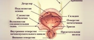
Structure of the throat
The throat is an organ that belongs to the upper respiratory tract and helps move air into the respiratory system and food into the digestive tract. The throat contains many vital blood vessels and nerves, as well as muscles of the pharynx. There are two sections in the throat: the pharynx and the larynx.
The trachea is a continuation of the pharynx and larynx. The pharynx is responsible for moving food into the digestive tract and air into the lungs. And the larynx bears responsibility for the vocal cords.
The role of the nasopharynx in the body
Connective. Transporting air flow not only through the nose, but also through the mouth due to the connection of the oral cavity with the nasal sinuses. Warming. The mucous membranes covering the cavity are penetrated by blood vessels that organize active heat exchange. Thus, the temperature of the air entering the throat rises to values sufficient for the safe functioning of the lower parts of the respiratory system (trachea, lungs). Protective.
The presence of tonsils makes it possible to bind viruses and pathogenic bacteria entering the human body through airborne droplets. The cilia of the ciliated epithelium intensively remove associated microbes. Olfactory. The mucous membrane contains special surfaces that are highly sensitive and capable of distinguishing the odors of aromatic substances even in small quantities (several molecules).
What is the throat made of?
The pharynx, or as it is otherwise called “ pharynx ,” is located behind the oral cavity and extends down the neck. The shape of the pharynx is a cone turned upside down. The upper part of the cone, wider, is located at the base of the skull - this gives it strength. The lower part, narrower, is connected to the larynx. The outer layer of the pharynx is a continuation of the outer layer of the oral cavity. Accordingly, this layer has numerous glands that produce mucus. This mucus helps keep the throat moist during eating and speaking.
Nasopharynx
The pharynx consists of three parts. These parts have their own location and perform certain functions. The uppermost part is the nasopharynx . From below, the nasopharynx is limited by the soft palate and when swallowing, the soft palate moves upward and covers the nasopharynx, thereby preventing food from entering the nose. The upper wall of the nasopharynx has adenoids. Adenoids are a collection of tissue located on the back wall of the nasopharynx. The nasopharynx also has a passage that connects the middle ear and throat - this is the Eustachian tube.
Oropharynx
The oropharynx is the part of the pharynx that is located behind the oral cavity. The main function of the oropharynx is to promote air flow from the mouth to the respiratory organs. The nasopharynx is less mobile than the oropharynx. Therefore, as a result of contraction of the muscle mass of the oral cavity, speech is formed. In the oral cavity there is a tongue, which, with the help of the muscular system, helps move food into the esophagus and stomach. But the most important organs of the oropharynx are the tonsils, which are most often involved in throat diseases.
The lowest part of the throat performs the function of swallowing. The movements of the throat must be very clear and synchronized to simultaneously ensure the penetration of air into the lungs and food into the esophagus. This is achieved through a complex of nerve plexuses.
The larynx is located opposite the 4th -6th cervical vertebrae. The hyoid bone is located above the larynx. In front of the larynx is formed by a group of hyoid muscles, the lateral parts of the larynx are adjacent to the thyroid gland, and the laryngeal part of the pharynx is located in the posterior region of the larynx.
The skeleton of the larynx is represented by a group of cartilages (paired and unpaired), which are connected to each other by muscles, joints and ligaments.
Unpaired cartilages include:
- Cricoid
- Thyroid
- Epiglottic
Paired cartilages include:
- Arytenoids
- Corniculate
- Wedge-shaped
No human organ can function without muscles. The muscular system of the larynx is divided into three groups: muscles that narrow the glottis, muscles that dilate the vocal cords and muscles that tense the vocal cords. The muscles that narrow the glottis can be divided into several groups: cricoarytenoid, thyroarytenoid, transverse and oblique arytenoid muscles. The only muscle that widens the glottis is the paired posterior cricoarytenoid muscle. The cricothyroid and vocalis muscles are considered muscles that tense the vocal cords.
Possible diseases
It is susceptible to various diseases due to its location and its functions. All diseases can be divided into groups:
- inflammatory;
- allergic;
- oncological;
- injuries.
Table of diseases.
| Diseases | Symptoms | Predisposing factors |
| Inflammatory | 1. Deterioration of general condition, malaise, weakness, fever. | 1. Hypothermia. |
| 2. Sore throat. | 2. Reduced immunity. | |
| 3. Redness of the throat, enlarged tonsils. | 3. Contact with sick people. | |
| 4. Sore throat. | 4. Being in a large crowd of people during the high morbidity season. | |
| 5. Congestion, nasal discharge. | ||
| Allergic | 1. Itching. | 1. Contact with an allergen. |
| 2. Redness. | 2. Burdened heredity. | |
| 3. Nasal discharge. | 3. History of allergic reactions. | |
| 4. Sore throat. | 4. Flowering season. | |
| 5. Watery eyes. | ||
| Oncological | 1. Presence of a neoplasm. | 1. Burdened heredity. |
| 2. Difficulty breathing. | 2 Smoking. | |
| 3. Difficulty swallowing. | 3. Contact with a source of gamma radiation (work in an X-ray room, etc.). | |
| 4. Drastic weight loss of more than 7-10 kg per month. | ||
| 5. General malaise, weakness, enlarged tonsils and lymph nodes. | ||
| 6. Temperature around 37°C for more than 2 weeks. | ||
| Injury | 1. Sharp pain. | 1. History of trauma. |
| 2. Bleeding. | ||
| 3. Crepitation of bones. | ||
| 4. Swelling of the affected area. | ||
| 5. Redness of the affected area. |
How does the human throat work?
The internal structure of the human throat has a number of the same features as the part of the neck in front of the spinal column in some mammals, but, of course, there are differences, and there are many of them. The vagus nerves, carotid artery and other vital systems pass through the area from the hyoid bone to the manubrium of the clavicle. This part of the human body is the object of close study in otorhinolaryngology.
The human throat consists of two interconnected sections: the pharynx and larynx. The anatomical structure of these parts of the human throat is directly related to the functions they perform.
How the human throat works is described in detail on this page.
Traditional methods
Inflammation of the nasopharynx, the treatment of which always involves complex therapy, can also be treated at home with the help of alternative medicine. Treatment with folk remedies suggests that the above drugs can be supplemented with drugs prepared according to the prescriptions of alternative medicine, but they should not be completely replaced by them.
So, for internal use, an infusion can be prepared from herbs such as:
- marshmallow;
- sage;
- chamomile;
- calendula;
- plantain.
Structure of the human pharynx
The pharynx is the “gate” leading to two of the most important systems of our body - the respiratory and digestive. This tube, as if “suspended” from the base of the skull, connects the nasal cavity with the larynx and is divided into three parts: nasal, oral and laryngeal.
These photos show the structure of the human throat:
The nasopharynx can be safely called a “crossroads”. The choanae (openings of the nasal cavity) emerge there, and on the sides (at the level of the inferior turbinates) the entrances to the auditory tubes are visible, leading directly to the tympanic cavities of the right and left ears. All openings are “protected” by accumulations of special lymphoid tissue - pharyngeal - and tubal tonsils.
Below the nasopharynx is attached to the oropharynx, connected to the oral cavity by the arch of the pharynx. The upper borders of the pharynx are the soft palate and the uvula, and the lower border is the root of the tongue (it is adjacent to the anterior wall of the pharynx with another “hidden” tonsil - the lingual). On the sides you can see the palatine arches, in the niches of which paired palatine tonsils “sit in ambush”. The posterior wall of the pharynx is also covered with lymphoid tissue and closes the so-called lymphatic pharyngeal ring. The laryngopharynx is adjacent to the epiglottis and the root of the tongue, gradually narrows and passes into the esophagus.
This shows how the human throat works from the inside:
Tonsils get their name because of their resemblance to almond seeds, due to the loose structure of the lymphoid tissue.
In newborns, the tonsils are not developed; their formation, depending on the individual characteristics of the child, is completed by approximately six months or one year.
Below are photos and a description of the structure of the larynx as part of the human throat.
Pharynx
Nasopharynx, oropharynx, hypopharynx
The pharynx (lat. pharynx) is a funnel-shaped canal 11-12 cm long, with its wide end facing upward and flattened in the anteroposterior direction. The upper wall is fused with the base of the skull.
At the back, the pharynx is attached to the pharyngeal tubercle of the basilar part of the occipital bone, on the sides - to the pyramids of the temporal bones, then to the medial plate of the pterygoid process.
At the level of the VI cervical vertebra, the pharynx, narrowing, passes into the esophagus.
The pharynx represents that part of the digestive tube and respiratory tract, which is the connecting link between the nasal cavity and mouth, on the one hand, and the esophagus and larynx, on the other.
The cavities of the pharynx: upper - nasal, middle - oral, lower - laryngeal.
The nasal part (nasopharynx) communicates with the nasal cavity through the choanae, the oral part communicates with the oral cavity through, and the laryngeal part communicates with the larynx through the entrance to the larynx [1].
Functions of the pharynx[ | ]
Functions of the pharynx: moving the bolus of food from the mouth into the esophagus, carrying air from the nasal cavity (or mouth) to the larynx. Thus, the respiratory and digestive tracts intersect in the pharynx.
Transfer from the larynx to the esophagus of a layer of mucus along with retained particles of dust and dead cells, which remove cilia from the respiratory system and move them towards the mouth, where they are swallowed and thus eliminated.
Sagittal section of the nasal and oral cavities, pharynx and larynx. Drawing from Grey's Anatomy
In humans, the pharynx extends from the base of the skull to the 6-7 cervical vertebrae. The internal space of the pharynx makes up the pharyngeal cavity.
The pharynx is located behind the nasal and oral cavities and larynx, in front of the main part of the occipital bone and the upper six cervical vertebrae.
According to the organs located in front of the pharynx, it can be divided into three parts: the nasopharynx, the oral part and the lower part of the pharynx.
The nasopharynx consists of the upper part of the pharynx and the nasal cavity. It is there that most of the muconasal secretion is produced; the Eustachian tubes and canals from the paranasal sinuses (Highmore sinuses) exit there.
Compression of the pharynx is carried out by the constrictor muscles (compressors) of the pharynx. There is an upper constrictor (lat. musculus constrictor pharyngis superior), a middle constrictor (lat. musculus constrictor pharyngis medius) and a lower pharyngeal constrictor (lat. musculus constrictor pharyngis inferior).
The pharyngeal levator muscles: the stylopharyngeus muscle (lat. musculus stylopharyngeus) and the tubopharyngeus muscle (lat. musculus salpingopharyngeus).
Literature[ | ]
The structure of the human larynx
The larynx is attached by muscles to the hyoid bone and connects the nasopharynx with the lower respiratory tract - the trachea and lungs. The shape of this organ is provided by a system of cartilages that form a flexible, movable tube. The cricoid cartilage underlies the larynx, the thyroid cartilage serves as a frame, and the epiglottic cartilage acts as a lid, protecting the airways from chewed food during swallowing. Paired cartilages (cuneiform, arytenoid, corniculate) strengthen the larynx, help it narrow and expand.
Look at the photo of how the human throat works:
Inside, the larynx is like an hourglass, in the middle of which there are elastic vocal cords that form an opening for the passage of air - the glottis.
The tone of the voice and its individual color are regulated by the length of the chords according to the principle: the shorter the length, the higher the timbre. The larynx is in constant motion: when exhaling and swallowing or singing, it rises, and when inhaling, producing low sounds, it lowers.
The larynx and pharynx are associated with the breathing process: inhaled air from the nose passes through these sections and rushes further into the trachea, to the lungs. Together they participate in the reflexive process of swallowing. The tissues of the pharynx protect against infection, and the structure of the larynx protects the airways from food getting into them. The larynx “gives birth” to the voice, and the pharynx strengthens it.
Here you can see a diagram of the structure of the human throat:
If you have questions for your doctor, please ask them on the consultation page. To do this, click on the button:
Anatomy of the human pharynx and larynx: photo with description
Anatomical structure of the pharynx
Nasopharyngeal region:
- Beginning of the department. Soft palatal tissue protects the nasal passages from food particles getting into them
- At the top are the adenoids - tissues that accumulate on the back.
- The nasopharynx, throat and middle ear are connected by the Eustachian tube.
- The nasopharynx is almost motionless.
Oropharynx:
- Middle of the department. It is located in the mouth - behind, deeper than the nasopharyngeal region.
- Promotes air to the pulmonary and bronchial tubes.
- The mouth contains the tongue, which pushes food into the esophagus.
- The tonsils are the most important organ of this department. They protect against infections, but they themselves are most often exposed to diseases.
Swallowing department:
- The lower part of the pharyngeal region. Equipped with nerve roots that help in the work of both breathing and the esophagus.
- Thanks to this department, everything happens correctly: pieces of food enter the esophagus, and air enters the lungs, and all this at one moment.
Anatomical structure of the larynx
It has a skeleton with cartilage, which is held together by articular and muscle ligaments. The larynx consists of the hyoid bone, adjacent to the thyroid gland. It functions by contracting the hyoid muscles. The larynx is a complex department that is responsible for the important process of functioning of the body in this area. Each part of this department is responsible for the functionality of one or another part of the throat.
The laryngeal muscles are responsible for the following work:
- Narrowing and increasing the diameter of the glottis with the help of the thyroarytenoid, cricoarytenoid, oblique arytenoid and transverse muscles.
- The ligaments work with the help of the vocal and cricothyroid soft tissue.
Inlet section of the larynx:
- Behind the inlet section are the arytenoid cartilages, consisting of small tubercles.
- Anteriorly, the epiglottis is located.
- On the sides there is aryepiglottic folded tissue consisting of blade-shaped tubercles.
Cavity region of the larynx:
- Origin - extends from the vestibular fold tissue to the epiglottis. This tissue consists of a moistened shell.
- The interventricular section is the narrowest part of the larynx. It starts from the vocal cords and ends at the top, near the vestibular cords.
- Subvocal section - located below, near the slit, which is responsible for the voice. At the end it has an extension from which the trachea begins to extend.
Laryngeal membranes:
- Mucous membrane - consists of a covering with many nuclei and a prism.
- Fibrous-cartilaginous - delicate, soft, hyaline cartilage. They are surrounded by fibers. Together all this forms the laryngeal frame.
- Connective tissue - connects the laryngeal region and other parts of the neck from the inside.
The anatomy of these two sections is related to their functional characteristics.
What parts does the nasopharynx consist of?
Thanks to the large number of vessels, this organ warms the air, which subsequently enters the human lungs. With the help of olfactory receptors, the patient can detect various compounds that are present in the air.
First you need to understand where the nasopharynx is located and what parts this organ consists of. The nasal, oral and laryngeal regions can be distinguished.
Moreover, the pharynx is not only the upper part of the respiratory tract. This organ is the beginning of the digestive tract. Cold air constantly enters the nasopharynx, which can contain dangerous bacteria. Low temperature weakens the body and can cause inflammation.
To understand the causes of diseases, you need to know the cross-sectional structure of the human nasopharynx. When considering the diagram, you can determine the composition of this body.
The nasal part of the pharynx consists of small bundles of muscle fibers that are covered with a layer of epithelium. It includes several types of walls:
The upper wall (vault) is adjacent to the occipital part . The lower part of the nasopharynx is located next to the soft palate. During swallowing, it blocks the oral cavity. The posterior wall is located next to the cervical vertebrae. It is separated only by a layer of connective tissue. The anterior part of the pharynx is adjacent to the nasal cavity, in which there are openings (choanae).
With their help, air enters the human nasopharynx. You can understand how this process occurs in the photo, which clearly shows the holes in the nasopharynx.
Structure of the human throat: pharynx and tonsils
To study the causes of throat diseases, you need to know its structure. Otolaryngologists deal not only with diseases of the throat, but also with underlying structures: the larynx, pharynx and trachea.
Speaking about the anatomy of the human throat, we should start with the pharynx, which is a cavity in which there are three sections: nasal, oral and laryngeal. Hidden under its mucous membrane are the so-called “balls” of muscles that help the pharynx perform the functions of swallowing, breathing and voice formation.
The upper part of the pharynx, which communicates with the nasal cavity through the choanae, is called the nasopharynx. On its side walls, the mouths of the eustachian tubes open, located at the level of the posterior ends of the inferior nasal concha. On the posterior upper wall of the nasopharynx there is an accumulation of lymphadenoid tissue that forms the nasopharyngeal, or third, tonsil, which looks like 5-6 ridges, which seem to emanate from one center. In the structure of the throat, this tonsil in children starting from 2-3 years old is often in a hypertrophied state; with age, it begins to decrease and by the onset of puberty it takes the form of diffuse lymphadenoid tissue, which protrudes slightly above the surface of the mucous membrane of the nasopharynx dome.
Pay attention to the photo of the structure of the human throat: the middle part of the pharynx is limited by the lateral and posterior walls, which are a continuation of the corresponding walls of the nasopharynx, and communicate anteriorly with the oral cavity through the pharynx. The cavity of the pharynx is limited from above by the soft palate, from the sides by the anterior and posterior palatine arches, and from below by the root of the tongue.
Lymphadenoid tissue is well developed in the pharynx. It forms significant accumulations between the palatine arches, forming the palatine tonsils (first and second).
and on the free surface, which faces the pharynx, they have numerous slits, or lacunae, penetrating the entire thickness of the tonsil. Squamous stratified epithelium covers the free surface of the tonsil and lacuna. At the root of the tongue there is a similar accumulation of lymphadenoid tissue. It forms the lingual, or fourth, tonsil. These four tonsils, as well as the lymphatic follicles, form a chain in the form of a ring in the thickness of the mucous membrane, which is called the pharyngeal lymphadenoid ring.
Next in the structure of the human throat is the oropharynx; it is separated from the laryngopharynx located below, which passes directly into the esophagus, by a plane that is a posterior continuation of the root of the tongue. The lower part of the pharyngeal cavity has the entrance to the larynx. The mucous membrane of the nasopharynx is lined with stratified columnar ciliated epithelium, and the remaining two sections of the pharynx are lined with stratified squamous epithelium. The mucous membrane of the pharynx contains numerous mucous glands. Under the mucous membrane of the pharynx there are muscles called pharyngeal constrictors. With their help, food is pushed into the esophagus.
Treatment and prevention
The doctor makes prescriptions depending on the nosology. If this is an inflammatory disease, then the treatment looks like this:
to reduce the temperature "Aspirin", "Paracetamol"; antiseptics: "Septefril", "Septolete"; gargling: "Chlorphilipt", soda with iodine; nasal drops ("Galazolin", "Aquamaris"); if necessary - antibiotics; probiotics (Linex).
Hypothermia is contraindicated. It is worth keeping your immune system in good shape, and during the “dangerous” seasons (autumn, spring) to stay in large crowds as little as possible. If this is an allergic disease, then you should take the following medications:
antiallergic (“Citrine”, “Laratodin”); nasal drops (“Galazolin”).
Prevention is to take antiallergic medications during the flowering season and avoid contact with allergens.
If it is oncology, then self-medication is contraindicated and an urgent consultation with an oncologist is needed. Only he will prescribe the correct therapy and determine the prognosis of the disease. Prevention of cancer is considered to be quitting smoking, maintaining a healthy lifestyle, and avoiding stress as much as possible.
The injury is treated as follows:
cold on the injured area of the body; pain relief; in case of bleeding - tamponade, drug control of bleeding (hemostatic therapy, transfusion of blood substitutes); further assistance will be provided only in a hospital.
What role does the nasopharynx perform?
Although the structure of the human nasopharynx is very simple, this part performs a number of important functions. The main functions of the cavity are:
- pneumatic - air is transported not only through the nose, but also through the mouth, due to the fact that they are connected to each other;
- warming - the cavity of the nose and pharynx is practically penetrated by blood vessels, thanks to which active heat exchange occurs in the cavity. This is why the air is warmed to an acceptable temperature before it enters the lower respiratory tract;
- protective - there is a lot of lymphatic tissue in the pharynx, which performs a protective function. Pathogenic viruses and bacteria are retained by lymphoid tissue and then, together with mucus, are removed from the respiratory organs.
If a person is often sick, the tonsils increase in size and become a constant source of infection. In this case, drug treatment using antibiotics and anti-inflammatory drugs is indicated.
- Olfactory - in the mucous membrane there are special olfactory receptors that help to acutely sense all odors.
Thanks to the nasopharynx, a person can breathe, eat and smell. In addition, such a cavity protects a person from pathogenic microorganisms that can cause various diseases.
Anatomical structure
Studying the structure of the nasopharynx is necessary in order to understand the principle of operation of this organ and know how to deal with certain diseases.
The nasopharynx, through special quadrangular openings called choanae, communicates with the oral and nasal cavities.
The upper wall of the nasopharynx is adjacent to the occipital and sphenoid bones. The posterior part borders the first and second cervical vertebrae. There are openings in the side walls through which the nasopharynx communicates with the middle ear through the auditory tubes.
The nasopharynx contains:
- olfactory receptors;
- mucous membrane;
- coatings for filtering air from foreign substances;
- nasopharyngeal tonsils and tonsils, which protect the body from viruses and harmful microorganisms.
The structure of the muscular system of the nasopharynx is represented by small bundles with numerous branches. The nasal mucosa has goblet cells and glands that are responsible for humidifying the inhaled air and secreting mucus. Due to one of its functions, warming, this organ has many vessels that help warm cold air.
The structure of the organ is unique, and the functions of the nasopharynx are extremely important for the normal functioning of the body.
Oropharynx
Below the nasopharynx, at the level of the third and fourth cervical vertebrae, is the oropharynx, which is limited by the lateral and posterior walls. The oropharynx is the intersection of the digestive and respiratory systems. The oral cavity is isolated from the nasopharynx cavity by the arches of the soft palate and the root of the tongue. It has a mucous fold that isolates the nasopharynx when swallowing food or straining the speech apparatus (conversation).
Organ diseases, their diagnosis and prevention
All parts of the pharynx are examined and treated by a specialized specialist - an otolaryngologist (ENT doctor). Considering that the upper part of the pharynx takes the first blow from pathogens, its ailments are very common. There are about ten most characteristic diseases of this organ, of varying complexity and origin.
To get a full clinical picture of the disease, the doctor conducts professional diagnostics. Three main methods are used for this:
Visual examination with the use of a nasal speculum, on the basis of which a preliminary diagnosis is established, which can subsequently be clarified taking into account test data or more detailed studies. Laboratory research. They are carried out to determine the causative agent of the disease, the reaction of the main body systems to it and the selection of drugs for the most effective suppression of the pathogen, in particular antibiotic therapy. Basic studies - urine and blood analysis, mucosal smear. Endoscopic examination. It is carried out using a special device - an endoscope (fiberscope), which is a flexible thin tube. Before the procedure, pain relief is performed by injecting a lidocaine solution. Endoscopy is indicated for everyone, even small children; it allows the doctor to identify the source of inflammation and determine its degree. In this case, it is possible to display the results on a computer monitor. During endoscopy, the nasal cavity, the mouths of the Eustachian tubes, the pharyngeal tonsil, cartilaginous ridges, and epithelial membranes are examined. Sometimes a biopsy of biological material is also necessary.
The most common diseases of the nasopharynx:
Laryngitis. This is an acute inflammation that affects the mucous membrane of the throat. It usually heals quite easily and quickly, but it causes pain, often severe. Pharyngitis. Respiratory disease with spreading inflammatory process. Treated with conservative methods. Acute sore throat (tonsillitis). Damage to the tonsils by pathogenic bacteria. It is treated with antibiotics and goes away quite quickly if therapy is started in a timely manner. In terms of frequency of patient requests, it is second only to ARVI and influenza. Peritonsillitis. It is mainly a complication of tonsillitis, when the infection penetrates into the thickness of the soft palate. The most common pathogen is streptococcus. Retropharyngeal abscess. Purulent inflammation of loose tissue and lymph nodes, developing against the background of various infectious diseases (angina, measles, scarlet fever, diphtheria) or injury to the membranes. In most cases, children get sick. Treated with antibiotics, rinses and thermal procedures.
Adenoiditis. Overgrowth of the adenoids, which leads to difficulty or even complete cessation of nasal breathing. This mainly occurs in children; people often pay attention to this disease only when it enters the chronic stage. To restore breathing, surgical excision of overgrown tissue is used, which normalizes the situation, but to some extent weakens local immunity. Hypertrophy of the palatine tonsils. Preschool children suffer the most from it; it often develops in parallel with adenoiditis. It is treated medicinally with antiseptic, astringent and cauterizing agents. If the growth covers more than two-thirds of the pharynx, surgical removal of the affected glands is recommended. Injuries, wounds, foreign bodies. The severity of the damage determines the treatment options - medication or surgery.
The main symptoms of most organ diseases are increased body temperature, general intoxication, and pain in the affected area.
Sometimes a cough and increased mucus production are associated. In addition to medications (antibiotics, antipyretics and vasoconstrictors, mucolytics), bed rest, plenty of fluids, a special diet, compresses, rinses and physiotherapy are prescribed.
Preventive measures aimed at preventing throat diseases:
Healthy lifestyle (sports and physical exercise, hardening, regular ventilation of premises). Strengthening the immune system (rational and healthy nutrition, courses of vitamin and mineral complexes in the autumn and spring seasons, inhalations). Avoiding respiratory diseases, the symptom of which is rhinitis. If they occur, timely treatment and consultation with a doctor to avoid complications, relapses or the development of a chronic form of the disease. Avoiding hypothermia of the body, especially the legs.
The cavity that connects the nasal passages and the middle part of the pharynx is the nasopharynx. Anatomists simultaneously attribute it to the upper respiratory tract and the beginning of the digestive tract. Because of this location, it is indispensable in the body and is often susceptible to various diseases.
Diseases, pathologies and injuries
The following problems exist:
- Laryngospasm
- Insufficient hydration of the vocal cords
- Tonsillitis
- Angina
- Laryngitis
- Laryngeal edema
- Pharyngitis
- Laryngeal stenosis
- Peritonsillitis
- Pharyngomycosis
- Retropharyngeal abscess
- Scleroma
- Parapharyngeal abscess
- Damaged throat
- Hypertrophied palatine tonsils
- Hypertrophied adenoids
- Injuries to mucous membranes
- Burns of mucous membranes
- Throat cancer
- Injury
- Cartilage fracture
- Injury to the junction of the larynx and trachea
- Suffocation
- Tuberculosis of the larynx
- Diphtheria
- Acid intoxication
- Alkali intoxication
- Phlegmon
Related problems that cause sore throat:
- Smoking
- Smoke inhalation
- Inhaling dusty air
- acute respiratory infections
- Whooping cough
- Scarlet fever
- Flu
To determine the exact cause of your throat pain and irritation and to prescribe appropriate treatment, consult your doctor immediately.
Popular video on the structure and functions of the larynx:
The pharynx is the area that intersects with the respiratory and food tracts. Food from the oral cavity goes straight into the pharynx, then into the esophagus. Through the choanae or oral cavity, air enters the throat, then into the larynx . Structure of the pharynx: nasopharynx, oropharynx and hypopharynx.
Inflammation of the nasopharynx treatment, complications, prevention
At the initial stage of nasopharyngitis, you can practice treatment with traditional methods. The most effective (time-tested) of them are:
1. Use of beet juice. They need to put five drops in their nose twice a day. You can also soak a cotton swab in this juice and insert it into both nostrils.
2. Rinsing the nasopharynx with a solution of calendula. To do this, you need to dilute the juice a little in water and suck the product in through your nose and release it through your mouth. This procedure is unpleasant, however, it will quickly help get rid of acute inflammation of the nasopharynx.
3. Remedy from pine buds. To prepare it you need to take 1 tbsp. pine buds and pour 200 ml of boiling water over them. Leave in a thermos for two hours, strain and drink in small sips. The product will help relieve dryness and sore throat.
In the absence of timely treatment, nasopharyngitis can cause the following problems in the patient’s condition:
1. Development of secondary bacterial damage, which spreads to the sinuses, lungs and bronchi. Thus, a person can easily develop pneumonia, sinusitis, various forms of sinusitis and bronchitis.
2. Transition of nasopharyngitis into a chronic form.
3. Hearing impairment due to severe damage to the auricle.
4. Severe weakening of the immune system, which will lead to a greater risk of other diseases joining the inflammation. In addition, if the patient’s immune condition worsens, existing chronic pathologies may worsen.
5. The appearance of bronchial asthma.
Fortunately, in most cases, nasopharyngitis can be prevented. To do this, you need to follow these tips:
1. Temper yourself (take a contrast shower, wipe yourself with a damp towel, do rubbing). This will significantly reduce the risk of inflammation of the nasopharynx due to hypothermia or drinking cold drinks.
2. Play sports. It is best if it is fitness, swimming or running.
3. Take vitamin complexes to strengthen the immune system.
4. Give up bad habits, as smoking and alcohol greatly irritate the nasopharynx.
5. Dress according to the weather and make sure that there is no hypothermia in your legs, throat and back.
6. If your throat is very dry, it is useful to drink a spoonful of olive oil instead of plain water. It will better moisturize dry throat tissues.
7. As a preventive measure, it is recommended to rinse the nasal cavity with herbal tinctures once a week. This procedure will reduce the number of pathogens that can collect in the nose and cause inflammation.
8. If pain, soreness or other unpleasant symptoms occur, you should immediately consult a doctor, since a diagnosed disease in the early stages will greatly facilitate the course of treatment.
9. Treat in a timely manner those diseases that can cause inflammation of the nasopharynx.
10. If you are prone to respiratory and infectious diseases, especially in children, it is very useful to consume freshly squeezed homemade juices from fruits and vegetables. They will strengthen the immune system and give the body strength to fight and resist infections.
Anatomy of the larynx and its purpose
The larynx is located opposite the IV, V and VI vertebrae of the cervical spine, under the hyoid bone in the front of the neck. The outlines of this organ are clearly visible from the outside. Behind the larynx is the lower part of the pharynx. Important blood vessels lie on either side of the larynx, and the anterior wall of the organ is covered by muscles located under the hyoid bone, the fascia of the neck and the upper part of the lateral segments of the thyroid gland. The lower part of the larynx ends at the base of the trachea.
The larynx is protected by a kind of frame made of hyaline cartilage. The anatomical diagram has 9 elements:
- single: cricoid, epiglottis, thyroid;
- paired: wedge-shaped, corniculate and arytenoid.
Natural musical instrument
The human larynx is often compared to a musical instrument, both stringed and wind. When you exhale, air passes through the larynx, causing vibrations in the vocal cords, which are stretched like strings. This produces sound. The degree of tension on the laryngeal ligaments can vary, as can the size and configuration of the plane in which the air circulates. The latter is achieved thanks to the motility of the muscles of the oral cavity, tongue, pharynx and larynx itself, which is controlled by the transmission of nerve impulses from the brain to these structures.
Only humans have the ability to control and change their voice. Anthropoids completely lack the ability to regulate the flow of exhaled air, which is why they cannot sing and talk the way people do. The only exception is the gibbon, which can produce some musical sound. In addition, in the anatomy of monkeys there was a strong separation of the vocal sacs, which act as resonators. In the human throat they are present as rudimentary formations - laryngeal ventricles.
In the process of voice formation, a large role is given to a pair of arytenoid cartilages, between the processes of which the vocal cords are stretched. The triangle-shaped opening between them is called the glottis. There are true and false vocal cords. The latter are folds of glandular epithelium that secrete mucus. To avoid drying out, the vocal cords are regularly moistened with the secretion of the Morganian ventricles located on each side of them. The formation of sound occurs when the degree of tension of the ligaments changes, which leads to an increase or decrease in the glottis when air is exhaled through it. A person can consciously exercise control over this process.
The structure of the larynx is comparable to the motor apparatus. It also contains a skeleton made of cartilage tissue, parts of which are attached through joints and ligaments, and muscles that allow you to change the size of the glottis and the level of tension of the vocal cords.
Structure of the hypopharynx
The hypopharynx is the lower part of the pharynx, which is located behind the larynx itself on the 4-5 vertebrae and runs from the very beginning of the larynx to the entrance to the esophageal system. Its anterior part forms the root of the tongue, where the lingual tonsil can be found. Food entering the mouth turns into a lump, enters the laryngopharynx, esophagus and then only to the final authority - the stomach.
The base of the wall of the larynx is a fibrous membrane that adheres to the bones of the skull and is covered with a mucous veil on top and a muscular veil on the outside. Epithelium with cilia is the covering of the nasopharynx, and multilayered and flat epithelium is the covering of the oropharynx and hypopharynx. Thanks to the muscular membrane, food moves through the esophagus. The muscle covering has two directions – transverse and longitudinal. When swallowing food, the longitudinal muscle lifts the pharynx, and the transverse muscle pushes through the bolus of food.
The anatomy of the pharynx is such that each division performs its own task. Thanks to the ideal structure of the pharynx in its system of interaction between departments, it provides a person with the following properties:
- air intake through the nasal cavity;
- ensures that food passes from the mouth to the stomach;
- speech educator;
- protector from bacteria and dust.
Most common diseases
Diseases of the nasopharynx can be divided into 4 large groups:
- Inflammatory. These diseases are characterized by symptoms of intoxication (apathy, sleep and appetite disturbances, fever, chills), and with tonsillitis - enlarged tonsils.
- Allergic. Most often they are characterized by the following symptoms: itching, soreness and/or redness in the throat, nasal discharge, lacrimation.
- Oncological. Symptoms possible for this pathology: the presence of a tumor, difficulty breathing or swallowing, a sharp decrease in body weight by more than 7–10 kilograms per month, low-grade fever (37 C) for more than 2 weeks, general weakness, enlarged lymph nodes and/or tonsils.
- Traumatic: bleeding, sharp pain, swelling and redness of the affected area, bone crepitus.
A little about the most common diseases:
- Nasopharyngitis is a pathological lesion of the membranes of the nasopharynx. It is typically characterized by lightning-fast onset, febrile temperature, pain in the temple area, difficulty breathing through the nose, runny nose, and sore throat.
- Tonsillitis is an inflammatory process in the tonsils of the pharyngeal ring. Accompanied by complaints of pain and difficulty eating, a clinic of general intoxication.
- An abscess of a purulent nature, which begins to form as a result of damage to the lymph nodes and nearby tissues. In this case, there will be unpleasant pain when swallowing, choking, often food gets into the nose, impaired nasal breathing, nasal voice, hyperthermia. A characteristic feature is the patient’s posture: the head is thrown back with an inclination towards the painful side, swelling at the back.
- Adenoids are a pathological enlargement of the pharyngeal tonsil, leading to difficulty in nasal breathing and even decreased hearing.
- Nasal polyps. It is a proliferation of the nasal mucosa and paranasal sinuses. The main symptoms are nasal congestion, loss of smell, sinusitis, and secondary infection.
If symptoms of a pathological process appear, you should consult a doctor. He will prescribe appropriate treatment. Self-medication is extremely undesirable - this can only aggravate the process.
Structure of the nasopharynx
As mentioned above, the nasopharynx is a small cavity. It has a direct connection with the nasal cavity through special internal openings. Also, the anatomy of the nasopharynx is such that from above it connects with the sphenoid and occipital bones, and the posterior wall of the cavity adjoins the first and second vertebrae of the human neck. Also in the structure of the nasopharynx, you can include the lateral auditory openings or Eustachian tubes. The anatomy of the tubes, in turn, is such that they are surrounded by cartilage tissue from below and above.
Through the pharyngeal openings, the nasopharynx cavity is connected to the tympanic cavities. Thus, it turns out that the structure of the nasopharynx actually connects all the cavities located in the human skull with each other.
Despite the fact that the structure of the nasopharynx can be called elementary, it is in this cavity that most colds are localized. Some of them are familiar to us, like common colds, but some of them can be very dangerous for human health and life.
Anatomy of the throat
Structure of the human throat
The throat, like the pharynx and larynx, consists of osteomuscular and visceral parts. The osteomuscular apparatus provides movement of the head in three planes. Visceral consists of:
- swallowing, respiratory, thyroid and salivary glands;
- pairs of large nerve and vascular bundles;
- lymphatic centers.
The throat is cranially limited by the lower edge of the jaw and the external protrusion of the occipital bone. It is bounded caudally by the margin of the sternum, the clavicle, and the C7 protuberance.
The tissues of the throat are vertically and horizontally divided by three leaves of the cervical fascia. They provide plasticity and mobility of the cervical muscles and organs during movements of the head and neck without obstruction in the vascular, swallowing and respiratory tracts.
The superficial and pretracheal sheet of the cervical fascia is connected to the clavicle and manubrium of the sternum, which prevents the spread of inflammation caudally. The space between the pretracheal and vertebral sheets, which contains the cervical internal organs and the neural vascular bundle, is weakly connected caudally to the intercostal space. The blood supply to the throat is provided by the branches:
- carotis ext.;
- thyroidea inf.;
- A. vertebralis.
The main venous outflow is directed to v. Jugularis int., the smaller one goes to the vertebral and inferior thyroid veins. Motorically, sensitively and vegetatively, the organs of the throat are moved by n. VII, IX, X, XI, XII (nerves 2–5 arterial arch), 8 laryngeal nerve and cervical sympathizer, slightly - with the help of n. V.
Laryngeal cartilages
Epiglottis
(Fig. 2,
a, 4
) consists of elastic cartilage, which enters the upper notch of the thyroid cartilage with the so-called stalk and is attached from the inside to the plates of this cartilage, forming
the tubercle of the epiglottis
(
b, 1
). The posterior surface of the epiglottis is covered with numerous pits in which the grape-shaped mucous glands are located. Inflammation often develops in these glands, ending in an abscess of the epiglottis.
Rice. 2.
Rear view of the larynx: a - muscles of the larynx: 1 - uvula; 2 - palatine tonsil; 3 - root of tongue; 4 - epiglottis; 5 - aryepiglottic muscle; 6 - oblique arytenoid muscles; 7 - cricothyroid muscle; 8 - posterior cricoarytenoid muscle; 9 — plate of the cricoid cartilage; 10 - transverse arytenoid muscle; 11 - lateral lingual-epiglottic fold; b — laryngeal cavity: 1 — tubercle of the epiglottis; 2 - ventricular fold; 3 - vocal fold; 4 - external thyroarytenoid fold; 5 - cricoid cartilage; 6 - thyroid gland; 7 - cricothyroid muscle; 8 - vocal muscle; 9 - ventricles of the larynx; 10 - thyroid cartilage
The internal structure of the larynx is shown in Fig. 3. The anterior surface of the epiglottis through the broad ligament ( a, 7
) connected to the body and horns of the hyoid bone. In children and some adults, the epiglottis is presented in the form of a semi-folded sheet covering the entrance to the larynx. Such an epiglottis is a significant obstacle when examining the larynx using indirect laryngoscopy.
Rice. 3.
The internal structure of the larynx with the right plate of the thyroid cartilage removed: a - elastic cone and quadrangular membrane: 1 - hypoglottic ligament; 2 - medial cricothyroid ligament; 3 - quadrangular membrane; 4 - thyroid cartilage; 5 - fold of the vestibule; 6 - vocal fold; 7 - elastic cone; 8 - cricoid cartilage; 9 - sublingual-thyroid membrane; 10 - lateral hyoid-thyroid ligament; b — muscles and ligaments of the larynx (right side; sagittal midline section): 1 — lateral hyoid-thyroid ligament; 2 - medial cricothyroid ligament; 3 - cricothyroid muscle; 4 - thyroarytenoid muscle; 5 - vocal fold; 6 - fold of the vestibule; 7 - thyroid epiglottis muscle; 8 - median hyoid-thyroid ligament
Thyroid cartilage
located on the cricoid cartilage.
Its plates, connecting in front at an angle of 38°, protect the internal structures of the larynx from external mechanical influences. At the upper edge of the angle of the thyroid cartilage there is a superior notch ( a, 10
).
sternothyroid
and
thyrohyoid
are attached to the outer surface of the plates of the thyroid cartilage , the first of which lower the larynx, the second raise it.
The posterior edges of the plates of the thyroid cartilage pass into the superior and inferior horns. The superior horns ( a, 1
)
are connected to the horns of the hyoid bone
(
a, 14 the hyoid-thyroid ligaments a, 13
).
, the median hyoid-thyroid ligament
runs upward (
a, 12
).
Anteriorly and laterally, the lower edge of the thyroid cartilage is connected to the arch of the cricoid cartilage via the broad cricothyroid ligament
(
a, 9
).
Cricoid cartilage
serves as the base of the larynx;
from below it is firmly connected with the trachea, and above and in front - with the thyroid cartilage through the ligamentous apparatus and corresponding joints. These joints are formed by the articular surfaces of the cricoid cartilage and the lower horns of the thyroid cartilage (see Fig. 1, a, 4
).
Arytenoid cartilages
got their name from the shape of their movement, reminiscent of the counter-movement of oars during rowing.
These cartilages have the shape of a triangular pyramid and are located on the superoposterior edge of the plate of the cricoid cartilage, to which they are connected by the cricoarytenoid joints.
On each arytenoid cartilage there is a vocal process, to which a vocal fold is attached, which converges anteriorly at the angle of the thyroid cartilage with the vocal fold of the opposite side.
A number of muscles of the larynx are attached to the vocal processes and cricoid cartilage (see Fig. 1, a, 5-8
)
All cartilages of the larynx, consisting of hyaline cartilage (except for the epiglottis), begin to become saturated with calcium salts from the age of 25-30. The process of ossification of the cartilage of the larynx progresses steadily, and by the age of 65 the ossification of the larynx becomes complete. Partially, this process may also affect the ligamentous apparatus, as a result of which the cartilages of the larynx become inactive, its acoustic properties “fade”, the voice weakens, becomes muffled and rattling (senile voice)
Prevention of throat diseases
During the cold season in countries with temperate climates, it is very easy to get a cold or sore throat. To avoid sore throat and viral diseases, you should:
Clear your throat with gargles. For rinsing, you need to use warm water, gradually reducing its temperature. Instead of water, you can use a decoction of medicinal plants - calendula or sage, pine cones, eucalyptus. Change your toothbrush once a month and after illness, to avoid becoming re-infected by germs remaining on the brush, visit the dentist. Constantly strengthen your immune system with a varied and nutritious diet, drink not too hot tea with lemon or fruit juice made from wild berries and fruits. For preventive purposes, you can use rosehip decoction and syrup, propolis, and garlic. If possible, limit contact with sick people and use gauze bandages. Avoid hypothermia and getting your feet wet in cold weather. Periodically ventilate the room and carry out wet cleaning. At the first symptoms of a throat disease, protect it from cold and take antiviral drugs. The ideal medicine for the throat is honey - a natural antiseptic. Honey should be consumed not only during illness, but also for prevention every day. Seek medical help promptly. Only after consulting a doctor and on his recommendation can you take antibiotics. If the course of the disease is favorable, it is better to complete any course of treatment to avoid complications.
Do not forget that the throat and larynx must be carefully protected, since their diseases, especially in acute form, are fraught with serious consequences. If you cannot avoid the disease, you should visit a doctor, because self-medication and uncontrolled use of folk recipes can undermine your health.
The complex structure of the throat is due to many interacting and complementary elements that perform important functions for the human body. Knowledge in the field of throat anatomy will help you understand the functioning of the respiratory and digestive systems, prevent throat diseases and select effective treatment for emerging diseases.
Role and functions of the organ
The cavity in the form of a so-called channel, which is the nasopharynx, is responsible for supplying and filtering air that comes from the nasal passages. The tonsils and mucous membranes, which are also located in this section of the pharynx, are responsible for important functions of the human body. In particular, there are several key tasks of the nasopharynx.
The path of oxygen into the respiratory system from the outside.
Warming . A large number of tiny vessels and capillaries located in the inner shell provide heat exchange and heat the air coming from outside. Thanks to the heated air, the functioning of the lower parts of the respiratory system is safer, and they are not irritated and are protected from many diseases.
Protective . Thanks to the excellent blood supply and the presence of mucus in the membrane of the nasopharynx, the inhaled air is cleaned and filtered. Lymphoid formations that are located in the nasopharynx, such as tubal, pharyngeal and lingual tonsils, perform a protective function. Together, these tonsils form a lymphatic pharyngeal ring, which protects the lungs from various infections.
Smell . Receptors located in the nasopharyngeal organ are responsible for the perception of odors.
The auditory tube passes air from the nasopharynx into the middle ear cavity, which maintains atmospheric pressure at equilibrium pressure.
The connection of the nasal passages with the oropharynx through the nasopharynx allows a person to breathe through the nose and mouth.
Structure
What is the nasopharynx and what is its structure? As described above, this organ is a kind of cavity. The anatomy of the nasopharynx is formed in such a way that it has specific openings through which it communicates with the nasal cavity. at the pharynx, almost at the level of the skull bones (cheekbones), a small cavity is localized by the nasal root and temporal bones. The walls that form the cavity consist of muscles of small size and volume. They are not located in one place, but are located everywhere and spread to all the walls. The surface is covered with epithelium, which has many layers. There are such walls:
- Lateral.
- Bottom.
- Upper.
- Front.
- Rear.
There are some holes on the side walls. This is the exit point of the Eustachian tubes - they are also called auditory tubes. The holes around them are enveloped with special rollers, which consist of cartilage, thereby connecting the ear of the middle section with the external environment. This is necessary to regulate pressure and evacuate liquid and humidity. There is also another feature of these holes - they are needed to connect to the cavity where the eardrum is located. Thanks to this, normal conduction of various sounds and noise occurs.
Moving to the lower wall, the nasopharynx is limited by the soft palate, the function of which is necessary during the act of swallowing. The palate rises, thereby blocking communication with the oral cavity. This is necessary to prevent food from entering the respiratory tract. During the act of breathing, the palate is tightly adjacent to the tongue, namely to the root.
The upper wall of the cavity connects the sphenoid and occipital bones, thereby forming an articulation. The openings, which are the communication between the nasopharyngeal cavity and the nasal cavity, create the anterior wall. In medicine they are also called choanae.
But the back wall is formed and adjacent to the spine, namely to the first and second vertebrae of the cervical spine. The wall is delimited from the spine by a special layer consisting of connective tissue of loose origin. Thanks to this anatomical structure of the chamber, the walls have special mobility.
In general, all cavities in the human skull communicate with each other directly through the nasopharynx.
Tonsils
Human anatomy is so uniquely structured that at the entrance gate (and this is the nasopharynx) there are “indicators” of the immune system as a defense device. These are tonsils and multiple formations of the lymphatic system.
There are quite a lot of nasopharyngeal tonsils on the walls of the cavity:
- The amygdala is located in the lower part (lingual).
- Two tonsils located on the lateral walls (palatine).
- The only tonsil on the upper wall (adenoids).
All tonsils form a kind of protective gate, preventing any penetration of microbes, viruses and infections into the body.
Nasopharynx in babies
As for newborn children, their anatomical structure of the nasopharynx is not fully formed. Babies have much smaller organ volume parameters, height and width. There is also a certain vault missing that adults have.
The openings that communicate with the nasal cavity are small in size, and upon examination one can observe that they have the shape of a circle or triangle. In children aged 2–3 years, they increase in size and take on an oval shape, like in adults.
The oropharynx is...
Another element in the structure of the human throat and larynx is the oropharynx.
This fragment is located behind the oral cavity. Its main function is to conduct air flow from the mouth to the respiratory organs. This part is more mobile compared to the nasopharynx. Thanks to this, when the muscle tissue of the oral cavity contracts, a person can speak.
We already know that the structure of the throat has certain components, but they will also include other, even smaller components. Among them is the tongue, which helps, by contracting the muscular systems, move food into the esophagus. There are also tonsils, which are very often involved in throat diseases.