The large intestine is the part of the digestive system in which the digestion process ends and undigested residues are excreted. The large intestine begins from the ileocecal angle (the transition of the ileum to the cecum) and ends with the anus. The Bauginian valve, located at the beginning, allows the food bolus to pass in only one direction.
Intestinal structure
Being one solid organ, the intestine consists of several sections that pass into each other, these are:
- duodenum;
- small intestine;
- colon;
- rectum.
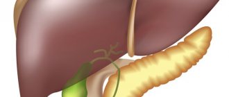
The human intestine, the photo of which is presented above, has a complex anatomical structure. All the main departments are clearly visible here.
If we consider in more detail, the anatomy of the human intestine consists of smaller sections:
- duodenum;
- jejunum and ileum;
- cecum;
- ascending transverse and descending colon;
- sigmoid and rectum;
- anus.
The human intestine begins immediately after the stomach and joins it. And it ends with the anus - the anus. Being an integral part of the digestive tract, the intestines closely interact with all organs included in it. It is into the intestinal sections that bile comes from the gallbladder, while it itself supplies the stomach with hydrochloric acid for the primary decomposition of incoming food. Having a complex, varied structure and purpose, it plays one of the most important functions in human life.
Thus, the total length of the intestine in an adult is about 7-9 meters, while in a newborn it is 3.5 meters long. Since it grows with a person, its location may change depending on age. The diameter and shape of the intestines also changes, increasing and expanding with age.
The structure of the human cecum
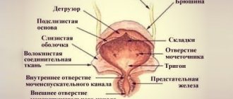
The cecum (caecum), as a section of the large intestine, is the initial part of the large intestine below the point where the ileum enters the colon. The length of the cecum is 6-8 cm, the diameter is 7.0-7.5 cm. The cecum is located in the right iliac fossa, on the iliacus and psoas major muscles. The cecum is covered with peritoneum on all sides, but does not have a mesentery. One of the structural features of this section of the large intestine is that on the posteromedial side of the cecum below, all three bands of the colon converge at one point. appendix (appendix vermiformis) , departs from the cecum .
At the point where the ileum enters the cecum there is an ileocecal opening (ostium ileocaecale) , which looks like a horizontal slit. This opening in the structure of the cecum is limited above and below by two folds (lips) protruding into the cavity of the cecum, forming the ileocecal (ileocecal) valve (valva ileocaecalis). Anteriorly and posteriorly, the folds (lips) come together and form the frenulum of the ileocecal valve (frenulum valvae ileocaecalis) in the anatomy of the large intestine. In the thickness of the folds of the valve there is a circular layer of muscles, the contractions of which prevent the return of food masses from the cecum to the ileum. Somewhat below the ileocecal valve, on the inner surface of the cecum there is an opening of the appendix (ostium appendicis vermiformis) .
How does the human intestine work?
Just like the esophagus and stomach, the intestines work through peristaltic contractions, pushing the contents towards its end, that is, the anus. During this movement, chyme is processed by intestinal juices and broken down into amino acids and other simple compounds. In this state, they can be absorbed into the intestinal walls and enter the blood, through which nutrients and energy are distributed throughout the body. The intestinal wall consists of four layers:
- serous outer lining of the intestines;
- muscle layer;
- submucosa;
- intestinal mucosa.
These layers are conductors of valuable nutrients for the body, and also play the role of an energy exchanger. The intestine is the largest organ in the human body. Just as the lungs supply the body with oxygen from the outside world, the human intestines serve as a conductor between the blood and the energy consumed. The photo below shows that the blood supply to this organ is through three main branches of the abdominal aorta.
Peristalsis is very diverse; contractions can be rhythmic, pendulum, figurative peristaltic and antiperistaltic, tactical. Such movements of the intestinal muscles allow not only to move the masses towards the exit, but also to mix, grind and compact them together.
What symptoms should alert you to a malignant tumor of the colon?
- As a rule, in the early stages of the disease, symptoms are scant: chronic fatigue, lethargy, loss of interest in life, general abdominal discomfort, unexplained weight loss, hypochromic normocytic anemia.
- In later stages, persistent constipation, aversion to food, incontinence of gas or feces, blood in the stool (there may be either streaks or a significant amount of scarlet blood), and a painful urge to have ineffective bowel movements (tenesmus) may appear.
- With advanced tumors, chronic or acute intestinal obstruction may develop. Symptoms of this life-threatening complication include pain in the abdomen, nausea, vomiting, sometimes even stool, inability to defecate, combined with a painful urge to defecate.
- When the process spreads throughout the peritoneum – peritoneal carcinomatosis – ascites develops. At the same time, fluid accumulates in the abdominal cavity, which compresses the internal organs and aggravates the serious condition of the patient.
At the age of over 50 years, a combination of even mild discomfort in the abdominal area with hypochromic anemia (decrease in hemoglobin and red blood cells in a general blood test) in combination with an increase in ESR and leukocyte levels, as well as a positive stool test for occult blood and increased blood clotting should be mandatory order to entail endoscopic examination of the intestine.
Duodenum
The duodenum is one of the shortest sections, but it is not the least important in the entire digestive system. The length of the human intestine in this section is about 21-25 centimeters. It is here that the incoming food is broken down into its components: carbohydrates, proteins and fats. The duodenum is also responsible for controlling the release of the required amount of hydrochloric acid that enters the stomach and helps break down food into smaller fragments. Through the production of various enzymes and the flow of bile, it sends signals to the rest of the intestines to begin moving food from the stomach, facilitating the beginning of the release of secretions for further processing of chyme.
Are there methods that are 100% accurate in diagnosing colon cancer?
The modern level of endoscopic technology, when carried out in a timely manner, allows diagnosing and typing intestinal tumors at the earliest stages; if necessary, data from sigmoidoscopy or colonoscopy with biopsy of suspicious areas can be supplemented by rectal ultrasound, irrigoscopy, ultrasound of the abdominal cavity and pelvis. These studies make it possible to clarify the location of the tumor, the degree of its growth into nearby organs and tissues, and the presence of inflammation in the surrounding tissues. To get a complete picture, MRI and computed tomography are performed. Modern technologies allow, if it is psychologically impossible to perform a colonoscopy, to conduct a so-called virtual colonoscopy. To diagnose the presence and localization of distant metastases, PET-CT (positron emission tomography) is used.
Contact your doctor
Small intestine
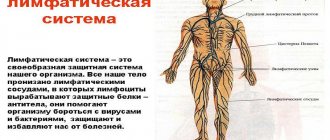
Immediately after the end of the duodenum, sections of the small intestine join it, the first of which is the jejunum, and then it smoothly passes into the ileum. Thus, this department consists of two parts. The length of the human small intestine, including all its sections, ranges from 5 to 7 meters. The processes of digestion and absorption of nutrients occur in it. Energy exchange occurs by transferring nutrients and microelements through the walls into the blood. The walls of the small intestine secrete special enzymes called enterocytes, which are capable of breaking down food into simple amino acids and glucose from fatty acids. Subsequently, through absorption into the intestinal mucosa, these substances enter the body. Glucose and amino acids are transmitted through the blood. Fatty acids, in turn, enter the lymphatic capillaries, passing through them to the liver.
The small intestine is very important for humans and, despite the fact that the entire intestinal system is long, it is precisely without this section that a person cannot exist. The bauhinium valve is located between the small and large intestines. It is a muscle fold and serves to prevent the movement of feces from the large intestine back to the small intestine.
The human small intestine has connecting fasteners of various widths and shapes, ensuring the position of the intestine and its rounded loops, as well as its fixation. With their help, it is fixed to the posterior abdominal wall. The small intestine contains a lot of blood and lymphatic vessels, as well as nerve endings.
Small intestine and its functions
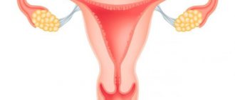
The diagram clearly demonstrates the location of the small intestine between the stomach and large intestine.
The small intestine is responsible for the digestive process, and it is named so because of its comparatively smaller diameter and thinner walls, unlike the large intestine. But in its size it is not inferior to any organ of the gastrointestinal tract, capturing almost the entire lower space of the peritoneum and part of the pelvis.
The overall work of enzymes in the small intestine, gallbladder and pancreas promotes the breakdown of food into individual components. Here, the absorption of vitamins, nutrients, and active components of most medications necessary for the human body takes place.
In addition to digestive and absorption functions, it is responsible for:
- movement of food masses further through the intestines;
- strengthening the immune system;
- hormonal secretion.
This segment is divided according to its structure into three sections: duodenum, jejunum, and ileum.
Colon
The large intestine is located around the perimeter of the relatively small intestine and has a frame-like shape, located closer to the abdominal cavities. After food passes through the jejunum and ileum, broken down to the simplest amino acids, and after they are absorbed into the intestinal walls and blood, the rest of the mass, which is based on fibers and cellulose, enters this section. The main function of the large intestine is to absorb water from the remaining mass and form solid feces for removal from the body. Nevertheless, digestion processes continue to occur in it.
The human large intestine is saturated with various microorganisms that help process substances that cannot be absorbed into the human body. Various types of lactobacilli, bifidobacteria and some types of E. coli live here. The content and concentration of such bacteria is responsible for the health of the intestine and its microflora. If any of the types of microorganisms decrease in number or disappear altogether, then dysbacteriosis develops in the body. It can occur in quite severe forms and promotes the development and proliferation of pathogenic microbes and fungi, which not only reduces the level of immunity in general, but can also have serious consequences for the health of the body.
The structure of the human intestine of the large section includes the following intestines:
- blind;
- ascending colon;
- right flexure of the colon;
- transverse colon;
- descending colon;
- sigmoid colon.
The large intestine is much shorter than the small intestine and ranges from one and a half to two meters in length. In diameter it ranges from 7 to 10 centimeters.
Colon
Colon
(lat.
intestinum crassum
) - the lower section of the gastrointestinal tract, starting after the small intestine and ending with the anus. Together with the small intestine it makes up the intestine.
Colon Anatomy
There are three sections in the large intestine: the cecum (No. 6 in the figure on the right; lat. caecum
) with the vermiform appendix (No. 8), the colon (Latin
colon
) with four subsections (ascending colon, transverse colon, descending colon (No. 7 ) and sigmoid colon) and rectum (No. 9; lat.
rectum
) with a wide part - the ampulla of the rectum and a terminal tapering part - the anal canal (No. 10), ending in the anus. The length of the colon in an adult is on average 160 cm, the internal diameter is on average 5 to 8 cm and decreases in the direction from the cecum to the rectum. The thickness of the wall of the colon is 2–3 mm, during contraction it is 4–5 mm, the thickness of the wall of the rectum is 2.4–8 mm. The large intestine is separated from the small intestine by the ileocecal valve.
In the colon, the bulk of water, electrolytes, glucose, vitamins and amino acids produced by symbiotic bacteria are absorbed and feces are formed from chyme, as well as the accumulation and retention of the latter until it is excreted.
The most numerous endocrine cells of the intestine are located in the mucous membrane of the colon - L-cells, which produce the hormones enteroglucagon (glucagon-like peptide-1) and peptide YY.
The normal residence time of the contents (chyme and feces) in the large intestine is about 26 hours.
Acidity in the lower colon
I.A. Churkin studied the acidity in the lower parts of the colon in healthy people using endoscopic pH-metry. He found that the average acidity value for the entire rectum is 8.1 pH, for the sigmoid intestine - 8.1 pH, and also determined the following average acidity values for the lower parts of the colon (in pH):
- lower ampullary rectum: pH=7.3 (item 1 in the figure on the right)
- mid-ampullary rectum: pH=7.7 (position 2)
- upper ampullary rectum: pH=8.5 (position 3)
- supramullary rectum: pH=8.7 (position 4)
- distal third of the sigmoid colon: pH=8.7 (position 5)
- middle third of the sigmoid colon: pH=7.9 (position 6)
- proximal third of the sigmoid colon: pH=7.9 (position 7)
Colon microbiota
The large intestine, to a much greater extent than the small intestine, is populated by various microorganisms, the number of species of which approaches 500. In the large intestine, microorganisms make up 30% of the dry mass of the luminal contents.
The most common and physiologically significant are anaerobes: bifidobacteria, lactobacilli, bacteroides, veillonella, fusobacteria, eubacteria, peptostreptococci, clostridia and aerobes and conditional anaerobes: Escherichia coli, lactose-negative enterobacteria (Enterobacter, Citrobacter, etc.), Proteus, Enterococcus ki, klebsiella , staphylococci, yeast-like fungi. The number of microorganisms increases towards the distal parts of the colon, more in the luminal rather than in the parietal zones (Dobrovolsky O.V., Serebrova S.Yu.). Qualitative and quantitative composition of the main microflora of the colon in a healthy person in colony-forming units (CFU) in terms of 1 g of feces (according to OST 91500.11.0004-2003 “Protocol for the management of patients. Intestinal dysbiosis”):
| Types of microorganisms | Age, years | ||
| less than 1 | 1–60 | more than 60 | |
| Bifidobacterium ( Bifidobacterium ) | 1010–1011 | 109–1010 | 108–109 |
| Lactobacilli ( Lactobacillus ) | 106–107 | 107–108 | 106–107 |
| Bacteroides _ _ | 107–108 | 109–1010 | 1010–1011 |
| Enterococci ( Enterococcus ) | 105–107 | 105–108 | 106– 07 |
| Fusobacterium ( Fusobacterium ) | <106 | 108–109 | 108–109 |
| Eubacterium ( Eubacterium ) | 106–107 | 109–1010 | 109–1010 |
| Peptostreptococcus ( Peptostreptococcus ) | <105 | 109–1010 | 1010 |
| Clostridia ( Clostridium ) | ⩽103 | ⩽105 | ⩽106 |
| E. coli ( Escherichia coli ) typical | 107–108 | 107–108 | 107–108 |
| Lactose-negative E. coli | <105 | <105 | <105 |
| Hemolytic Escherichia coli | 0 | 0 | 0 |
| Other opportunistic bacteria: Klebsiella , Enterobacter , Hafnia , Serratia, Proteus , Morganella , Providencia , Citrobacter and others | <104 | <104 | <104 |
| Staphylococcus aureus (S taphylococcus aureus ) | 0 | 0 | 0 |
| Staphylococcus saprophyticus ( Staphylococcus saprophyticus ) and epidermal ( Staphylococcus epidermidis ) | ⩽104 | ⩽104 | ⩽104 |
| Yeast-like fungi of the genus Candida | ⩽103 | ⩽104 | ⩽104 |
| Non-fermenting bacteria: pseudomonas ( Pseudomonas ), acinetobacter ( Acinetobacter ) and others | ⩽103 | ⩽104 | ⩽104 |
In addition to those listed, bacteria of the following genera are present in the human colon in varying quantities: Actinomyces, Bacillus, Corynebacterium, Peptococcus, Acidaminococcus, Anaerovibrio, Butyrovibrio, Acetovibrio, Campylobacter, Disulfomonas, Propionibacterium, Roseburia, Ruminococcus, Selenomonas, Spirochetes, Succinomonas, Coprococcus
.
In addition to these groups of microorganisms, you can also find representatives of other anaerobic bacteria ( Gemiger, Anaerobiospirillum, Metanobrevibacter, Megasphaera, Bilophila
), various representatives of non-pathogenic protozoan genera (
Chilomastix, Endolimax, Entamoeba, Enteromonas
) and more than ten intestinal viruses (Ardatskaya M.D., Minushkin O.N.).
According to some data, approximately 1% of the total microbiota consists of the bacteria Christensenella minuta.
A decrease in the amount of
Faecalibacterium prausnitzii
in the normal microflora may be associated with Crohn's disease.
The figure above shows the species and quantitative composition of the dominant microflora of the human colon, obtained by the most modern method - using the analysis of the 16S rRNA gene (may vary in different populations, but not in a very significant way). Slightly less than half of the total number of bacteria in the colon are bacteria of the following species: Faecalibacterium prausnitzii, Eubacterium rectale, Collinsella aerofaciens, [Clostridium] clostridioforme, Bacteroides vulgatus, Anaerostipes hadrus, Ruminococcus bromii, Eubacterium hallii, Blautia wexlerae, Bacteroides dorei, Roseburia faecis, Dorea longicatena, Subdoligranulum variabil, Bacteroides uniformis, Blautia obeum, Bacteroides ovatus, Blautia luti, Parabacteroides distasonis, Lachnospira pectinoschiza Dialister invisus, Roseburia inulinivorans, Ruminococcus callidus
.
Large intestine in children
A child's colon has a length equal to his height.
The sections of the large intestine are developed to varying degrees. The newborn has no omental processes, the bands of the colon are barely visible, and haustra are absent until six months of age. The anatomical structure of the colon after 3–4 years of age is the same as in an adult. The cecum, which has a funnel shape, is located higher, the younger the child. In a newborn it is located directly under the liver. The higher the cecum is located, the more underdeveloped the ascending colon is. The final formation of the cecum ends by the age of one year.
The appendix in a newborn has a cone-shaped shape, a wide open entrance and a length of 4–5 cm, by the end of 1 year - 7 cm. It has greater mobility due to the long mesentery and can be located in any part of the abdominal cavity, but most often occupies a retrocecal position.
The colon in the form of a rim surrounds the loops of the small intestine. The ascending part of the colon in a newborn is very short (2–9 cm), and begins to increase after a year.
The transverse part of the colon in a newborn is located in the epigastric region, has a horseshoe shape, length from 4 to 27 cm; by two years she is approaching a horizontal position.
The descending colon in newborns is narrower than the rest of the colon; its length doubles by 1 year, and by 5 years it reaches 15 cm. It is poorly mobile and rarely has a mesentery.
The sigmoid colon is the most mobile and relatively long part of the colon (12–29 cm). Until 5 years of age, it is usually located in the abdominal cavity due to an underdeveloped small pelvis, and then descends into the small pelvis. Its mobility is due to the long mesentery. By the age of 7, the intestine loses its mobility as a result of shortening of the mesentery and the accumulation of adipose tissue around it.
The rectum in children of the first months is relatively long and, when filled, can occupy the small pelvis. In a newborn, the ampulla of the rectum is poorly differentiated, the fatty tissue is not developed, as a result of which the ampulla is poorly fixed. The rectum reaches its final position by the age of two years. Due to the well-developed submucosal layer and weak fixation of the mucous membrane, young children often experience its loss.
The anus in children is located more dorsally than in adults, at a distance of 20 mm from the coccyx (Bokonbaeva S.D. et al.).
Large intestine in newborns
The colon in a newborn has an average length of 63 cm, and by the end of the first year of life - up to 83 cm. Subsequently, the length of the colon is approximately equal to the height of the child.
By birth, the colon has not completed its formation. The newborn does not have omental processes (they are formed in the 2nd year of the child’s life); the ribbons of the colon are barely outlined; colon haustra are absent (appear after 6 months). The bands of the colon, haustra and omental processes are finally formed by 6–7 years. The large intestine provides water resorption and evacuation-reservoir function. It completes the breakdown (both under the influence of enzymes coming from the small intestine and bacteria inhabiting the large intestine) and the absorption of nutrients, and the formation of feces occurs.
The mucous membrane of the colon in children is characterized by: deepened crypts, flatter epithelium, and a higher proliferation rate. Juice secretion in the colon is insignificant under normal conditions, but it increases sharply with mechanical irritation of the mucous membrane (Geppe N.A., Podchernyaeva N.S.).
Some diseases and conditions of the colon
Certain colon (including rectal and anorectal) diseases and syndromes:
| Three stages of colon cancer |
- colon cancer
- functional dyspepsia, including dyspepsia in children
- irritable bowel syndrome
- irritable bowel syndrome with diarrhea
- irritable bowel syndrome without diarrhea
- pseudomembranous colitis
- Crohn's disease
- nonspecific ulcerative colitis
- enterocolitis
Symptoms and conditions that may be associated with diseases of the colon (including the rectum):
- stomach ache
- nausea
- vomit
- constipation, including constipation in children
- dyschezia
- flatulence
- diarrhea (diarrhea), including diarrhea (diarrhea) in children
- anismus
- gastrocolic syndrome
- encopresis
- tenesmus
- melena
On the website GastroScan.ru in the “Literature” section there is a subsection “Diseases of the colon and anus”, containing medical articles concerning the treatment of the rectum and diseases of the anal canal. Back to section
Appendix
The appendix is a vermiform appendage of the cecum, located in the large intestine, which can be located towards the bottom or upward, towards the liver. The appendix carries out the function of storing lymphoid tissues that are part of the immune system. Beneficial bacteria of the microflora of the large intestine accumulate here, which serves as a reserve storage for them in the event of dysbacteriosis. During the use of antibiotics that kill the bacterial environment of the large intestine, the microflora of the appendix is not affected. Thus, it is much more difficult for people with a removed appendix to experience the state of dysbiosis. It is a kind of incubator for the development of E. coli, bifidobacteria and lactobacilli.
The vermiform appendix does not have standard sizes and can vary depending on the individual structure of the digestive tract. The length of the intestine in an adult in the appendix is 7-9 centimeters, and in diameter up to 1 centimeter. However, its length can range from 1 centimeter to 23, which will be the norm. At the junction with the large intestine, the appendix has a small fold of mucous membrane, which serves as a barrier against chyme entering it. If this flap is not large enough and does not protect it from moving masses, it becomes full and inflamed, which is a disease called appendicitis. In this case, surgical removal of the appendix is used.
What you need to know before having a colonoscopy
Health officials recommend that anyone over 50 with an average risk of colorectal cancer have a colonoscopy every 10 years or a flexible sigmoidoscopy every five years.
The main instruments used when examining the colon for cancer are flexible sigmoidoscopes and colonoscopes. These expensive pieces of equipment are not designed for single use, which means they must be thoroughly cleaned inside and out and sterilized before each use. This is where the problem lies.
Earlier in the year, another medical tool, the duodenoscope, used to treat cancer, gallstones, biliary and pancreatic duct diseases, was linked to at least 25 outbreaks of drug-resistant bacteria, sickening 250 people.
This is especially concerning because this endoscope was recalled in 2016 after it was discovered that a small mechanism on the endoscope was causing the bacteria to be transmitted between patients.
The company has reportedly corrected the problem, but now Sen. Patty Murray, D-Wash., is asking for evidence that the endoscope can be properly disinfected, as the company claimed.
Rectum
At the end of the large intestine there is another section - the rectum. Through it, feces accumulate, form and are expelled. The exit from the rectum is located in the pelvic area and ends at the anus. The length of the human intestine in this lead ranges from 13 to 23 centimeters, and in diameter from 2.5 to 7.5 centimeters.
The rectum, despite its small size, consists of several sections:
- supramullary;
- rectal ampulla;
- perineal section;
- anal columns;
- internal, then external sphincter;
- anal sinuses and valves.
Is it possible to do anything to prevent colon cancer?
To prevent the development of a malignant tumor of the colon, especially for people in the older age group, it is recommended to lead a healthy lifestyle and adhere to universal preventive measures - undergo an annual examination (rectal examination, stool occult blood test, colonoscopy). We have posted detailed material on colon cancer screening on our website.
Detection of precancerous changes and malignant tumors of the colon in the early stages allows timely treatment to begin. Early diagnosis and active treatment, widely introduced into widespread clinical practice, have made it possible to achieve high five-year survival rates - with the initial detection of colorectal malignancy at stage 1, this figure is 90-93%, at stage 2 - about 70-75%, at stage 3 - th stage – 40-48%. And despite the improvement of treatment methods, survival rate when cancer is initially detected at stage 4 does not exceed 5-9%.
Colonoscopy allows you to diagnose precancerous conditions and tumors in the early stages of development, timely identify malignant tumors using biopsy and histological examination, and also quickly and minimally traumaticly remove polyps that are prone to “malignancy” (malignization). The European Oncology Clinic actively uses chromoscopy and NBI endoscopy techniques, which make it possible, even during diagnostic endoscopy, to diagnose precancerous changes at the initial stage with a high degree of certainty and to carry out endoscopic intraluminal surgery for resection of small tumors without metastases. The removed macroscopic specimen will necessarily undergo histological and, if necessary, immunohistochemical examination. Such diagnostic and treatment options can quickly return patients to active life, as well as convince high-risk patients to undergo timely testing, since many people are frightened by the prospect of a colostomy and delay seeking help even if they have symptoms.
Find out the exact cost of treatment
The structure of the intestinal wall
The human intestine has a layered structure, which ensures its functions of peristalsis, secretion of enzymes and juices, and interchange of substances with the rest of the body. The walls consist of four layers:
- mucous membranes;
- submucosa;
- muscle layer;
- outer serous layer.
The mucous membrane of the small intestine consists of villi that provide interaction with the surface of the intestines and the circulatory system.
The muscle layer consists of an inner circular, round layer and an outer longitudinal layer.
The mucous membrane of the large intestine does not have villi, but consists of scripts and mucous folds.
The structure of the human intestine can be easily recognized by color. The large intestine is gray in color, while the small intestine is pink in color.
Diagnostics
A targeted diagnostic search for suspected colon cancer includes clinical, radiological, endoscopic and laboratory examination. Valuable information can be obtained from an objective examination, palpation of the abdomen, percussion of the abdominal cavity, digital examination of the rectum, and gynecological examination.
X-ray diagnostics involves plain radiography of the abdominal cavity, irrigoscopy with the use of a contrast agent. In order to visualize the tumor, take a biopsy and smears for cytological and histological examination, rectosigmoidoscopy and colonoscopy are performed. Informative methods of topical diagnosis include ultrasonography of the large intestine and positron emission tomography.
Laboratory diagnosis of colon cancer involves the study of a general blood test, stool for occult blood, and determination of carcinoembryonic antigen (CEA). In order to assess the prevalence of the malignant process, liver ultrasound, MSCT of the abdominal cavity, pelvic ultrasound, chest radiography are performed, and, if indicated, diagnostic laparoscopy or exploratory laparotomy is performed.
Colon cancer requires differentiation from many diseases of the intestine itself and adjacent organs, primarily chronic colitis, ulcerative colitis, Crohn's disease, actinomycosis and tuberculosis of the colon, benign tumors of the colon, polyposis, diverticulitis, cysts and ovarian tumors .
Sections of the colon
Features of the anatomical location of the intestine in the abdominal cavity made it possible to divide it into 4 sections:
- Ascending colon.
- Transverse colon.
- Descending colon.
- Sigmoid colon.
The total length of the four parts reaches 1.5 -2 meters.
Ascending colon
The intestine is located to the right of the midline of the abdomen (right flank) in the abdominal cavity. Being a continuation of the cecum, it rises up to the lower edge of the liver. At this level, it forms the right flexure of the colon (hepatic flexure) and passes into the transverse part of the large intestine. The length of the ascending part is about 15-20 cm. The ascending part is topographically limited from behind by the quadratus dorsi muscle and the right kidney, above by the right lobe of the liver and gall bladder, in front by the anterior abdominal wall, and inwardly by loops of the small intestine. In a small number of people, the intestine has its own mesentery, which ensures its mobility and the development of volvulus of the cecum and colon (in rare cases).
Transverse colon
The ascending and descending sections of the large intestine are connected to each other by the transverse colon. The intestine is located in a horizontal plane, slightly sagging downwards. It originates from the hepatic flexure and reaches the left hypochondrium, forming the splenic flexure (left flexure of the colon). The left flexure is located above the right flexure of the colon. When palpating the abdomen, it can be detected above the navel in the form of a horizontal elastic cord.
The length of the transverse colon varies from 25 cm to 65 cm in an adult. The transverse colon is bounded on the right by the liver, on the left by the stomach and spleen. The duodenum and pancreas are located behind the intestine, and the loops of the small intestine are adjacent below. The anterior part is covered by the anterior abdominal wall. In the abdominal cavity, it is attached to the walls using the mesentery.
Descending colon
It starts from the left flexure of the colon and descends to the left iliac fossa, passing into the sigmoid colon. Posterior to the intestine lies the left kidney and the quadratus dorsi muscle. Covered in front and left by the abdominal wall. The right side of the descending colon is adjacent to the loops of the small intestine. The length of an adult varies from 10 to 30 cm.
Sigmoid colon
It is located in the left iliac region and forms 2 loops: proximal and distal, which lie on different muscles. The proximal part is supported by the iliacus muscle, and the distal part by the psoas major muscle. The length of the sigmoid colon can be from 15 cm to 50 cm in an adult. Near the intestine there is the left ovary, uterus, and bladder.
Human gastrointestinal tract
Any organism needs energy to maintain its vital functions. It can be obtained in different ways. For example, in humans, like many other animals, there is a gastrointestinal tract and a digestive system in general for this purpose.
This part of the body is quite complex and is responsible for the complete processing of all incoming substances, maximum extraction of useful substances and disposal of residues. It all starts in the oral cavity, because this is where food initially enters. First, it is thoroughly crushed and mixed with saliva, with the help of which the primary breakdown immediately begins, and then enters the stomach. Here, with the help of various substances, the incoming food is further processed and carbohydrates, part of the water, ethanol and some salts are absorbed.
The next stage is the small intestine. This is where the main digestion of food occurs, the breakdown of carbohydrates, proteins and fats into simpler components and their absorption for transport and delivery to cells. This is possible due to the special structure of the mucous membrane of this organ. The fact is that the inner surface of the intestine is covered with microgrowths - villi, which significantly increases the absorption area. This section is also an important part of the hormonal system, since the synthesis of some peptide hormones that regulate the activity of the entire gastrointestinal tract and immune processes occurs here.
And finally, the last stage of the gastrointestinal tract is the large intestine. The structure and functions of this organ are worth considering separately and in more detail, because they are no less interesting than those of the other parts of the digestive system. And, of course, they are also very important.
General characteristics of the digestive system
Its work can be compared to a giant factory for processing food, breaking it down, assimilating and recycling substances. In each section of the system, specific biochemical reactions occur with the participation of an arsenal of enzymes and biologically active substances, such as vitamins.
The large intestine, the structure and functions of which we are studying, is physiologically considered as an organ involved in the secretion, digestion, absorption and removal of substances from the overlying sections. To understand the functions, first consider how the large intestine works.
Location
The large intestine, the general anatomy of which was described a little earlier, begins after the bauhinian valve, which separates the ileum and cecum. This structure does not allow the reverse movement of chyme - the pulp obtained as a result of digestion of food.
Next, the intestine passes up and to the left, encircling the previous section of the gastrointestinal tract, and then descends again, ending with the anus. Through it, feces are evacuated, that is, the body gets rid of unnecessary residues. However, sometimes the rectum is separated from the colon. In this case, its last section becomes the part called the sigmoid.
Diseases of the small intestine
There are disruptions in the production of enzymes necessary for the complete breakdown of food. Insufficiency of digestive function - maldigestion. The condition in which absorption is impaired is called “malabsorption.” As a result, the body does not receive the substances it needs. The following processes may develop: destruction of bone tissue, splitting of nails and hair loss.
Symptoms of diseases of the small intestine:
- pain in the navel area;
- bloating, heaviness in the abdomen;
- loose stools, light-colored feces;
- "boiling" in the stomach;
- weight loss.
Inflammation of the small intestine - enteritis - can be caused by bacteria. The production of enzymes and digestion in general are disrupted. In the absence of enzymes responsible for digesting carbohydrates, intolerance to this food component develops. For example, lactase deficiency is the inability to break down the milk sugar lactose. Celiac disease is the absence of enzymes that break down gluten in cereals. Undigested substances become toxic products that poison the intestines.
To restore microflora, it is recommended to take probiotics along with prebiotics. In case of enzyme deficiency, the patient is prescribed medications that contain the missing substances. Treatment of intestinal dysbiosis is carried out with antibiotics and probiotics.
Parameters of different parts of the digestive tube
The small intestine (intestinum tenue) has a length of 1.6 to 4.3 meters. In men it is longer. Its diameter gradually decreases from the proximal to the distal part (from 50 to 30 mm). Intestinum tenue lies intraperitoneally, that is, intraperitoneally, its mesentery is a duplicate of the peritoneum. The leaves of the mesentery cover blood vessels, nerves, lymph nodes and vessels, and fatty tissue. The cells of the intestinum tenue produce a large number of enzymes that take part in the process of digesting food along with pancreatic enzymes; in addition, all medications and toxins, when taken orally, are absorbed here.
The length of colon is comparatively smaller - 1.5 meters. Its diameter decreases from beginning to end from 7-14 to 4-6 cm. As described above, it has 6 divisions. Caecum has an outgrowth, a vestigial organ, the appendix, which, according to most scientists, is an important component of the immune system.
Throughout the colon there are anatomical formations - bends. This is the place where one part of it transitions to another. Thus, the transition of the ascending to the transverse colon is called the hepatic flexure, and the splenic flexure is formed by the transverse descending sections.
The intestines are supplied with blood through the mesenteric arteries (superior and inferior). The outflow of venous blood is carried out through the veins of the same name, which make up the portal vein basin.
The intestine is innervated by motor and sensory fibers. The motor branches include the spinal and branches of the vagus nerve, and the sensory fibers include the fibers of the sympathetic and parasympathetic nervous system.
Appearance
The length of the large intestine is about 1.5 m and the diameter is 5-8 cm. It runs around the small intestine in the form of a frame.
Important macroscopic features of the walls of the large intestine are the haustra or group of pouches. If they are located in the inner wall of the intestine, they are called plicae semilunares coli.
Appearance of the large intestine
Colon segments:
- Cecum (cecum) with appendage.
- Hind intestine.
- Large intestine: ascending, colon, descending, sigmoid.
- Rectum.
Parts of the human colon
Avoid eating antibiotic-fed CAFOs and processed meats
Processed meats and red meat CAFOs have been linked to colon cancer. It is important to understand that many of these meat products contain antibiotic residues and other compounds that may increase the risk of cancer.
Processed meats, such as bacon, ham, pastrami, salami, pepperoni, hot dogs, and some sausages , are foods that use smoking, aging, curing, or chemical preservatives as preservatives. Nitrates in processed meats are often converted to nitrosamines, which are directly linked to an increased risk of certain types of cancer.
A 2007 analysis by the World Cancer Research Fund (WCRF) found that even eating just one sausage each day could increase the risk of developing bowel cancer . Specifically, they found that eating 1.8 ounces of processed meat (equivalent to one sausage or three pieces of bacon) every day increased the risk of developing cancer by 20 percent.
Research also shows that the risk of colorectal cancer among people who eat red meat (in one study, five ounces a day) is 24 percent higher than those who eat less meat. However, red meat does not appear to be a problem in itself; the process of preparing it and the source of the meat itself likely also play a role. Meat from grass-fed animals, for example, contains anti-cancer compounds.
When it comes to meat, I recommend eating organic meat from grass-fed animals. In addition, such meat does not need to be subjected to severe heat treatment (in rare cases).
For the record, I believe that many people require animal protein to maintain optimal health, although most consume much more protein than is necessary (or healthy).
Digestion and absorption of nutrients
Chemical changes in food components mainly occur in the lumen of the small intestine. The same processes occur inside the epithelial cells and near the villi. Numerous glands of the small intestine in the mucous layer secrete up to 2 liters of digestive juice per day with enzymes that decompose food into its component parts. Proteins and peptides are broken down into amino acids. Fats are broken down into fatty acids and glycerol. The main product of digestion of complex carbohydrates is glucose.
The functions of the small intestine are not only the breakdown of food. Another important process occurs - the absorption of final products into the blood and lymphatic capillaries in the villi. Water, nutrients, vitamins and mineral components pass from the intestinal lumen into the blood and lymph and can participate in metabolism. From them, like from construction kit parts, the body creates its own proteins, fats and carbohydrates.
Intestinal absorption is a complex chemical and physiological phenomenon. Amino acids and glucose directly enter the blood of the capillaries of the intestinal villi. Fats are absorbed into the lymphatic capillaries and then enter the bloodstream. Not only the diffusion of molecules through the mucous membrane occurs. Some particles are actively transported from the intestine due to the coordinated work of ions.
Important! Malabsorption in the intestines is a serious problem for the entire body. Metabolism worsens, a deficiency of vitamins, microelements, and iron appears.
The intestine is commonly called the “second brain” of the human body. The upper sections produce hormonal substances that are necessary for the intestines themselves and the entire body for normal activity and the functioning of the immune system. Most of the cells that produce such compounds are located in the walls of the duodenum.
Functions
The large intestine is not a physiologically active organ. Usually the main purpose of this organ is considered to be the formation of feces and disposal of them from the body. In fact, the functions of this organ are broader.
Firstly, this is where the extraction of nutrients from food is completed. Everything useful that was impossible to digest before is processed. For example, only at this stage can fiber be extracted. Also, the remaining water and salts are almost completely sucked out of the chyme.
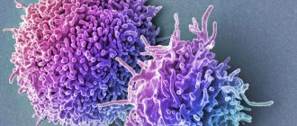
Secondly, the human large intestine is an important part of the immune system. Colonies of bacteria of different types are located here, mostly anaerobic. Some of them help digestion, others prevent the spread of pathogenic microbes, others produce enzymes that promote the proper functioning of the organ, as well as vitamins K, E, B6 and B12, which are necessary for the entire body. In short, the microflora of the large intestine is an important part of the protective barrier of the human body. And it is so powerful that it can cope with even single cancer cells, completely destroying them.
Thirdly, it is the structure of the large intestine, in particular its muscle layer, that ensures the constant movement of digested food. At the same time, a person cannot even control it. The rate of peristalsis is usually constant and increases after a new portion of food enters the stomach. Thus, the muscles of the colon are responsible for maintaining a more or less constant speed of the food "conveyor".
Despite the fact that the gastrointestinal tract can cope with almost everything that a person consumes as food, you should not mindlessly throw anything into yourself. It’s not difficult to disrupt your intestines, but you may not notice it right away. And when symptoms of its malfunction appear, it can be very difficult to restore balance, so it is better to stick to a healthy diet with a sufficient amount of fiber, which perfectly stimulates the gastrointestinal tract.
Diseases
In the vast majority of cases of intestinal problems, we are talking about stool disorders. Diarrhea, constipation, flatulence - most often the cause of these unpleasant phenomena is errors in nutrition. In this form, intolerance to lactose, gluten and some other substances may occur. Dysbacteriosis can also cause problems with stool disorders or constant exacerbations of allergies. At the same time, you should not self-medicate or take advertised medications without consulting a doctor, especially if bowel dysfunction occurs on an ongoing basis. This can only make the problems worse.
Inflammation in any part of the intestine can also be quite dangerous. If it is not treated, the mucous membrane becomes covered with ulcers, and then the processes of decay begin. Even more dangerous is the appearance of formations in the intestinal lumen. These could be malignant tumors or hemorrhoids, but both require medical attention. Fortunately, almost always formations are located in the last section, which greatly facilitates access and diagnosis. And, as doctors tend to believe, diseases of the intestine, as well as the entire gastrointestinal tract, are almost always the result of poor nutrition and lifestyle. Fortunately, modern medicine is able, if not to completely cure a person, then to alleviate his condition as much as possible and maintain quality of life with minimal intervention.
Be healthy!