- Causes
- How does the disease manifest itself?
- What complications can there be?
- Types of testicular tumors
- Stages of tumor development
- How to make a diagnosis
- Treatment methods
- Postoperative rehabilitation and disease prevention
- Prognostic factors and survival statistics
Primary testicular tumors are the most common solid malignancy in men aged 20 to 35 years.
For unknown reasons, the incidence of this cancer—mainly testicular seminoma—increased in the 20th century. Over the past decade, the incidence of testicular cancer has increased by about 1.2% per year, although the rate of increase has been slowing. Germ cell cancer accounts for more than 90% of all testicular cancer cases. Approximately 9,000 new cases are diagnosed in Europe each year. And the overall incidence in Europe at the beginning of 2019 was 137 patients per 1 million population.
Characteristics of the disease
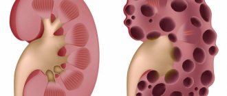
Testicular cancer is a malignant neoplasm; it is characterized by unpredictability of development and rapid growth of tumor cells.
This cancer is more common among other malignant tumors. And since it is considered very aggressive, statistics call it one of the most common causes of death among cancer patients under 35 years of age.
This form of cancer is considered the most common form of cancer among men aged 15-35 years. Typically, this cancer pathology is unilateral, although there are cases of a bilateral form of the cancer process (about 2% of diseases of this form).
This male tumor forms and quickly develops in the male gonads (testicles), but then begins to spread throughout the body by hematogenous and lymphogenous routes (usually to the brain and bone structures, lungs, liver).
Testicular tumors
The number of types of neoplasms of male testicles is large. This is explained by the fact that this organ of the male reproductive system consists of many membranes, each of which differs in tissue and composition. And from each such membrane different tumors can develop.
Classification of tumors
Modern medicine distinguishes three groups of testicular tumors:
- germinal;
- gonadal stroma;
- rest.
The most frequently diagnosed tumors of the first group are germ cell tumors. They, in turn, include tumors such as seminoma, teratoma, choriocarcinoma, embryonal carcinoma, yolk sac tumors, as well as some mixed tumors. As for cancerous tumors, they are quite rare compared to all others. Their frequency rate is two percent. Tumors of this type are diagnosed mainly in early male age. However, modern medicine has identified three periods with a high risk of cancer. These are boys under ten years old, young men from 20 to 40 years old, as well as older men over 60 years old.
Reasons for the development of testicular tumors
Factors that can provoke the occurrence of tumors are the following:
- Cryptorchidism. This is a pathology characterized by undescended testicles into the scrotum, which occurs from the very first weeks of a child’s life or during the development of the fetus in the womb. The testicle remains in the inguinal canal or in the abdominal cavity. This reason increases the risk of developing a tumor tenfold. Moreover, if the testicle remains in the abdominal cavity (a syntrabdominal testicle), the risk of tumors becomes greater than if it were retained in the scrotum. The risk increases many times (by about 30 percent) if both testicles are retained in the abdominal cavity, rather than just one. The appearance of cancerous tumors on a healthy testicle (lowered into place) is observed in twenty percent of men with this pathology. All other cases are the development of neoplasms on an undescended testicle. This is explained quite simply. The fact is that in the abdominal cavity, where the undescended testicle is located, the temperature is higher than in the scrotum. It is two to three degrees higher. And exposure to higher temperatures has a negative effect on the male reproductive organs. Moreover, if this effect is long-term, which causes the occurrence of certain mutations in the organ. In turn, these mutations can cause rapid cancer development.
- Hereditary factor. If a father, brother or grandfather was diagnosed with cancer, the risk of developing a cancer tumor in a child increases several times.
- Testicular injuries. More recently, this factor was also considered to predispose to the appearance of cancer. However, some modern scientists have a slightly different opinion on this matter. They believe that because a man consults a doctor for any testicular injury, a diagnosis is made, as a result of which the tumor (if any) is detected.
Signs of tumor development
The most common reason why men seek medical advice and help is the feeling that there is some kind of foreign formation in the scrotum. This can be discovered completely by accident. It should be noted that it is very important to identify such a tumor in a timely manner. In most cases, it is early diagnosis, leading to timely treatment, that helps save the patient’s life. But doing this on your own at very early stages of tumor development is quite difficult, one might say almost impossible. Because this is a very small nodule (or slight swelling of the scrotum), which is absolutely painless. That is why independent detection of a newly emerging cancer tumor is a very rare occurrence.
The only option to detect a tumor in its very first stages of development is a routine examination by a doctor or seeking medical help for some other reason, during which the tumor is discovered.
It must be said that the detection of malignant tumors in approximately ten percent of cases occurs due to the appearance of certain metastases, which are characterized by a certain distance. For example, metastases can appear in the retroperitoneal lymph nodes and compress the ureters. This, in turn, disrupts to one degree or another the functioning of the urinary system. And a violation of the outflow of urine is one of the reasons for the development of pyelonephritis, acute hydronephrosis and other pathologies. A man seeks the help of a doctor for the above reasons, and as a result, a cancerous tumor of one stage or another is discovered. Metastases can also occur in the lymph nodes of the cervical region. They contribute to compression of the upper respiratory tract, which causes shortness of breath, and in some cases, cough. Metastases that appear in the brain due to cancerous tumors can have a negative impact on the psyche. They cause neurological disorders, which manifest themselves in the development of paresis, and in some, more rare cases, paralysis. Testicular tumors can also metastasize to the lung area, which disrupts the normal functioning of the respiratory system. Metastases also appear in the skeletal system. This entails severe pain, as well as the risk of multiple fractures.
There are cases when a sign of the development of testicular tumors is an increase in the size of the male mammary glands. This phenomenon is called gynecomastia.
So, to summarize, it must be said that the most obvious primary symptom of the development of testicular tumors in the male body is an increase in size of either one testicle, or both, the appearance of areas of compaction, nodules of various sizes. As for advanced cases, which occur when a cancerous tumor is not detected in time, they are characterized by severe general weakness of the body, nausea, accompanied by loss of appetite and some other symptoms of a nonspecific nature.
Diagnostics
Diagnostic measures aimed at identifying a tumor include several procedures. Let's look at each of them.
- Examination of the patient: general condition of the body, scrotum with its careful palpation. At the same time, an examination (palpation) of areas of the body where metastases are most likely to develop is also carried out.
- Ultrasound, aimed primarily at examining the testicle, in which the development of a tumor is suspected. This is necessary both to identify the tumor itself and to determine its connection with possible other formations in the scrotum area. In addition, ultrasound examination is carried out for all organs of the peritoneum in order to confirm the presence or absence of metastases. An ultrasound is performed to exclude tumor metastasis to the lymph nodes and retroperitoneal region.
- Blood tests: general and so-called important. A standard blood test is aimed at identifying proteins that are characteristic only of cancerous tumors. These are so-called tumor specific markers. An important blood test helps determine the amount of alpha-fetoprotein, human chorionic beta subunit, lactate dehydrogenase. If the level of these substances in the blood is elevated, this may indicate the development of a cancer process in the body.
- X-ray of the lungs, carried out in order to exclude metastases in them.
All of the above tests and examinations must be carried out after treatment. This must be done in order to determine the degree of effectiveness of treatment, as well as to exclude the possible occurrence of relapses.
Treatment of tumors
It must be said that the choice of treatment is directly determined by the root cause of the pathology, and also depends on the stage of its development.
If the cancerous tumor (seminoma) is at the first stage of its development and is not accompanied by damage to the lymph nodes, then in most cases the testicle is completely removed. And then the iliac and retroperitoneal nodes are irradiated. In the second stage of seminomas, which occurs in parallel with damage to the lymph nodes, but provided that there are metastases in them no more than 5 centimeters, treatment consists of using the same measures. That is, this means removal of the testicle and further irradiation of the lymph nodes. But treatment of the third stage of a cancerous tumor is already more complex. It involves removal of the affected organ, radiation treatment of the lymph nodes, after which strong chemotherapy is prescribed.
In the case of non-seminoma tumors, a surgical intervention called orchiectomy is prescribed. It involves the complete removal of the organ affected by the pathology. If nearby lymph nodes are affected, standard chemotherapy may be prescribed. In some cases, this is not enough and a decision is made to perform a second operation, during which partial or complete removal of the affected retroperitoneal lymph nodes occurs.
Preventive measures
The most important preventive measure for detecting testicular cancer is a thorough independent examination of the scrotum area. It is recommended to carry out this procedure approximately once a month. If you have even the slightest suspicion of the development of some pathology, you should immediately go to the doctor. It must be remembered that it is the timely diagnosis, and, therefore, earlier treatment, that in most cases helps to achieve a complete cure. How to perform a self-examination? This must be done while standing in front of a mirror. First you need to carefully examine the scrotum. Detection of redness of various localization and distribution on the skin is already a reason to seek medical advice. Moreover, you should see a doctor if one of the testicles or both have increased in size. Therefore, careful palpation of the scrotum is necessary. To do this, taking it in your hand, you need to use your fingers quite slowly and, most importantly, carefully, without pressing too hard, to feel each testicle, supporting it at the same time. The epididymis (a fairly thin, elastic tube) is also palpated. Everything needs to be done carefully.
So, if, upon self-examination, a man reveals such signs as an increase in the size of one testicle, detection of a tubercle of any size, redness of any area of skin in the scrotum area, then, without delay, you need to contact the clinic for examination. In addition, you need to consult a doctor if, during an independent examination of the testicles, in particular during palpation, a man experiences some painful sensations. If there is blood in the urine, you should also consult a specialist. A feeling of heaviness in the scrotum area, the appearance of pain in the lower abdomen, as well as in the entire groin area of varying degrees of manifestation and of a permanent nature, is also a reason to visit a doctor. Consultation is also needed in the case of causeless enlargement of the mammary glands in a man.
Leading clinics in Israel
Assuta
Israel, Tel Aviv
Ikhilov
Israel, Tel Aviv
Hadassah
Israel, Jerusalem
Prognosis and prevention
Modern methods of treating cancer involve therapy while preserving reproductive functions. But still, various consequences may appear after such a disease, including male infertility.
Based on statistical data, in half of the cases, patients after chemotherapy treatment can restore erectile and reproductive functions, even if the tumor masses are removed along with the affected organ. The remaining second testicle can fully fulfill the task, and the patient will be able to have children in the future. The worst prognosis for such a pathology as testicular cancer is possible only with angiolymphatic invasion of the tumor and the presence of metastases.
Methods for preventing testicular cancer include timely treatment of cryptorchidism, as well as preventing trauma to the scrotum and avoiding radioactive exposure to the genitals. You can identify the disease at an early stage if you regularly conduct independent examinations, and in case of any changes, immediately undergo examination by a urologist-andrologist.
Classification of testicular cancer (testicular cancer)
This type of cancer is divided into germ cell, non-germ cell and mixed types:
- Germ cell tumors are the most common type of testicular cancer (95% of all cases); they develop from seminal germ cell structures;
- Non-germinogenic forms of the tumor are formed from the testicular stroma;
- Mixed species consist of cells of non-germinogenic and germinogenic formations.
Germ cell tumors are further subdivided into:
- Seminomas; Seminoma
- Non-seminomas (teratoma, chorionic carcinoma, embryonal carcinoma or embryonal carcinoma, etc.).
Non-germinogenic types of tumors are very rare (about 5% of cases) and are divided into the following types of tumors:
- Leydigoma;
- Sertoliomas;
- Dysgerminomas.
Types of malignant neoplasms on the testicle in men
The classification of testicular cancer into stages is based on the prevalence of the disease. Important criteria are damage to the retroperitoneal lymph nodes and the presence of metastases in other organs - most often the liver and lungs. The first stage of testicular cancer is characterized by the presence of a neoplasm on the male testicular gland without signs of metastasis. If the retroperitoneal lymph nodes are affected, the condition is clinically regarded as the second stage of cancer. Stage III testicular cancer is characterized by damage to the mediastinal lymph nodes or distant metastases (to bones, liver, lungs, brain).
According to histological structure, the following types of testicular tumors in men are distinguished:
- Germinogenic (coming from the seminiferous epithelium);
- Non-germinogenic (arising from the stroma of the gonad);
- Mixed neoplasms.
Testicular germ cell tumors occur in 95% of cases in men. They can be represented by the following neoplasms:
- Seminoma;
- Embryonic cancer;
- Chorionic carcinoma;
- Malignant teratoma.
40% of cases of germ cell tumors are seminomas, 60% are non-seminoma tumors. Non-germinogenic tumors of the sex cord stroma include sertolioma, sarcoma, and leydigoma.
Signs of testicular cancer
The first signs and symptoms of such a malignant process are the appearance of dense formations in the tissues of the scrotum, which contribute to the enlargement of the organ.
These lumps may or may not be painful. Typically, patients feel some swelling on the man's testicle, pain usually manifests itself in the abdomen and scrotum. Men may experience the following symptoms:
- Dramatic weight loss;
- Fast fatiguability;
- Low constant temperature (37.5%).
A further sign of the formation of a cancerous tumor is the feeling that the testicles are very swollen - they are greatly increasing in size. Then breathing difficulties arise, shortness of breath appears, lymph nodes become enlarged, back pain appears, and general weakness is felt.
Another sign is a noticeable decrease (or absence) of sexual desire, soreness and enlargement of the mammary glands. If the tumor metastasizes, a cough appears, right costal pain, and shortness of breath occur.
If the tumor grows into the epididymis, the following symptoms may occur:
- Deformation and enlargement of the testicle;
- A painful lump is felt;
- Swelling of the scrotum;
- Painful sensations along the spermatic cord and lower abdomen;
- Other symptoms (chest pain, back pain, swollen lymph nodes, difficulty breathing).
Epididymal cancer, in turn, can provoke the formation of secondary sexual characteristics and thyroid diseases, which can change the patient’s appearance. In adults, a decrease in libido is observed, impotence and feminization are possible. Boys often experience gynecomastia and premature masculinization (hirsutism, voice mutation, frequent erections, macrogenitosomia).
Stages
Stage 0 (carcinoma in situ)
The testicle has a precancerous condition called germ cell neoplasia in situ. This is also called intratubular germ cell neoplasia.
Stage 1A - The tumor is in the testicle and epididymis, but may have spread to the inner layer of the membrane that surrounds the testicle (tunica albuginea). All levels of tumor markers are normal.
Stage 1B - One of the following is true.
- It is found in the testicle and epididymis and has spread to the blood vessels or lymphatic vessels of the testicle.
- It has grown into the outer layer of the membrane that surrounds the testicle (tunica vaginalis).
- It has grown into the spermatic cord or scrotum and may have spread to blood vessels or lymphatic vessels in the testicle.
All levels of tumor markers are normal.
Stage 1C —The tumor is found anywhere in the testicle, spermatic cord, or scrotum, and 1 or more tumor marker levels are above normal.
Stage 2A —The cancer has spread to 1 or more lymph nodes in the groin, and they are no larger than 2 cm. Tumor marker levels may be slightly higher than normal.
Stage 2B - The cancer has spread to 1 or more lymph nodes in the groin, and they are between 2 and 5 cm. Tumor marker levels may be slightly higher than normal.
Stage 2C - Cancer has spread to 1 or more lymph nodes in the groin and they are larger than 5 cm.
Stage 3A—The cancer has spread to lymph nodes outside the groin or to the lungs (called distant metastasis). One or more levels of tumor markers may be slightly higher than normal.
Stage 3B—The cancer has spread to 1 or more lymph nodes in the groin, and 1 or more tumor marker levels are moderately higher than normal.
Stage 3C - any of the following are true:
- The cancer has spread to the lymph nodes in the groin, and one or more levels of tumor markers are significantly higher than normal.
- The cancer has spread to lymph nodes outside the groin or to the lungs. This is also called metastatic testicular cancer. One or more levels of tumor markers are much higher than normal.
- The cancer has spread to a distant part of the body other than the lymph nodes or lungs. One or more levels of tumor markers may be higher than normal.
Differences between right and left testicular cancer
Despite the fact that they are paired organs, cancer of the left testicle is different from cancer of the right. The causes of the appearance of a cancerous tumor, the form of growth and the course of the disease of both testicles are similar, but metastases spread differently.
When localized in the right testicle, the tumor metastasizes to the lymph nodes, which are located along the aorta and in the space between the aorta and the inferior vena cava. In left testicular cancer, nodes located around and in front of the aorta are more affected. From a cancerous tumor of the right testicle, one can expect metastases to the retroperitoneal lymph nodes on the left side, which is unusual for tumor localizations on the left side. If a neoplasm is diagnosed in one testicle, then there is a possibility of appearing in the other.
Diagnostics
The evaluation of patients with suspected testicular cancer begins with a history and physical examination.
Laboratory tests and imaging studies include:
- determination of serum alpha-fetoprotein level;
- determination of the level of serum β-subunit of human chorionic gonadotropin (beta-hCG);
- determination of lactate dehydrogenase (LDH) level
- Ultrasound of the testicles;
- high-resolution computed tomography (CT) of the abdomen and pelvis;
- chest x-ray;
- Head magnetic resonance imaging (MRI) bBrain should be performed if brain metastases are suspected after clinical examination.
Stages of the disease
Staging of testicular cancer occurs according to the international TNM system, where T denotes the size of the tumor:
- T-1 – the formation does not extend beyond the boundaries of the tunica albuginea;
- T-2 – sizes are also limited, but there is deformation of the scrotum and an increase in the size of the testicle;
- T-3 – the tumor extends beyond the boundaries of the tunica albuginea and grows into the adnexal tissues;
- T-4 – the tumor extends beyond the boundaries of the egg and grows into the scrotum or testes.
N - indicates the size of regional metastasis:
- N-1 – regional metastases are detected by X-ray and radioisotope diagnostics;
- N-2 – enlarged lymph nodes with metastases are easily palpable.
M – indicates the presence of distant metastases.
Another system for determining the stage of development of this type of cancer is also used:
- I – the tumor is located within the egg;
- II – the tumor begins to spread to the lymph nodes;
- IIa – lymph nodes with metastases no more than 2 cm;
- IIb – the size of lymph nodes with metastases ranges from 2 to 5 cm;
- IIc - the size of lymph nodes with metastases exceeds 5 cm;
- III – 0 – the lymph nodes of the neck and chest are involved in the process;
- IV – metastases spread to distant organs.
Don't waste your time searching for inaccurate cancer treatment prices
*Only upon receipt of information about the patient’s disease, a representative of the clinic will be able to calculate the exact price for treatment.
Classification
Germ cell tumors
More than 90% of all testicular cancers are germ cell tumors. This type of cancer starts in the cells that produce sperm.
The two main types of germ cell tumors that develop in the testicles are seminomas and nonseminomas. Each tumor type grows differently and is treated differently.
Germ cell tumors may consist of a mixture of seminoma and nonseminoma. These tumors are called mixed germ cell tumors. They are usually considered nonseminomas.
Sometimes germ cell tumors start outside the testicle. These tumors are called extragonadal germ cell tumors. Extragonadal germ cell tumors can start anywhere in the body, but they usually start in the area between the lungs (mediastinum), abdomen (retroperitoneum), lower spine, or pineal gland in the brain.
Seminomas grow more slowly than non-seminomas. Seminomas most often develop in men aged 40 years.
Nonseminomas account for about 55% of all germ cell tumors. Nonseminomas most often occur in men aged 0 to 30 years.
The cells in nonseminomas are very different from seminomas.
There are 4 main types of nonseminomas. They are named and described based on how cancer cells look under a microscope.
- Fetal carcinoma is a type that tends to grow quickly and spread beyond the testicle. On biopsy, these tumors look like very early embryonic cells.
- Yolk sac carcinoma is rare in adults, but it is the most common form of testicular cancer in children and infants. When children develop yolk sac carcinomas, they usually respond very well to treatment.
- Choriocarcinoma is a rare and aggressive type of testicular cancer. It can quickly spread to distant organs, including the lungs, bones and brain. Choriocarcinomas are usually part of a mixed germ cell tumor.
- A teratoma has areas similar to each of the 3 layers of the developing embryo (endoderm, mesoderm, and ectoderm). Pure teratomas are rare. This type of cancer is most often part of a mixed germ cell tumor.
Rare testicular tumors
Tumors that do not originate from germ cells (nongerm cell tumors) can also develop in the testicle, but they are rare.
Sex cord stromal tumors are also called gonadal cord stromal tumors. They begin in the cells of the stroma, which serves as the supporting tissue in the testicle. There are 2 main types of stromal tumors:
- Leydig cell tumors
- Sertoli cell tumors
Most of these tumors are benign, but some may be malignant.
Testicular non-Hodgkin's lymphoma begins in the epithelium of the testis. Learn more about non-Hodgkin's lymphoma.
Testicular adenocarcinoma begins in a duct that helps transport new sperm to the epididymis, where sperm mature.
Paratesticular rhabdomyosarcoma is a type of soft tissue sarcoma that begins outside the testicle in the spermatic cord.
Diagnosis of testicular cancer
To make such a diagnosis, it is necessary to undergo an examination by a specialist who will palpate the scrotum and conduct a general examination to find out why it is swollen. Sometimes even at this stage there is a suspicion of the presence of a malignant tumor. Lymph nodes in the groin, supraclavicular and peritoneal locations are also examined.
After such an examination, the doctor gives a referral for diagnostic tests. Usually prescribed:
- Ultrasound. This test helps determine the presence or absence of a tumor with almost 100% accuracy;
- MRI and computed tomography. Similar to ultrasound, but is more informative about the disease;
- Osteoscintigraphy. She clarifies whether there are metastases or not;
- Tests for tumor markers;
- Morphological diagnosis of tumor fragments (this study is prescribed after removal of the testicle to determine the risk of metastases).
Analysis for tumor markers is of great importance in identifying the degree of tumor development, choosing therapy and monitoring the response to prescribed treatment. Tumor markers (ACE, LDH, hCG) are characteristic substances that are produced by malignant neoplasms. For example, ACE increases in 70% of patients with testicular cancer. HCG increases in patients with testicular choriocarcinoma. Testicular cancer can be diagnosed at the initial stage of the disease using a pregnancy test. The fact is that this test will immediately show an increase in the level of hCG in the urine.
The final diagnosis is made after a testicular biopsy through an inguinal approach. As a rule, during such a diagnosis, an urgent morphological examination of the biopsy is carried out, and if the presence of cancer is confirmed, then excision of the gonad with the spermatic cord is performed (orchifuniculectomy).
Based on all studies, the most appropriate therapy is prescribed.
Causes of development and risk factors
The specific etiology of testicular cancer is not known, but various risk factors have been associated with it quite reliably.
Cryptorchidism
Patients with cryptorchidism have a four to eightfold increased risk of developing a germ cell tumor. The risk of developing a germ cell tumor when the testicle is inside the abdominal cavity is about 5%. The risk is 1% if it persists in the inguinal canal. Surgical placement of an undescended testicle in the scrotum—orchiopexy—when the patient is younger than 6 years of age further reduces the risk.
Approximately 5%-20% of patients with a history of cryptorchidism develop tumors of the normally descended testis.
Genetic factors
Patients with Klinefelter syndrome (47XXY) have a higher incidence of germ cell tumors, especially primary mediastinal germ cell tumors. Family members of patients with Klinefelter syndrome have a 6- to 10-fold increased risk of developing a germ cell tumor. Patients with Down syndrome are also at increased risk of germ cell tumors.
An increased risk also occurs in patients with hereditary diseases such as:
- cutaneous ichthyosis;
- Müller's syndrome;
- androgen insensitivity syndrome;
- mixed gonadal dysgenesis.
Family history
First-degree relatives have a higher risk of developing the disease than the general population, although the incidence remains low. Brothers are at particularly high risk, with a relative risk of 8–10. A two- to six-fold increase in testicular cancer has been reported among the sons of affected men.
Infertility
Walsh et al reported that men with male factor infertility were nearly 3 times more likely to develop subsequent testicular cancer. Intra-abdominal germ cell neoplasia has been found in 0.4–1.1% of men undergoing testicular biopsy for infertility.
Chemical carcinogens
Exposure to diethylstilbestrol (DES) in utero is associated with cryptorchidism. Increased risk has been suggested with exposure to Agent Orange and numerous industrial occupations.
Testicular cancer treatment methods
Common methods used in the treatment of testicular cancer:
- Surgical intervention;
- Chemotherapy;
- Radiation therapy.
Surgical intervention involves removal (orchifunnectomy) of a testicle that has undergone a tumor process. Sometimes this operation also involves the removal of lymph node structures (retroperitoneal lymphadenectomy). After surgery, a course of chemotherapy or adjuvant radiation therapy is usually prescribed.
The disadvantage of complex treatment of testicular cancer (removal, chemotherapy and radiotherapy) can be temporary or long-term infertility and impotence. That is why, before starting therapy, it is advised to preserve the seed fund using cryopreservation if a man plans to have a child in the future.
Causes
Modern means of self-defense are an impressive list of items that differ in their operating principles. The most popular are those that do not require a license or permission to purchase and use. In the online store Tesakov.com, you can buy self-defense products without a license.
According to statistics, testicular cancer occurs mainly between the ages of 20 and 40 years. The peak incidence occurs at the age of 32 years. After 40 years, the likelihood of developing the disease decreases significantly.
The exact causes of carcinoma are not known. There are several risk factors for the development of this pathology:
- genetic predisposition;
- cryptorchidism;
- radiation education;
- living in an environmentally unfavorable area.
The influence of factors such as testicular trauma, instrumental interventions and infertility has not yet been proven.
Cryptorchidism: 1 - scrotum; 2 - normal testicle; 3 - undescended testicle
Cryptorchidism (undescended testicle into the scrotum) is considered one of the leading risk factors for testicular cancer. Normally, the testicles should descend into the scrotum by the time the baby is born or within two years after the baby is born. The testicles cannot exist in the retroperitoneal space: too high a temperature changes the structure of the testicles and provokes the growth of a malignant tumor. To prevent this from happening, after the age of 2 years, doctors suggest performing an operation to lower the testicle into the scrotum and thereby save the boy from the risk of developing cancer.
Some statistics:
- With cryptorchidism, the likelihood of developing testicular cancer increases by 3-10 times (according to various sources).
- With unilateral cryptorchidism, the tumor most often occurs in the undescended testicle.
- Surgery to lower the testicle into the scrotum reduces the risk of developing cancer, but does not completely eliminate the possibility.
Hereditary predisposition is another reason for the development of testicular cancer. Scientists were able to find out: there are certain gene regions responsible for the occurrence of carcinoma. The presence of testicular cancer in a grandfather, father or brothers is a reason for a thorough examination and constant monitoring of the condition of the testicles.
If there is a family predisposition, the likelihood of developing cancer increases several times.
Disease prognosis
The successful outcome after treatment is influenced by many factors:
- Early detection of cancer (survival rate is about 90%);
- Detection of cancer at stages 2-3 with metastases (complete cure is impossible, but 5-year survival rate is about 50%).
If a man plans to become a father in the future, it is recommended to cryopreserve the semen before starting treatment. Unfortunately, it is impossible to completely exclude the likelihood of developing this type of cancer, since in most cases it begins in the prenatal period. But timely treatment of urological pathologies and abnormalities (dropsy, cryptochism) will help greatly reduce the risk.
Considering the rapid growth rate of the tumor, you should be more attentive to any suspicious changes in your body and promptly contact a specialist for consultation. Then the risk of catching the disease in the initial stage and completely getting rid of it is quite high.
Symptoms
Manifestations of testicular cancer include:
- the appearance of a palpable tumor in the scrotum (a round, painless formation);
- increase in size of the scrotum (due to tumor growth);
- pain in the testicles (in later stages of the disease).
All symptoms of testicular cancer are not specific and can occur in many other diseases. For an accurate diagnosis, you need to consult a doctor (urologist or oncologist).
Testicular cancer develops from a single cell that can divide uncontrollably. In the initial stages, the tumor is invisible and cannot be detected by known methods. From the moment of the birth of the first atypical cell until the appearance of a space-occupying formation, about 5-7 years pass. Subsequently, the tumor begins to grow rapidly, increasing in size by 2-3 times every 2-3 months. There are cases where testicular carcinoma reached very large sizes, interfered with walking and caused significant discomfort by its very existence.
If at least one of the symptoms occurs, you must immediately consult a doctor!
Testicular cancer is an aggressive tumor with rapid growth and the appearance of distant metastases. During metastasis, carcinoma cells spread through the blood and lymph to other organs. Metastases are most often found in the retroperitoneal lymph nodes and lungs.
Symptoms associated with testicular cancer metastases:
- shortness of breath, cough and difficulty breathing (if the lungs are affected);
- swelling of the lower extremities (due to compression of the inferior vena cava);
- pain in the lumbar region (as a result of enlarged retroperitoneal lymph nodes);
- renal colic (due to compression of the ureter);
- kidney damage and development of renal failure;
- intestinal obstruction (due to compression of the intestine by altered lymph nodes).
The appearance of metastases indicates a significant spread of cancer and requires a special approach to treating the disease.
Prevention
Women discover the first signs of breast cancer themselves, so it’s worth adopting the habit of taking your health more seriously from them. The first signs can be detected by self-examination. Self-examination is best done in the shower with both hands. Carefully feeling the testicles, the skin near them, the surface of the body under the testicles and above the testicles.
Such an examination helps to detect swollen testicles or that a lump has appeared on the scrotum, the presence of a nodule at the bottom of the scrotum, any growths on the testicles, thickening of the skin - any deviation from the normal appearance.
In any case, further examination will be carried out by a specialist who will check whether your concerns are justified. But early detection of unpleasant symptoms will help you begin treatment earlier and avoid major health problems in the future.
Treatment
After confirming the diagnosis of testicular cancer, treatment is carried out surgically, as well as with radiation and chemical therapy. Typically, treatment involves a complex of techniques, starting with surgery, after which the use of chemotherapy drugs is necessary. Organ-preserving surgery can be performed either in a single case or in the presence of bilateral tumors in the testicles. Cancer is highly treatable when diagnosed at an early stage.
We recommend reading: Signet ring cell cancer of the stomach - causes, types, diagnosis
Surgical treatment
The operation most often involves removal of the affected testicle, but sometimes it is also possible to remove the affected retroperitoneal lymph nodes and spermatic cords. All surgical procedures are performed under general anesthesia. After surgery, the patient is prescribed radiation and chemotherapy to prevent tumor recurrence.
Chemotherapy
At an advanced stage of the disease, chemotherapy is prescribed, which involves completing a course according to the BEP regimen using special drugs for 4 episodes of treatment every three weeks. If the patient is in remission, but examination reveals secondary lesions, the patient is prescribed surgical therapy with excision of the tumor. The group with a possible unfavorable outcome includes people with high levels of tumor markers.
Self-examination
A man himself is able to identify testicular cancer in its first stages. This can be done by ordinary palpation. If it begins to look something different, becomes too hard or soft, swollen, lumpy, or changes in shape or size. The disease can make itself felt by painful sensations. At the first suspicion of this insidious disease, you should contact a specialist for diagnosis. When a man goes to the clinic on time with his problem, it is much easier to solve it, but if suspicious symptoms of the disease are ignored, the outcome can be quite disastrous.
We have looked at the symptoms of testicular cancer in men and will deal with the consequences.
Who is at risk?
Like any other disease, testicular cancer has its own characteristics and has its own risk factors that increase the possibility of manifestation.
First of all, these include the following:
- Gene system . If very close relatives suffered from this terrible disease, then this fact significantly exceeds the possibility of developing cancer.
- Bad habits : drinking alcohol, smoking.
- Cryptorchidism. Congenital pathology. In this case, one, or maybe both testicles do not move into the scrotum, but freeze in the abdominal cavity. According to experts, with a unilateral disease, in 2 cases out of 10, the disease will definitely develop, and with a bilateral disease, the chance of getting the disease increases by another one.
- Infertility . People who suffer from infertility are more susceptible to getting cancer.
- Diseases carried by viruses. Mumps brings with it extreme danger, which after exposure gives its own complications, orchitis being the main one.
- HIV-infected patients have a greater chance of contracting cancer. We present to your attention an article about the first symptoms of HIV infection in men here.
- Age restrictions . The maximum number of cases observed in patients is between 35 and 55 years of age.
- Body type . According to medical observations, thin people who are tall are more susceptible to the disease.
- Previously suffered cancer . In many cases, if a person has already suffered from such ailments and the lesion was removed (testicular amputation), then there is a possibility of developing the disease on the second, remaining testicle.
People who are at risk need to be especially careful about their well-being and respond to any changes, especially to factors that cause cancer. In an article about throat cancer and its symptoms in the early stages, a similar question was considered.
How to get rid of prostatitis without the help of doctors, at home?!
- so that the pain stops
- normalize urination
- so that sexual desire appears and the opportunity to have sexual intercourse. Shaken men's health can and should be restored! with a timely course of treatment. Read more "
Symptoms of benign tumors
The most common symptoms of a testicular tumor are its focal compaction in the form of a node, usually unilateral, an increase in the size of the testicle, a change in its shape, or swelling of half of the scrotum. The listed manifestations, as a rule, are not accompanied by pain, however, as the tumor grows, pain occurs in the testicle itself and along the spermatic cord.
Rare primary symptoms may be a simple increase in size of the testicle, which was previously small and atrophied, the appearance of pain in the lower abdomen due to enlargement of the retroperitoneal lymph nodes.
Systemic metastatic damage to the lymph nodes of the retroperitoneal space and lungs causes an increase in lymph node packets or pain in the back, abdomen, chest, difficulty breathing, signs of ileofemoral thrombosis, pneumonia, etc.
Acute pain in the testicle and its enlargement simulate the clinical picture of acute epididymo-orchitis; the addition of hydrocele of the testicular membranes in 10% of cases is characteristic of a tumor lesion.
Hormonal activity of some testicular tumors causes changes in secondary sexual characteristics. Germ cell tumors (chorionepithelioma) in 5-10% can manifest as gynecomastia, non-germ cell tumors (leydigoma) - precocious puberty, hirsutism and pronounced development of secondary sexual characteristics in boys. In children, tumors are most often diagnosed at the age of 1-2 years and during puberty.
Without urgent specific treatment, the disease usually progresses rapidly and leads to death.
If the symptoms of epididymal cancer are extremely poorly expressed, which complicates the timely detection of a dangerous disease, then benign neoplasms become noticeable at an early stage of their development. This, of course, is a huge plus.
Initially, a small tubercle appears, for example, in the form of a pea, which is easily palpated and has a dense structure. Over time, and also depending on the nature of the pathological nature, the neoplasm can increase, and to completely obscene sizes.
A feeling of constant discomfort, the appearance of persistent pain, as well as swelling of the scrotum are the main symptoms characteristic of the vast majority of tumors that are not classified as malignant.
A man who suspects the development of such changes should pay attention to the following symptoms:
- visual deformation of the testicles - for example, one testicle becomes much larger than the other, their rounded structure also changes;
- an increase in the contents of the scrotum is easy to determine by simultaneous palpation of two testicles;
- swelling of the scrotum - it becomes as if filled with fluid, significantly increasing in size;
- pain syndrome, which is localized not only in the external genitalia, but also in the lower abdomen;
- swollen lymph nodes;
- exacerbation of existing chronic diseases, including those with a history;
- blood or blood in urine and semen;
- difficult or painful urination, an increase or, conversely, a decrease in the number of urges;
- deterioration of health;
- the appearance of pain in the back and chest;
- decreased body endurance;
- unnatural hormonal activity of the body, which increases or, conversely, decreases the development of secondary sexual characteristics. As a result, the patient's appearance may change beyond recognition. Often a man loses his libido, essentially turning into a woman.
Each type of benign testicular tumors has its own symptoms that are unique to it. Determining it is very important, since in some cases this is the only way to establish an accurate diagnosis and, accordingly, prescribe adequate treatment.
Varicocele, for example, is characterized by expansion of the pampiniform venous plexus inside the testicle. You can see it both visually and by palpation, depending on the stage of development of the disease.
The cyst is usually small in size, but sometimes it can grow to such a size that it is difficult to distinguish it from a hydrocele. In this case, you can find out that we are talking about cystic formations using a characteristic symptom - the cysts seem to be floating inside the scrotum.
As for hydrocele, an important symptom of this disease is the appearance of significant swelling that envelops the entire appendage. An overgrown tumor can make palpation of the testicle impossible - only huge swelling will be felt.
Acute pain, radiating not only to the lower abdomen, but also to the lower back and even the leg, as well as fever, are symptoms characteristic of epididymitis. This disease belongs to the group of those that are easiest to diagnose.
The spermatocele is usually located on the upper part of the testicle. If this pathology is not treated, it can grow to the size of a hydrocele. Spermatoceles are often confused with cystic formations. Given the mild symptoms, this disease is mainly diagnosed only on the basis of appropriate clinical studies.
Hematocele manifests itself as a significant increase in the size of the testicle itself. The fact is that blood accumulates inside it, and not outside. Therefore, if a testicle suddenly begins to grow, this should cause serious concern in a man.
A disease in which the contents of the peritoneum can enter the scrotum is called an inguinal hernia. In this case, the neoplasm will be very large - its upper limit is extremely difficult to determine, since it ends in the abdominal cavity itself.
If you have symptoms such as nausea, vomiting, or sudden attacks of severe pain, you must call an ambulance.
A benign testicular tumor is a neoplasm in the scrotum, which can have different origins, but almost always indicates the presence of a specific disease. Contrary to the popular belief, which was formed under the influence of the word “benign”, that it does not need to be treated, the help of a specialized specialist must be immediate.
Some types of tumors can lead to such serious consequences as erectile dysfunction, infertility, and hormonal imbalance. The man eventually ceases to feel that he belongs to the stronger sex.
Modern medicine makes it possible to get rid of benign tumors mainly through surgery. In this case, which is very important, the affected testicle is preserved, unlike cancer, when it is removed along with the tumor.
Etiology
Based on the fact that a tumor of the testicle, or rather its epididymis, is a rare disease, modern medicine has studied it relatively superficially. But nevertheless, a certain clinical picture is known, due to which the diagnosis, as a rule, is not delayed.
From a visual point of view, a benign formation is a compaction of various shapes and dimensions, which is localized in the tail of the appendage. In rare cases, it may appear directly in the appendage itself.
In almost 80 percent of cases of tumors found in the testicles, they are benign. All the rest are malignant. Every man needs to remember this.
It should be noted that this disease, based on its specificity, is very difficult to treat even with surgical treatment, not to mention conservative treatment, that is, with the help of various medications.
We suggest you read: Pancreatic tumor stages 4 and 3, and how long they live with it
Predisposing factors traditionally include:
- chronic diseases of the endocrine system;
- cryptorchidism;
- hormonal disorders in the male body;
- bad habits – smoking, alcohol abuse, taking drugs;
- mechanical injuries and microtraumas of the scrotum;
- radiation exposure;
- hereditary factor - if someone in the family has previously had similar problems, then the risk of developing tumors in future generations increases significantly.
Not all benign tumors are capable of developing into classic cancer. But still, a number of diseases can provoke this. The slightest signs of the development of a neoplasm in the scrotum should be the reason for immediate contact with a specialized specialist.
Numerous studies have proven the predisposing influence of cryptorchidism, Klinefelter's syndrome, testicular trauma, infertility, microwave and ionizing radiation. There are indications of chemical mechanisms, with the exogenous intake of estrogens and dysfunction of the hypothalamic-pituitary part of the endocrine system.
Pathogenesis
There are tumors of germ cell and non-germ cells. In adults, the percentage of these tumors is 95:5, in children - 76:24. Additionally, mixed tumors are isolated. The growth of malignant testicular tumors of germ cell origin begins from the intratubular focus of spermatogenic epithelium. The tumor enlarges as it grows, becomes palpable and can replace testicular tissue.
Local spread of the tumor consists of its invasion into the epididymis, its membrane and the spermatic cord. When the lymphatic system is affected, the retroperitoneal (rarely pelvic and inguinal) lymph nodes are primarily affected.
Hematogenous spread occurs directly during invasion and indirectly through the lymphatic duct and veins. Among the distant organs, the lungs are primarily affected by metastases. The skeletal system is rarely affected.
Why does the disease develop?
The main causes of the disease have not been fully studied to date. It is generally accepted that the occurrence and development are influenced by some congenital factors, disorders in the reproductive system that arose in the uterine state.
As already described above, the reasons for the occurrence of this insidious disease in a particular man are unknown.
But it is generally accepted that some factors that are directly related to the disease are to blame:
- Heredity. People whose relatives suffered from similar diseases are at great risk.
- Changes occurring in the reproductive system . If patients, even before they reach 11 years of age, do not have their testicles surgically returned from the abdominal cavity to the scrotum (this disease occurs at birth), then their risk of getting cancer is much higher than for others.
- Previously suffered cancer.
- Significant deviations in the genitals . With them, the opening of the urethra can only open in the lower part of the genital organ. For people who have these symptoms, the likelihood of getting cancer increases significantly.
- Testicular torsion. 64% of patients have a chance of developing cancer.
Forecast
The prognosis for benign testicular tumors is favorable. Removing the tumor and/or the organ itself eliminates the problem. The second testicle completely takes over all the functions of producing sperm and hormones. Removing one testicle does not affect a man’s sexual activity, does not interfere with fatherhood, and does not interfere with a full life.
The prognosis for malignant tumors depends on the stage of the disease, the presence of metastases and the level of tumor markers. The appearance of multiple metastases is an unfavorable sign.
Clinical manifestations: how to recognize the disease?
The presence of a tumor in the scrotum is the first symptom with which a man consults a doctor. The tumor is painless to the touch, with a soft-elastic or dense-elastic consistency. At the initial stages, the skin of the scrotum is not changed. Often, pathology is detected after injury.
- pain in the scrotum, radiating to the perineum and groin (only in 30% of men);
- discomfort when walking (with large tumor sizes).
It is important to understand: the tumor itself does not lead to unpleasant sensations. This is its danger, because pain and discomfort arise only when the pathological process moves to the spermatic cord. Pronounced changes occur during metastasis. Tumor cells spread through the lymphatic vessels, resulting in the following symptoms:
- Impaired urine outflow, acute urinary retention (due to compression of the ureter).
- Development of secondary pyelonephritis and hydronephrosis.
- The appearance of a painful cough, shortness of breath (with metastasis to the cervical lymph nodes and compression of the respiratory tract).
- Mental changes, paralysis and paresis (with metastasis to the brain).
- Bone pain and fractures (when tumor cells spread into bone tissue).
- Swelling of the legs (due to compression of the inferior vena cava).
- Impaired defecation, intestinal obstruction (with localization of metastases in the lymph nodes of the gastrointestinal tract).
Benign testicular tumors do not metastasize, but can reach large sizes. Such formations interfere with walking and cause pain. If the tumor is large, it becomes difficult to remove sperm, which causes infertility.
Malignant neoplasms can also lead to infertility in men. This is not only a matter of physical compression of the vas deferens, but also a change in the chemical composition of sperm. It is not yet known exactly how the tumor affects this process. There is a version that the growth of atypical cells increases the local temperature in the scrotum, and this has a detrimental effect on sperm. It is also known that in testicular cancer the concentration of antisperm antibodies increases. Spermatozoa stick together and lose mobility, which significantly reduces their chances of successfully fertilizing the egg.
Testicular tumor: causes
Not every testicular lump indicates cancer. There are many diseases that cause swelling and orchitis. For example, a hydrocele, which is an accumulation of fluid in the scrotum between the testicle and the scrotum itself. Such swelling can be very large, but, unlike cancer, they are not dangerous and can be treated with low-traumatic surgical methods.
Hydrocele
If a hardening or enlargement of the testicle is detected, you must quickly contact a urologist who will make a diagnosis and choose therapy.
It is important to understand that tumors can cause different symptoms. Some of them enhance masculine characteristics: the skin becomes rougher, facial hair growth increases and muscle volume increases. Also, on the contrary, they can cause feminization of the body: the volume of the mammary glands increases, the pitch of the voice changes. Such changes also cannot be ignored.
How to treat?
We looked at the symptoms of testicular cancer in men. How long do they live? If left untreated, the average life expectancy is from 1 to 3 years.
Testicular cancer can respond well to therapy even when the tumor has begun to metastasize. But it is best to consult a doctor immediately if you notice any symptoms of the disease. By delaying going to the doctor, you may wait until it is too late to do anything during your appointment with the oncologist. Therefore, the sooner you consult a doctor, the higher the likelihood of success for a full recovery.
The main treatment method is surgery. The tissue where the cancer is spreading is removed. After removal of the testicle, the patient undergoes radiotherapy (radiation of the tumor) or chemotherapy (cure the patient with the support of anti-cancer drugs). This is why it is so important to identify epididymal cancer in men. The symptoms will help with this.
What could be the consequences?
If oncology is detected in the early stages, the patient has a great chance of being cured of the disease completely without any consequences.
But, unfortunately, according to statistics, most men do not immediately turn to a specialist, but only after a while. In such situations, the chance of success is significantly reduced. The patient may be offered to undergo an orchiectomy procedure, that is, removal of the affected testicle, in which case men develop an inferiority complex. But externally, the problem can be eliminated without problems by implanting a prosthesis in place of the organ that was removed. Removal of both testicles is accompanied by infertility. Some patients may develop a tumor on the other testicle. Chemotherapy can cause complications; in addition, it causes nausea, vomiting, and hair loss. Treatment of the disease should not be delayed, as it tends to progress quickly and spread to other organs, which can lead to death. This is the danger of testicular cancer in men.
The symptoms and consequences of the disease are, of course, terrible, but timely diagnosis and treatment are of great importance for human life.
Testicular cancer stages III and IV - treatment
At stages III and IV, combined chemoradiotherapy is used. If single massive metastases are identified, radiation therapy is used. In case of multiple metastases, chemotherapy is performed. It is also prescribed for the necessary rapid effect of treating anuria or oliguria, since retroperitoneal metastases compress the ureters. Such cases require the administration of loading doses (100-120 ml) of the drug Sarcolysin.
Treatment of seminoma at stages 3 and 4 of the malignant process
If a typical seminoma is identified, then at these stages retroperitoneal lymphadenectomy is not performed, since sufficient effect is obtained from radiation therapy and the use of antitumor drugs.
If the primary tumor is inoperable, or the patient refuses surgery, then radiation therapy will also be prescribed provided there is no:
- severe general condition of the patient due to extensive dissemination of the tumor;
- cachexia – severe weight loss;
- severe anemia (anemia);
- leukopenia – a decrease in the level of leukocytes in the blood.
External beam radiation therapy with megavolt sources of ionizing radiation is used when irradiating large volumes of tissue. A total focal dose of 3000-4000 rad (30-40 Gy) is prescribed for 4-5 weeks. For palliative (symptomatic) treatment, which provides temporary relief, a total focal dose of 2000-3000 rad (20-30 Gy) is prescribed.
Treatment of testicular cancer using radiation therapy
Complications after irradiation appear:
- leukopenia;
- dysfunction of the stomach and intestines;
- chronic gastroenterocolitis;
- radiation nephrosclerosis (if the kidneys were irradiated).
Among the antitumor drugs, treatment of late-stage seminoma is carried out with Sarcolysine and Cyclophosphamide. Chemotherapy is carried out for 2 years in courses once every 3-4 months.
When spermatocyte and anaplastic seminoma are detected, treatment is carried out as in the presence of a testicular tumor - dysgerminoma (embryonic carcinoma), since these types of seminoma are resistant (resistant) to drugs and radiation.
The 5-year survival rate for typical stage 3-4 seminoma is about 58%, and for stage 1-2 anaplastic seminoma it is 96-87% for 5-10 years. If at stages 3-4 human chorionic gonadotropin decreases after radiation or chemotherapy, the prognosis is comforting; if it increases, the prognosis will be unfavorable.
In leading clinics, in the presence of metastases in the retroperitoneal lymph nodes at stages 3-4 of seminoma, progressive induction chemotherapy is carried out, combining the EP and VER regimens for 4 courses every 3 weeks. First, orchidectomy removes the testicle, as well as metastases in the lymph nodes or lungs. If cancer returns after testicular removal, intravenous adjuvant chemotherapy is given on an outpatient basis in three-week cycles, which are prescribed depending on the patient's response to medications and the extent of cancer spread and metastases. Carry out 3-4 cycles of VER. If the tumor marker level is high, the number of cycles of bleomycin, etoposide and cisplatin is increased.
If there are breathing problems from the administered bleomycin, then 4 cycles of EP chemotherapy with etoposide and cisplatin or BEP are performed. If VER is ineffective and the cancer has returned again, the following combinations of chemo drugs may be prescribed:
- PEI (with cisplatin, etoposide, ifosfamide);
- VIP, TIP (with paclitaxel (Taxol), ifosfamide, cisplatin);
- VeIP (with vinblastine, ifosfamide, cisplatin).
With high doses of drugs in foreign clinics, blood stem cells are taken from patients before chemotherapy and frozen, since bone marrow cells die. After chemotherapy courses, the cells are returned to the patient, i.e. Autologous bone marrow stem cell transplantation is performed. Although such intensive cancer treatment is considered not yet fully understood.
Radiation therapy for seminoma when cancer has spread to the retroperitoneal lymph nodes is practiced abroad. It is carried out along the midline of the abdomen in short sessions daily for 5 days, with a course of 2-3 weeks.
Treatment of nonseminoma in late stages of the disease
At stages 3-4 of non-symenoma, the testicle is removed and chemotherapy is prescribed. Bleomycin, etoposide and platinum are combined (according to the BEP regimen) or etoposide and platinum (according to the EP regimen). This is followed by surgery to remove any remaining cancer cells in the lymph nodes in the lungs or back of the abdomen. Metastases in the lungs are also removed. Stage 3-4 germinal non-meminoma tumors with retroperitoneal metastases larger than 5 cm are treated with induction chemotherapy, 3 courses of chemotherapy (BEP regimen) or 4 courses of EP regimen.
When repeat chemotherapy is prescribed:
- etoposide and ifosfamide (effect 10-20%), as monochemistry;
- combinations according to the EP regimen (42%);
- PEI (with ifosfamide, cisplatin, etoposide), complete regression is 33%;
- VeIP (with cisplatin, vinblastine, ifosfamide), complete regression - 52%.
If, during primary chemotherapy, patients have resistance to cisplastin and a second relapse occurs, high-dose chemotherapy and subsequent autologous bone marrow transplantation are used.
If, after chemotherapy, solitary and single tumor foci remain, then surgery is used. Lung resection to remove remaining pulmonary metastases and mediastinal lymphadenectomy are often performed. Several areas with metastases were discovered, and simultaneous operations were performed for simultaneous correction of two or more organs with the presence of different diseases. Positive prognosis after removal of all tumor foci and metastases is 39%.
After using all treatment methods, patients may develop complications in the form of cardiovascular diseases, peripheral neuropathy, hematological complications, decreased fertility and the appearance of other types of neoplasms. Information. Standard induction chemotherapy cannot save 6-13% of patients, high-dose chemotherapy – 40%. When cisplastin is included, cancer is not completely cured in 15-30%. Therefore, medicine is looking for new schemes and optimal treatment regimens for testicular cancer.
Conclusion! To prevent testicular cancer, it is necessary to promptly eliminate cryptorchidism, prevent injuries to the scrotum, avoid irradiation of the genitals, palpate the scrotum yourself and contact a doctor if any lumps, swelling or any neoplasm are detected to prescribe early treatment.
Testicular self-examination
For the patient, testicular cancer is visually expressed in too active growth of gland cells - i.e. in tumor formation.
Every man should have his testicles checked every month and visit a urologist at least once a year for a professional examination. The self-examination only takes about 3 minutes.
Progress of testicular self-examination:
- Examine the testicles in the shower or while taking a warm bath. At such moments, the skin of the scrotum becomes soft and relaxes, so it is easier to feel any irregularities.
- Study each testicle separately.
- First, gently pass the testicle between your thumb and forefinger, making sure there are no foreign lumps under the skin.
- Make sure that the testes have not changed in appearance since the previous self-examination. Check to see if the skin is tight or rough.
- Gently press the testicle with your fingers. A healthy organ will be soft and smooth.
- Do not panic if you find a small hard lump on the back wall of the testicle - this is the epididymis and vas deferens.
If you notice something unusual, make an appointment with a urologist or andrologist. And remember that timely consultation with a doctor guarantees a complete cure of the pathology.
UP
Sign up
Ask a Question
If you find an error, please select a piece of text and press Ctrl+Enter.
Share link:
Stages of the malignant process - classification
The most common classification is 3 stages of testicular cancer with metastases. The third stage is divided into substages: A, B and C, which take into account the prevalence of metastases and the level of cancer markers.
The international TNM classification distinguishes 4 stages of testicular cancer according to the tumor site:
- T1 in the tunica albuginea without testicular deformation;
- T2 the affected testicle is enlarged, the testicle is deformed;
- T3 tumor involved the appendage;
- T4 tumor has spread beyond the testicle.
Lymph nodes with metastases are graded in three stages:
- N0 metastases are not detected;
- N1 metastases were detected by X-ray (radioisotope) examination;
- N2 metastases can be determined by palpation;
- M0: no distant metastases;
- M1 distant metastases identified.
To prescribe a treatment regimen and predict survival, the type of cells that a malignant tumor produces is taken into account:
- seminoma (form cells that produce sperm);
- non-siminal (form other cells).
Nonseminomas are divided into tumors:
- embryonal cancer;
- yolk sac;
- teratoma;
- choriocarcinoma;
- polyembryoma.
- Stage 1
The tumor does not extend beyond the testicle, there are no metastases, damage to the lymph nodes or other organs. It is removed without saving the testicle. If necessary, medication, radiation therapy, and chemotherapy are given before surgery to shrink the tumor.
In case of a non-simenomas tumor, it is possible to remove the lymph nodes of the abdominal cavity due to the high risk of disease progression.
- Stage 2
When cancer develops at stage 2, the retroperitoneal and para-aortic lymph nodes are damaged and metastases grow. At stage 2, surgical treatment is performed with removal of the testicle and, if necessary, the affected lymph nodes. Chemotherapy (3-4 courses) or radiation is mandatory.
- Stage 2
At stage 3, distant lymph nodes, tissues and internal organs are affected by metastases. Based on the level of markers and conditions of organs affected by metastases, substages are determined:
- 3A – in case of damage to the mediastinal lymph nodes located between the lungs and/or the lungs themselves;
- 3B – with the formation of metastases in the lungs, distant lymph nodes and a moderately high level of markers;
- 3C – with a high level of markers and spread of metastases to internal organs, for example, the liver and brain.
- Stage 4
Stage 4 is considered stage 3C, and surgery is prescribed: radical orchiectomy (the testicle affected by the tumor is removed). Antitumor drugs are then prescribed in combination with several courses of chemotherapy. After it, the prognosis for survival is about 48%, patients live 5 or more years.
For large metastases (from 3 cm), they are also removed. To detect them in time, patients undergo frequent, thorough examinations.