Osteogenic sarcoma, or osteosarcoma, is a tumor of bone tissue consisting of osteoclasts with osteoblasts. Active cell division occurs during malignant processes in the body. It often occurs as a primary lesion, but there are examples of development against the background of osteomyelitis in a chronic course. It occurs in rapid development and the occurrence of early metastases. Develops in the long tubular bone and the metaphysis. Early treatment is recommended to have a chance of recovery.
General information
ICD-10 code:
- C40 - Malignant neoplasm of bones and articular cartilage of the extremities.
- C41 - Malignant neoplasm of bones and articular cartilage of other and unspecified sites
The human skeleton plays an important role in all life processes of the body.
Bones not only support the body, creating support and being a kind of frame. For example, the skull and chest are protection for the brain and lungs, heart, which are vital organs. In medicine they are referred to as flat bones. The muscles that allow movement and locomotion are supported by the bones of the arms and legs. They are called long bones. Bones also have hematopoietic ability. Bone marrow, which is located in the inner part of some bones, produces blood cells - white blood cells and platelets. Bones contain calcium, a vital mineral for the body.
Types and features
Rib cancer is classified according to the type of tissue that is involved in the malignant process.
- Osteosarcoma occurs when there is a bone tumor. This species does not manifest itself in the early stages. Only as the process develops do pain sensations appear, which are initially dull and can be attributed to previous injuries. As the tumor grows, the pain intensifies, becomes noticeable when breathing, and there may be short-term increases in body temperature. Over time, the pathology develops, increasing in size. If growth occurs inward, then there will be no visible changes; if growth occurs outward, then compaction, thinning of the skin and a visual vascular network will be felt at this site.
- Chondrosarcoma is a disease that grows from the cartilage tissue of the ribs. Sarcoma is characterized by tissue edema and swelling. It is detected in the early stages, as the underlying disease is monitored. This type is a consequence of the development of other malignant processes in the body:
- ecchondromas - the growth of a malignant tumor occurs from the bone to the soft tissues;
- enchondromas - neoplasms located inside the bone;
- osteochondromatosis - a benign neoplasm located in long bones and easily diagnosed by ultrasound; the presence of benign chondroma - neoplasms in the cartilaginous tissues of the skeleton;
- solitary osteochondromas - single;
- Paget's and Ollier's diseases are types of chronic skeletal diseases in which growth and deformation of bone tissue occurs.
- Fibrosacroma - the tumor is localized in the tendons and ligaments of the chest and has specific symptoms. First, external signs appear in the form of a nodule colored bluish-brown. There is pain at the site of this type of rib cancer. The cancer grows rapidly, metastases appear, affecting nearby organs and tissues.
- Reticulosarcoma - the development of cancer begins from blood vessels and is extremely rare. Since sarcoma is characterized by a large number of capillaries, it quickly develops until metastases appear, which penetrate into neighboring organs and tissues.
ul
Leading clinics in Israel
Assuta
Israel, Tel Aviv
Ikhilov
Israel, Tel Aviv
Hadassah
Israel, Jerusalem
Bones, like other organs, contain cells that, by multiplying, enable the bones to maintain their shape:
- Osteoblasts – create the bone matrix, which performs the function of bone formation. It consists of connective tissue and minerals;
- Osteoclasts – contribute to the destruction of the bone matrix, thereby preventing their accumulation. Thanks to osteocells, the correct shape of the bones is not deformed.
These two types of cells also help ensure that minerals are present in the blood in proper quantities.
Disease prevention
Prevention of sarcomas involves identifying people who are at risk and conducting regular examinations of them. This is especially true for those who have a history of other benign and malignant tumors, who have previously undergone radiation, and those who have relatives with cancer.
To prevent malignancy of precancerous diseases, which often cause bone sarcoma, timely surgical treatment is recommended. You should also always remember about your immunity: strengthen it with a healthy diet, vitamins, exercise, etc.
Be healthy!
https://youtu.be/ueHqybFNGSg
Osteosarcoma: what is it?
Osteosarcoma is a bone tumor that is malignant and characterized by rapid spread and metastasis. There are not many diseases associated with bones, but osteogenic sarcoma is the most common bone disease.
There are many sarcomas: osteochondrosarcomas - cancer of cartilage tissue, myosarcomas - cancer of muscle tissue, fibrosarcomas - cancer of soft connective tissues, synovial sarcomas - cancer of joint structures, telangiectatic sarcomas - the formation of cysts in bone tissue, etc.
The disease originates from the long tubular bones in the metaphysis. But in 10% of cases it forms in the diaphyseal part. In places where osteosarcoma forms, there is an imbalance between osteoblasts and osteoclasts, and therefore the matrix formed is not able to protect the bone. The bones become deformed, the disease spreads to other parts of the skeleton and spreads to other organs.
There are three categories of osteosarcoma:
- Osteoblastic – healthy cells are replaced by a cancerous tumor, and bone tissue is formed that differs in structure from healthy tissue;
- Osteolytic - bone lysis occurs (an imbalance between the destruction and formation of bone tissue). In this case, bone tissue is not formed;
- Mixed.
Osteosarcoma is a disease of the young. The age limit is from 10 to 40 years. According to statistics, this disease occurs more often in men than in women. Adolescent children are at risk, since it is at this age that a growth spurt occurs, and therefore malfunctions in the functioning of bone cells may occur. For girls, the growth spurt occurs at the age of approximately 10-14 years, for boys - 15-19 years. It is in areas where bone grows quickly, such as at the ends of long bones, that osteosarcoma is most likely to form. Oddly enough, after the fifty-year mark, the disease occurs in rare cases. But such cases still happen.
In medical practice, bone sarcoma is most often localized in the bones of the knees, the lower part of the femur (right and left), the upper part of the leg, in the so-called proximal tibia. Most often, osteoblastoclastoma or giant cell tumor of bone is formed in these areas of the skeleton.
According to statistics, the disease occurs in the lower extremities six times more often than in the upper extremities. In 80% of cases, osteosarcoma affects the area of the distal end of the femur. The second place where the tumor forms the most is the part of the shoulder bone - the proximal humerus. But in addition to these localizations, osteosarcomas of the bones of the hip, shoulder and jaw, and frontal bone are also found in medicine. But, the bones of the skull are very rarely susceptible to osteosarcoma, and is mainly detected in children and the elderly.
Types of osteogenic sarcoma
The disease has an aggressive course with early occurrence of metastases. Based on the form of the neoplasm, they are divided into osteoplastic, osteolytic and mixed.
The osteolytic form of sarcoma looks like an irregular and blurred single formation. The oncological process proceeds quickly - tissues are destroyed in a short period. The tumor moves lengthwise and widthwise into the bone, affecting soft tissue through the bloodstream.
The osteoplastic form is characterized by the appearance of a tumor when bone tissue is destroyed. The neoplasm may dominate bone growth. The cancerous tissue transforms, filling the empty cavities of the cancellous bone substance. The affected areas of the body are modified internally and externally.
The mixed form occurs when several processes occur in the bone structure. This form is more common in children, adolescents and between the ages of 14 and 30 years.
Osteogenic sarcoma of the humerus
Classification by structural composition
Classification, according to histological examination of osteosarcoma, is divided into 12 types. Some are included in the WHO classification. Tumors are also divided into several types according to macroscopic and microscopic diversity. In osteogenic sarcoma, the node can develop without the stage of cartilage formation.
Histological classification identifies the following types of neoplasms: typical sarcoma and pathology associated with Paget's disease, well-differentiated intraosseous, periosteal with small cell osteosarcoma, intracortical and multicentric, post-radiation osteosarcoma, telangiectatic, parosteal (juxtacortical), jaw and small cell osteosarcoma.
For this classification, a gradation of two levels is used. Low malignancy of the node corresponds to stages 1 and 2 (1g and 2g), high malignancy corresponds to stages 3 and 4 (3g and 4g).
WHO classification
The proposed classification identifies over 20 bone tumors:
- Pathology of cartilage tissue - all types of chondrosarcoma (central with primary or secondary, extraskeletal with peripheral, etc.).
- Neoplasms in the bone area - osteosarcoma ordinary, small cell, periosteal, osteoblastic, telaniectatic, parosteal, central low-grade, conventional sarcoma, superficial high-grade and secondary.
- Fibrosarcoma.
- Fibrohistiocytic type nodules – malignant fibrous histiocytoma and Ewing's sarcoma.
- Formations in the circulatory system - myeloma (plasmocytoma and malignant lymphoma).
- Giant cell formations.
- Malignant notochord nodules.
- Leiomyosarcoma is a neoplasm of smooth muscle tissue.
- Liposarcoma is a neoplasm in the area of adipose tissue.
Based on the location of the primary focus, the following are distinguished:
Osteogenic sarcoma of the skull
- Lesions of the bones of the skull and jaw;
- Formations in the area of long bones of the upper and lower extremities;
- Malignant nodules in the sacrum, hip and spine;
- Tumor of the ilium.
Causes
Today there is no clear answer to the question of why cancer forms, including osteosarcoma. But there are medical observations according to which the causes of bone sarcoma are:
- It has been reliably proven that if a person has previously suffered from any other cancer and during treatment doctors resorted to radiation and (or) chemotherapy, then the likelihood of developing bone cancer several years after the use of these procedures is very high;
- Ordinary fractures, bruises and injuries in themselves cannot cause osteosarcoma, but if this occurs systematically in the area of tubular bones, the fracture site is painful, then there is a risk of osteosarcoma formation;
- Chronic osteomyelitis, in the absence of the necessary treatment, can become an impetus for the formation of neoplasms in the bones, in particular in the hip joints;
- Various kinds of diseases of the musculoskeletal system, such as deforming osteosis, osteitis chondroma, fibrous dystrophy, osteochondral exostoses can also become precursors of osteosarcoma;
- Like any other type of cancer, osteosarcoma can be genetic. If someone in the family has suffered from this disease, then the person has a predisposition to it;
- If, due to his professional activity, a person has to work with various kinds of chemical and radioactive substances, then it is necessary to strictly observe all protective measures, since prolonged contact with such substances can lead to the formation of cancer, including osteosarcoma.
- It happens that due to a decrease in the efficiency of the immune system, cancer cells can form. The reason for this may be chronic diseases, which often undermine the functioning of the immune system.
Reasons for the development of pathology
The neoplasm most often affects the skeleton in areas of rapid growth. Therefore, doctors came to the conclusion that the cause of the pathology is active bone growth. There are an additional number of factors that cause tumors.
Causes of the disease:
- Single exposure to ion radiation on the body;
- Injury to the bone skeleton - approximately 2% of all cases;
- A benign formation that mutates into cancer;
- Presence of retinoblastoma in relapse.
Signs
The disease does not immediately make itself felt.
In the initial stages, the patient feels mild pain in the joints. People often think that they have rheumatism, arthritis, myositis, they try to be treated with advertised ointments and do not see a doctor for a long time. This is wrong behavior. At the first vague sensation of pain, you should immediately contact a specialist. Osteosarcoma differs in that there is no fluid inside the joints. The pain intensifies, while the affected area grows, the soft tissues in this area look swollen. You can see small dilation of the veins, the so-called phlebectasia.
If the neoplasm is localized on the lower extremities, especially on the knee joints, the person practically cannot bend his leg and moves with a limp. When palpating the affected area, a person feels severe pain.
Over time, if no measures are taken for these symptoms, painkillers stop working. The pain becomes constant, regardless of what position the person is in: in motion or at rest.
Osteosarcoma is characterized by rapid growth and rapid spread to other organs. Cancerous osteogenic cells can be transported through the blood and settle on other organs, forming metastases. Very often, metastases form in the lungs and brain.
Bone sarcoma: signs of the disease
The first sign of bone sarcoma is a vague, dull pain at the site of the lesion. The reason for its appearance is compression of the nerves. At first, the pain is not constant, but as the tumor grows, it becomes intense and constant, not going away even at rest and after taking analgesics. These changes occur over different periods of time, from several months to several years, depending on the type of sarcoma. In the later stages, the patient becomes unable to work and has to stay in bed.
Along with pain, a person discovers a tumor. What does bone sarcoma look like? This is a lumpy formation of varying density, mobile or immobile, sometimes hot to the touch. Large-sized cancer leads to tension and discoloration of the skin, swelling of veins and lymphatic vessels, severe deformation and swelling of the limb. When pressed, varying degrees of pain are observed.
The symptoms of bone sarcoma differ, in contrast to the morphological appearance of the tumor. Osteosarcoma and Ewing's sarcoma are characterized by severe pain at night and pathological fractures in places where the bone is thinned. Such complications occur especially often with sarcoma of the femur or pelvis. Osteosarcoma in the first stages passes without a significant deterioration in the person’s condition, but with Ewing’s sarcoma there is an increase in temperature to 39º, general weakness, leukocytosis, and an increase in ESR. Also, this type of neoplasm occurs cyclically: periods of exacerbations are replaced by temporary remissions.
Manifestation of bone sarcoma of different localization
Tumors in the elbow area limit their movement. The same thing happens in the knee joints. The person may begin to limp.
Damage to the spine is accompanied by radicular pain, which can radiate to the lower back, pelvis, and legs.
Compression of the spinal cord manifests itself in the form of neurological disorders, such as paresis, sensory impairment, etc.
Signs of rib sarcoma are pain in the sternum, the presence of a soft tissue component, a crunching sound when palpated, and signs of jaw sarcoma are the presence of a visible tumor, pain in the face, loose teeth, problems with chewing food.
Sarcoma of the skull bone is very dangerous, as there is a risk of compression and growth into the brain. It grows quickly and causes severe pain and intracranial pressure.
Pain from pelvic sarcoma radiates to the buttocks, groin, and perineum. Large tumors can affect the functionality of the pelvic organs. There is a disturbance in the urinary system, and some experience sexual dysfunction.
To summarize, we can distinguish 3 main symptoms of bone sarcomas:
- pain;
- impaired limb function, lameness (if it is sarcoma of the leg bone or sarcoma of the arm bone);
- the presence of a visible tumor.
In advanced stages, the patient's general condition worsens. There is a loss of body weight, an increase in temperature, anemia, and sleep disturbances.
Stages
Experts divide bone cancer into several stages. Division into stages makes it possible to determine the extent of the spread of the disease and, based on this, treatment tactics are selected.
- Localized sarcomas - when malignant cells have not gone beyond the area of initial localization;
- Metastatic sarcomas are the stage when the tumor has spread to other organs.
With this disease, the first stage very quickly passes into the second. When a tumor is first detected, in 80% of cases osteosarcoma is already metastatic.
It is not uncommon for a malignant tumor to form in several places in the skeleton at the same time. This type of bone sarcoma is called “multifocal conventional osteosarcoma.”
Don't waste your time searching for inaccurate cancer treatment prices
*Only upon receipt of information about the patient’s disease, a representative of the clinic will be able to calculate the exact price for treatment.
The disappointing fact is that several years after recovery, the disease may return. In medical terms, the disease recurs. Recurrence of osteosarcoma is mainly detected in the lungs. After the first relapse, subsequent relapses are unlikely.
Bone sarcoma disease: what is it?
Sarcoma is a group of malignant tumors that arise from immature connective tissue. It includes: bone, cartilage, muscle, adipose, fibrous tissue, as well as nerves, vessel walls. Just like bone cancer, sarcoma grows infiltratively in tissues, can form metastases and recurs after treatment. Its peculiarity is rapid growth and early metastasis.
Bone sarcoma: symptoms, treatment and prognosis
Bone sarcomas are located primarily in:
- long tubular bones;
- thigh;
- tibia.
Sarcoma of the humerus, spine, pelvic bones, ribs, shoulder blades and other parts of the skeleton also occurs.
The tumor can be single or multiple. Sarcomas grow both inside the bone, spreading along the periosteum and into the medullary canal, and across it, infiltrating into the soft tissue. The growth rates for each type of these neoplasms are different. Some may not manifest themselves in any way for years, while others spread throughout the body after a few months.
Diagnostics
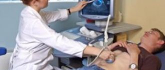
The diagnosis of osteosarcoma can be made after a thorough examination, laboratory and instrumental research methods. Osteosarcoma does not always manifest itself in the early stages of the disease as swelling in the area where the tumor is present. At the first examination, the doctor is alarmed by the presence of unexplained pain in the joints, which is a reason to refer the patient for additional examination. Today, there are several types of diagnostics. Let's look at them:
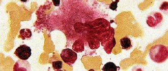
- General blood analysis. This fairly simple analysis allows you to identify pathological changes indicating a cancerous tumor in the bones. Thus, ESR and phosphatase levels increase significantly, and in 50% of cases the number of leukocytes is also increased. Low hemoglobin levels are also characteristic of osteosarcoma, like many other types of cancer;
- X-ray examination is one of the most effective diagnostic methods. In the picture, the specialist, in the presence of the disease, detects the so-called “Codman triangle”, which is a triangular visor. In addition, needle-like spicules are found;
- Biopsy (tissue sampling), as the most reliable method of examination, is done with great care for osteosarcoma. This procedure is performed by a surgeon. Today, trepanobiopsy has proven itself well, in which the likelihood of touching tissues adjacent to the tumor is minimized;
- Computed tomography (CT) is a modern diagnostic method that allows you to accurately determine the location of the tumor, its size and extent of spread. Unlike X-ray examination, CT can detect not only large metastases, but also micrometastases;
- Magnetic resonance imaging is an even more accurate type of diagnosis, which is often used to evaluate the results of the treatment methods used. A special contrast agent is injected into the body, which accumulates around the tumor. The device produces results by counting the number of cancer cells as a percentage, which allows you to evaluate the result of chemotherapy before and after its use;
- Osteoscintigraphy is another type of diagnosis of osteosarcoma, in which the presence or absence of cancer can be judged by the isotope content. Most often used to assess the effectiveness of chemotherapy. If the number of isotopes decreases after therapy, doctors state a positive histological result for chemotherapy treatment;
- Angiography is used to determine the level of tumor spread to blood vessels. Most often this is done before surgery. If cancer cells are present in the vessels, doctors resort to removing the organ.
Signs of pathology
Signs of the disease appear gradually - at an early stage of development they are almost invisible. There is still no fluid in the tissues, which does not reveal pronounced symptoms. The manifestation of the disease begins when pathogens spread to neighboring tissues:
- The width of the bone exceeds the normal size;
- There is a sign of swelling in the soft tissue area;
- A fine-vascular mesh pattern is noticeable on the skin;
- The sore leg or arm loses mobility and functions;
- Swelling of the legs is accompanied by noticeable lameness;
- There is constant pain, especially at the site of the lesion, which cannot be relieved by using analgesics.
Treatment
Therapeutic measures for osteosarcoma are selected individually, depending on the stage of the disease and the degree of prevalence. The most commonly used combination therapy is chemotherapy, radiotherapy and surgery. Let's look at them in more detail:
- Chemotherapy today makes it possible to reduce the size of the tumor before surgery and eliminate the remnants of cancer cells after surgery. When chemotherapy first began to be used, it did not give the desired results and was practically not used. To date, scientists have developed so-called polychemotherapy methods. The substances used in them, methotrexate, leucovorin, adriamycin, ifosfamide, effectively fight cancer cells. The tumor and the extent of its spread are noticeably reduced;
- Radiation therapy was widely used at a time when chemotherapy was not effective. But statistics showed that approximately six months after radiotherapy, metastases were detected. Today, radiation therapy as a method of combating osteosarcoma is used extremely rarely, only in cases where the localization of the tumor does not allow surgical intervention (spine, pelvic bones). Radiotherapy is also used to relieve pain;
- The surgical operation is carried out after a thorough study of all circumstances, such as the location, extent of spread, and the patient’s condition. Surgery is most often preceded by chemotherapy, which reduces the size of the tumor. Typically, after treating osteosarcoma with chemotherapy drugs, it is removed surgically. Today, organ-conserving surgery is possible if the tumor has not affected a large area of the organ. If the disease is found in the extremities, in particular in the lower leg or fingers, then amputation can be avoided. Doctors try to avoid radical amputation of the hip bone, shoulder girdle bones, and arms. However, in case of a pathological fracture, growth of a tumor into soft tissue or a neurovascular bundle, it remains the only measure. Subsequently, it is possible to replace the remote area with metal implants. If the spread of the tumor is great, then amputation must be resorted to. After surgery, an additional course of chemotherapy is advisable, which destroys any remaining cancer cells.
Bone sarcoma: prognosis
The prognosis for the patient depends on:
- the size of the tumor and its location. A small nodule in the tibia or arm is easier to remove than an iliac sarcoma, so the latter case will have a worse prognosis;
- stages of the disease. Before the tumor has time to spread throughout the body, a 5-year life expectancy after complex treatment is observed in 70-90% of patients. If stage 4 bone sarcoma is diagnosed, the prognosis is poor: 10-15% 5-year survival rate;
- histological type of sarcoma. As stated earlier, each species has a different growth rate. The most aggressive osteosarcomas are fatal within a year. Only 30% of patients are able to live with reticulosarcoma for 5 years. Parosteal osteosarcoma, which is characterized by a slow course, has a good prognosis. 5-year survival rate is 70-80%, and 10-year survival rate is 30%;
- possibilities of complex treatment and radical surgery. Thus, with Ewing's sarcoma, the 5-year survival rate after radiation therapy alone is only 10%, and after combined chemo-radiation treatment - 40%.
Forecasts
There is no clear answer to the question “can sarcoma be cured?” The prognosis for osteosarcoma is influenced by many factors: the stage of the disease, the degree of spread, the location of the tumor, the patient’s age, and concomitant diseases. For example, with osteosarcoma of the extremities, doctors give favorable prognoses, especially if the cancer cells quickly die after chemotherapy. If the disease develops in the bones of the body, in particular the ribs, as well as the spine and bones of the head, the disease is difficult to treat, and removing the tumor can be extremely difficult. In addition, the localization of this type of tumor rapidly metastasizes to other organs. In such cases, doctors' prognoses are disappointing.
There are also some types of osteosarcomas that are highly differentiated (similar to healthy cells) and spread slowly. This type of osteosarcoma is parosteal osteosarcoma. The prognosis is favorable, as the tumor responds well to treatment.
Thanks to modern methods of therapy and their complex use, the five-year survival rate has reached 70-80%. Although not so long ago, this figure did not exceed the ten percent mark. When the disease is detected at the stage of localized sarcoma, the survival rate reaches 90%.
Stages of bone sarcoma
Classification of bone sarcoma by stages helps to choose treatment tactics and prognosis for the patient.
- Stage 1 bone sarcoma is a low-grade tumor, without signs of distant metastases and damage to local lymph nodes:
- stage 1A - a tumor no more than 8 cm in greatest dimension;
- stage 1B – tumor larger than 8 cm.
- Stage 2 of bone sarcoma includes highly malignant tumors, also without metastases:
- stage 2A – no more than 8 cm in greatest dimension;
- stage 2B – tumor larger than 8 cm.
- Stage 3 bone sarcoma is a neoplasm that spreads throughout the bone. Moreover, it can have any degree of malignancy, but without metastases;
- Stage 4 bone sarcoma is characterized by the presence of metastases. The tumor can have any degree of malignancy and size. 4A indicates that there is a secondary lesion of the lungs, without metastases in regional lymph nodes, and 4B is a sarcoma with metastases in lymph nodes and other distant organs.
Characteristics of the disease
The disease can affect any part of the bone - part of the femur, ribs, ankle, in the frontal part of the skull, pelvis and others. Therefore, it is necessary to conduct a thorough examination of the body to identify tumors and metastases.
Diagnostics includes the following procedures:
- The doctor conducts a physical examination of the patient and collects anamnesis.
- An x-ray is prescribed to determine the location of the diseased area.
- Histology of biological material is carried out.
- Before surgery, the biological sample is examined for a biopsy - open, thick-needle or fine-needle.
- Secondary foci of malignant disease are detected using osteoscintigraphy.
- Computer (CT) and magnetic resonance imaging (MRI) make it possible to detect more accurately and in detail a neoplasm with metastatic growths.
- Vessels are examined for the presence of a malignant lesion using angiography.
- A study of the bone marrow is required to exclude pathology within the cells of the spine - trephine biopsy.
Osteosarcoma is treated with chemotherapy before surgery. When antitumor drugs are applied to the diseased area, the size of the tumor can decrease and the growth of metastases may be blocked. The following medications are used: Ifosfamide, Cisplatin, Carboplatin, Etoposide, Methotrexate, Adriblastine.
After chemotherapy, the malignant node is excised. On the skull and spine with the hip joint, removal is only possible in 50% or 80%. Sometimes a limb has to be removed. Radical resection is often used - this type is considered organ-preserving. After resection, it is necessary to replace the joints with blood vessels and perform plastic surgery of the nerve trunks, bones, etc.
Removal is carried out by capturing healthy tissue, highlighting the required area. After this, you need to eliminate the defect. For this purpose, autodermoplasty is used - healthy flaps of skin and muscles are transplanted using microvascular anastomosis. The relapse prognosis remains at 14-20%.
Cancerous foci in the tissues of the lungs and lymph nodes require complete removal. But you need to take into account the size of the plot.
In a child, such interventions are carried out in the following cases:
- The extraosseous concentration of cancer cells is less than 13%.
- The primary lesion is reduced in size.
- Neoadjuvant chemotherapy was administered to repair the pathological fracture.
- The disease is diagnosed at the second stage and the patient’s condition is satisfactory.
- There are no metastases in the lung.
- The parents insist on saving the limb.
Difficulties in surgical intervention are found in the treatment of organs of the hip region due to severe blood loss. For this purpose, interiliac-abdominal amputation with disarticulation is used.
After a successful operation, repeated courses of chemotherapy are prescribed to eliminate the remaining cancer cells. Recently, targeted therapy has been increasingly used - monoclonal antibodies are introduced to block the growth of cancer pathogens. To restore immunity, the immune drug Cytokine is prescribed.
Gamma ray irradiation is not used in the treatment of osteosarcoma. This is due to the fact that the cancerous lesion does not respond to radiation. In rare cases, external exposure to gamma rays is used on the diseased area. The procedure is performed for partial removal of the node, to relieve severe pain and in case of relapse.
Currently, this type of cancer is successfully treated in Israel using the latest equipment. Oncology in this country is studied in depth, the latest developments of scientists from all over the world are used.
Bone sarcoma is a rare type of malignant cancer that develops in the soft tissue area and in the bone cavity. The main difference is autonomous development, there is no connection to a specific organ. It affects the bony part of the body, but there are examples of development in tissue that does not have bone affiliation. The skeleton can be damaged in any area.
The tumor prefers to be localized in the area of long tubular bones and the junction. Cancer develops quickly, destroying bone and penetrating into the bone canal with muscle. The aggressive nature of the pathology is characterized by early metastasis to the tissue of the brain and lungs with the formation of secondary foci.
Previously, the disease was considered incurable - only 10% of patients survived. In recent years, with the introduction of the latest equipment and new treatment methods into medical practice, the chances for patients have increased.
According to medical statistics, men suffer from sarcoma more often than women - approximately 2 times. The peak occurs during adolescence. It is rare in adults over 50 years of age. Tall people are at risk.
The tumor most often develops on the leg - in the heel, femoral and hip areas. It is rare on the hands and affects the shoulder joint.
Sarcoma in the bones
How to recognize sarcoma
How to recognize bone sarcoma is a question many people ask. After all, it is necessary not only to catch the pathology in time, but also to distinguish it from other diseases of bones and tissues.
At an early stage, such tumors do not give any special symptoms. The only thing is that dull pain may occur, mainly at night. However, they cannot be removed with painkillers. Usually, many are reassured by the fact that such painful sensations can subside and disappear. However, as the tumor grows and it compresses blood vessels and nerves, the pain will intensify.
Then other symptoms begin to appear. For example, sarcoma of the femur can impair a person's mobility by affecting the hip joint. Impact on the vertebrae causes various neurological disorders. In addition, as the tumor develops, the bones become more fragile, as a result of which they can break literally out of the blue. Moreover, such unreasonable fragility of them manifests itself already at the initial stages.
Sacral sarcoma makes itself felt by pain, which doctors often call cauda equina syndrome.
The person will experience pain in the legs, which will be predominantly localized on the inside of the thigh, a loss of the knee reflex will be noted, and the situation will be complicated by weakness of the sphincter, anus and bladder. Men may develop problems with potency.
As the tumor grows, the bone deformations it causes will be noticeable without any additional equipment. In this case, the changes will affect the tissues around - they will be inflamed, the vascular network will clearly appear under the skin, the damaged part of the body will be swollen. It also happens that active tumor growth causes fever. Common symptoms of intoxication include:
- Severe and sudden weight loss without adequate reasons (diet, poisoning, etc.);
- Weakness;
- Anemia.
Metastases, which such a tumor actively produces, lead to the development of complications in various organs. The most common places for malignant cells to travel from bones are bones, bone marrow, liver, and lungs.
Stages of osteosarcoma
When a diagnosis of osteosarcoma is made, staging is performed to assess the extent of the process. Taking into account the prevalence, all osteosarcomas are divided into two large groups:
- Localized sarcomas. Tumor cells are present only at the site of its original localization or surrounding tissues.
- Metastatic sarcomas. Metastatic damage to other organs is detected.
It is assumed that 80% of patients have micrometastases that are so small that they are not detected during additional studies.
In addition, multifocal sarcoma is distinguished, in which tumors simultaneously arise in several (two or more) bones.
Osteosarcoma, like other malignant tumors, in some cases returns after treatment. A new tumor can arise either at the site of the previous one or in another part of the body (in the area of metastases). This condition is called relapse. Relapse in osteosarcoma most often develops 2-3 years after treatment and is found in the lungs. In the future, relapses are also possible, but the likelihood of their development decreases.
In osteosarcoma, the stages of tumor development are determined by the extent of the process. There are two groups of neoplasms depending on whether they have spread throughout the body:
- Localized - the localization of the tumor is noted only where it appeared initially, or in the tissues surrounding the bone;
- Metastatic - examination reveals metastases in the lungs, liver and other parts of the body.
Sometimes areas of metastases can be found in more than one organ, since osteosarcoma develops very quickly and metastasizes rapidly. In eighty percent of patients, oncologists find very small metastases. There is also multifactorial osteosarcoma. This is a form in which a tumor develops in several bones of the affected limb at once, for example, in the thigh and lower leg.
There are different classifications, most often doctors determine the stage of osteosarcoma in accordance with the MSTS system. Three indicators are taken into account:
- G - degree of malignancy. It can be low (G1), when the tumor tissue is similar to normal, and more aggressive high (G2), in which the cells have almost completely lost their original features.
- T—degree of tumor spread. It may be located within the bone (T1) or grow into adjacent tissue (T2).
- M—presence of metastases. M0 - no metastases, M1 - yes.
Another classification is the AJCC, which takes into account the spread of the primary tumor (T), metastases to nearby lymph nodes (N), metastases to other parts of the body (M) and the degree of malignancy (G).
Diagnosis of osteosarcoma
Diagnosis is carried out using a regenographic study. Initially, osteoporosis of the bone is diagnosed, since there are no clear contours of the neoplasms, they are blurred. Over time, the bones begin to deteriorate, and metastasis processes are observed.
To make a correct diagnosis, a number of additional studies are carried out:
- radiograph;
- morphological examination of the tumor (biopsy, knife biopsy, trephine biopsy);
- osteoscintigraphy (OSG);
- computed tomograph (CT);
- magnetic resonance imaging (MRI);
- angiography (vascular examination).
Symptoms of osteosarcoma
Point mutations in genes are considered the main cause of osteosarcoma. In combination with external irradiation, they can lead to the development of bone tumors.
Modern treatment of osteosarcoma necessarily involves:
- removal of a bone tumor;
- removal of identified metastases;
- carrying out polychemotherapy, i.e. not with one drug, but with a combination of them.
Only such treatment (surgery and chemotherapy) can lead to a complete cure. Otherwise, there is a very high risk of developing metastases associated with the rapid and unhindered spread of malignant cells throughout the body. Radiation therapy is not indicated in all cases, but only if surgery fails to completely remove the primary lesion.
The course of osteosarcoma is associated with many pitfalls. Therefore, such patients, especially those with tumor recurrence, should be observed only in a specialized center.
https://www.youtube.com/watch?v=j1-ABuxHAa0
Osteosarcoma is a malignant bone tumor that most often occurs in childhood and adolescence (2-3 new cases per 1 million children and adolescents), but can also occur in adults. In adults, it is common not only to damage the tubular bones of the extremities, but also to the vertebrae, jaw, and pelvic bones.
The causes of osteosarcoma are still completely unknown. But despite this, the symptoms of the disease are typical - increasing pain in the affected bone. Early diagnosis is very important in life prognosis. Late-stage bone sarcomas are characterized by rapid progression, and therefore treatment without chemotherapy is not possible.
Osteogenic sarcoma, or osteosarcoma for short, is the same malignant neoplasm as cancer or melanoma, but calling it cancer is incorrect and illiterate. Cancer develops exclusively from the cells of the mucous membrane, from no other tissues, and sarcoma arises only in connective tissue, and osteogenic sarcoma occurs from bone elements.
All of these processes have common features of malignant growth: indomitable cell proliferation, aggression towards surrounding tissues, the ability to form metastases and easy adaptability to serious changes in the body, and are treated using three methods: surgery, radiation and antitumor drugs.
The first symptoms are often associated with injuries, which are not uncommon in childhood. Initially, pain appears in the limb, which gradually increases. Later, swelling and limited mobility in the adjacent joint occur. Sometimes osteosarcoma can begin with a pathological fracture, i.e. not associated with significant mechanical damage.
At the earliest stage, when the tumor is still inside the bone, the disease does not manifest itself in any way, because its first symptom - pain - occurs only when the surface bone layer - the periosteum - is destroyed, since it is well innervated. The main bone substrate, where the malignant process develops, contains virtually no nerve endings, so for a long time there are no signs of trouble.
On the face, a neoplasm can be detected at an early stage, since the deformation of the jaw in this anatomical zone is more visible, and dental problems are often provoked. This localization is typical for young people who are more attentive to their own appearance.
Unlike other tumors, in osteosarcoma the general condition is disturbed only in advanced cases (at the last stage), which is why late diagnosis is associated. At this time, weakness, a sharp decrease in appetite, weight loss, and anemia appear.
In terms of the frequency of damage to anatomical areas, primacy belongs to the femur in the knee area, followed by the humerus in the upper third; adults are also characterized by damage to the vertebrae and pelvic bones, the facial skull - jaw.
In eight out of ten adult patients, osteosarcoma is diagnosed when presenting with severe deformity of the limb, when the pathological bone substrate grows into the soft tissues with the formation of a “bulge.” At this stage, pain appears, localized in one place, constant over time, but varying in intensity - sometimes a little more, sometimes a little less. Painkillers usually do not relieve pain. Limb mobility may be limited due to the involvement of muscles and nerves in the process.
Under axial load, the bone can fracture, which is not accompanied by intense pain typical of any traumatic fracture of a limb. Often patients simply complain of a “crunching” and “dull” soreness; the reason for seeking medical help is not an injury, but a limitation of movement. Often, vertebral sarcoma damage is mistaken for banal radiculitis, and the fracture is diagnosed late due to atypical low-intensity pain.
In the majority, bone lesions are detected simultaneously with metastases due to the latent course of the malignant process.
The main clinical manifestation of the disease is pain, constant and not going away even at night, not associated with physical activity or recent trauma. Trauma itself does not exclude the presence of osteosarcoma; often it is trauma that draws the patient’s attention to a dormant and clinically invisible problem.
Visible changes in the bone or adjacent soft tissue, as well as limited movement, are signs of a common process, but not necessarily metastatic.
The tumor leads to limitation of function, primarily movements, when nerve trunks are involved, as well as in case of a fracture, which is designated as pathological, that is, arising at the site of a bone defect.
Most often, osteosarcoma appears in the extremities, less often in the bones of the skull or pelvis. There are two forms of the tumor process: localized and metastatic. The stage of the disease consists of three intervals: stages I and II are characterized by osteosarcomas of varying degrees of aggressiveness, stage III is associated with the appearance of metastases.
Osteosarcoma occurs in various age groups, but most often in childhood and adolescence (up to 30 years of age), with girls predominantly affected between the ages of 10 and 14 years, and boys - from 14 to 19. Also, one tenth of all identified cases of the disease occur in elderly patients (over 60 years old).
- Height - tall boys and teenagers
The development of osteosarcoma can be caused by irradiation of the body, including during the treatment of other types of tumors. A special risk group is young people who have undergone radiation therapy during their growth period. But post-radiation osteosarcoma can only be caused by increased doses of radiation therapy - above 60 gray, while conventional x-rays are practically safe.
- Existing diseases of the skeletal system
In adulthood, a tumor may appear caused by diseases of the skeletal system suffered in childhood or adolescence.
This disease develops predominantly at an age close to old age and has a precancerous etiology. When it is severe, every tenth patient develops osteosarcoma.
- Presence of non-malignant tumors
Benign tumors of cartilage and bones, osteochondromas, under certain conditions can become malignant (turn malignant)
La Fraumeni syndrome is a genetic disease consisting of the inheritance of the pathological p53 gene, which causes tumor formation in 80% of patients. The p53 gene is responsible for controlling cell division. In the presence of such a syndrome, the risk of developing both osteosarcoma and other tumors is incredibly high.
The connection between the presence of this eye tumor and the occurrence of osteosarcoma has been scientifically proven. Also, when treating retinoblastoma using radiation therapy, the risk of developing osteosarcoma of the skull bones increases significantly.
If a person does not belong to a risk group, then in some cases osteosarcoma may still occur, and there may not be a reason that can be scientifically explained.
Osteogenic sarcoma, or osteosarcoma, is a tumor of bone tissue consisting of osteoclasts with osteoblasts. Active cell division occurs during malignant processes in the body.
It often occurs as a primary lesion, but there are examples of development against the background of osteomyelitis in a chronic course. It occurs in rapid development and the occurrence of early metastases. Develops in the long tubular bone and the metaphysis.
Early treatment is recommended to have a chance of recovery.
Description of the pathology
Tubular bones are more susceptible to the development of malignant neoplasms. In flat and short bones, osteosarcoma is recorded in isolated cases. Tumors are more common in the leg bones than in the arm area. In the area of the knee joint, the process of cancer formation is recorded in 80% of diseases.
It is quite rare for cancer to develop in the knee area.
Cancer diseases are very common today among people of all ages. Often the pathology can be observed in children and adolescents.
One of these malignant diseases is osteogenic sarcoma, which affects bone tissue and spreads metastases throughout the body, and this can threaten the patient’s life.
This disease is the most common among all bone cancers and accounts for 60% of all cases. Timely treatment makes it possible to achieve long-term remissions.
Prognosis and prevention
When treating the pathology at the initial stage, the prognosis can be favorable, as long-term remissions can be achieved. Today, about 90% of people who have sarcoma of the left humerus or hip joint can live more than five years after successful treatment. Chemotherapy makes it possible to avoid amputation of the affected limbs of patients.
Note! Chemotherapy is the most effective treatment for osteosarcoma; it successfully eliminates cancer cells. The only disadvantage is the occurrence of side effects.
( 2 ratings, average: 5.00 out of 5)
Diagnosis of a skull bone tumor
Diagnosis of cranial bone cancer includes:
- examination of the nasal cavity and ears using the endoscopic method;
- radiography of frontal and lateral projections of the head;
- CT and MRI with layer-by-layer radiological scanning of bone and soft tissue;
- PET – positron emission tomography with the introduction of glucose, containing a radioactive atom, to identify the oncological process in any area of the body and distinguish between benign and malignant tumors;
- PET-CT – for more rapid detection of sarcoma of the skull bone and other formations;
- osteoscintigraphy – scanning of the bone skeleton using radionuclides;
- histological examination of biopsy material after radical surgery (biopsy, puncture and/or surgical biopsy);
- urine and blood tests, including blood tests for tumor markers.
Diagnosis of a tumor of the skull bones is supported by collecting anamnesis and examining patients to establish all the symptoms of the disease and the general condition of the patient.
Features of therapy for bone sarcomas
In the treatment of osteosarcoma, the location, extent of the tumor, the presence or absence of metastases, as well as sensitivity to specific chemotherapy drugs must be taken into account. The main methods effective for this type of tumor are surgery and chemotherapy.
The treatment regimen for patients with osteosarcoma involves preoperative chemotherapy, then surgery, and then chemotherapy again. Preoperative treatment is intended to reduce the volume of neoplasia and destroy small foci of metastasis. In addition, this approach allows the oncologist to establish sensitivity to specific drugs, which will increase the effectiveness of subsequent chemotherapy after surgical treatment.
In the case of osteogenic sarcoma, methotrexate in high doses, ifosfamide, doxorubicin, cisplatin, adriablastin are prescribed. Preoperative chemotherapy is called neoadjuvant chemotherapy. If necessary, the names and dosages of the drugs may be changed in the future.
In the postoperative period, up to ten courses of treatment (adjuvant chemotherapy) are prescribed, including those drugs to which the best response was noted before surgery. Adjuvant chemotherapy is necessary to destroy microscopic metastases or treat larger lesions, if they have already formed.
Surgical treatment is the main stage, the effectiveness and timeliness of which determine the prognosis of the disease. Until recently, for osteosarcoma, most amputations and disarticulations were performed, after which the patient lost part or all of the limb. Such interventions were very traumatic and risky, because a large amount of tissue was removed.
Today, doctors try to preserve the patient’s maximum possible activity and functionality by performing gentle, organ-preserving resections. Of course, in advanced stages such operations may not be indicated due to active tumor growth and metastasis, but with timely diagnosis of the tumor they are the method of choice.
Organ-sparing operations for osteosarcomas include removal of the affected fragment of bone or joint, after which the defect is replaced with a prosthesis - made from artificial materials, natural or cadaveric bones. Thus, the limb can be saved, and the patient can lead a normal life. Moreover, even detected metastases cannot be an obstacle to organ-conserving surgery.
Organ-preserving surgery
It happens that the tumor has gone so far that the surgeon has no choice but to perform an extended radical operation, the indications for which are large osteosarcoma, its invasion of the main vessel and nerves, the extent of the lesion with necrosis and bleeding, the proliferation of microorganisms, and pathological fractures.
Amputations involving removal of the entire limb and disarticulation of joints are considered radical. Extended ones include interiliac-abdominal dissection and interscapular-thoracic amputation. They are very traumatic and require long-term rehabilitation during postoperative chemotherapy.
Secondary foci of osteocarcoma can also be subjected to surgical removal. They are most often found in the lungs. If chemotherapy is effective against the main tumor, then large metastases can simply be excised surgically.
Radiation therapy has no independent value in osteosarcoma, since the tumor is insensitive to the effects of radiation. However, this method is used in patients for whom surgery is impossible due to the localization of neoplasia, the general severe condition of the body, and concomitant pathology that prevents intervention.
Practice has shown that organ-conserving operations are, for the most part, not inferior in effectiveness to radical ones, and supplementing them with chemotherapy before and after the intervention significantly improves prognosis and survival. In addition, limb preservation is of great importance for the patient’s quality of life without compromising the radicalism of treatment, and this is possible in at least 80% of patients.
After treatment for osteosarcoma is completed, a follow-up examination is performed, including CT or MRI of the affected area, X-ray examination of the lungs, bone scintigraphy, and ultrasound of the abdominal organs to exclude metastasis.
The prognosis for osteosarcomas is quite good, given the aggressiveness of the disease. Thus, the sequential use of chemotherapy, surgery and chemotherapy again gives a 5-year survival rate of 70% for a limited form of the disease. If the tumor is sensitive to chemotherapy, then this figure reaches 80, or even 90%. Such high survival rates are associated with new approaches to treatment and the use of highly effective drugs, but until recently it involved amputation, and only every tenth patient survived.
If it was possible to completely remove the tumor during surgery, and the foci of metastasis decreased under the influence of chemotherapy, then the prognosis will be better.
To assess the prognosis, all patients with osteosarcoma are divided into three groups:
- The high-risk group is when the volume of neoplasia exceeds 150 ml, and during chemotherapy, the tumor cells either do not die, or more than half of them remain viable.
- Low-risk group - when osteosarcoma does not exceed 70 ml in volume, its sensitivity to chemotherapy does not matter, and survival rate reaches more than 90%.
- The standard risk group consists of all other patients, with survival rate being about 65-67%.
The intensity and duration of treatment is determined by the risk group to which a particular patient can be classified. Having gone through all stages of treatment, the patient does not disappear from the attention of oncologists, because the risk of metastasis and relapse of the disease remains. For monitoring, he will need to visit a doctor every three months in the first two years after treatment, for the third year the specialist will examine the patient once every four months, for the next two years - once every 6 months and then annually. For low-grade tumors, you can visit a specialist less frequently: once every six months for the first two years, then annually.
https://youtu.be/2uOT0ZRc0Ck
The most common locations
A malignant tumor primarily affects the bone tissue of long tubular bones. All other types of bones account for 20% of the damage. Osteosarcoma of the hip, i.e. femur, its distal end is the most common site of tumor formation. The disease occurs in flat bones, bones of the feet, hands, spine, and skull very rarely.
The area of the metaphysis of the bone is closer to the joint, this is where the neoplasm will be localized. But there are exceptions when, during the disease, the metaphysis remains unchanged, and it is the diaphysis that is affected. Also, doctors already know where exactly the metastases are located on a particular bone, for example, if the hip bone is affected, then the neoplasm should be looked for in the area of the distal end. With pain in the tibia, the internal condyle is affected, with pain in the humerus, where the roughness is located in the deltoid muscle.
Treatment of pathology
This pathological neoplasm is treated comprehensively. First, chemotherapy is used before surgery, the purpose of which is to remove metastases in a person’s lungs and reduce the size of the tumor. After chemotherapy, surgery is performed.
Note! Previously, the disease was treated only surgically, which often involved amputation of limbs.
Today there are many effective medications that inhibit the growth of cancer cells. Just a few decades ago, when a person was diagnosed with osteosarcoma of the femur, amputation of the limb was impossible due to the high risk of damage to important nerves. Modern treatment methods make it possible to preserve organs affected by metastases; they are removed during surgery.
During the operation, the tumor and part of the bone are removed. The removed area is replaced with an implant made of plastic or metal. This happens especially often with pathology of the pelvic bones.
If the tumor is located in hard-to-reach places, surgery will be difficult, so the doctor prescribes a course of radiation therapy. The doctor then prescribes a course of chemotherapy to eliminate any cancer cells that may remain after the surgery. In the postoperative period or after chemotherapy, doctors prescribe maintenance therapy with Cisplastin, Methotrexate or Carboplatin.”
Radiation therapy for the treatment of ODS cancer is ineffective, since cancer cells are practically insensitive to radioactive radiation. This treatment method is used when surgery cannot be performed for some reason.
Types of disease
In oncology, it is customary to distinguish three forms of this pathology:
- Osteolytic sarcoma, caused by the presence of a single tumor with unclear contours. In this case, bone tissue begins to actively deteriorate. As the tumor grows, the pathological area grows in length and width of the bone. Sarcoma begins to destroy the cortical layer of bone, then metastasizes to soft tissues and the entire human body, most often large metastases form in the lungs.
- The osteoplastic form of sarcoma is characterized by the predominance of the tumor over the process of tissue destruction. Tumor tissue begins to fill the space of the cancellous bone, forming fan-shaped growths. All this contributes to the deformation of pathological areas of the bone, which leads to irreversible changes in the human body.
- The mixed form involves a combination of equal growth of neoplasm and destruction of bone tissue.
Note! Mixed osteosarcoma is characterized by bone destruction with the formation of additional bone structures.
Rare types of disease
In oncological practice, there are also quite rare types of osteosarcoma:
- Telangiectatic tumor, which affects the lymph nodes in addition to the bones.
- A small cell neoplasm that is located in the femur of the lower limb. It is the most dangerous because it is combined with other types of tumors.
- Fatal multifocal tumor caused by a large number of metastases from one neoplasm. This type of pathology cannot be treated.
- An extraskeletal neoplasm, which is a rather rare type of disease. In this case, the legs are affected, making treatment difficult.
- Periosteal tumor, which is localized on the surface of the bone without spreading to the bone marrow.
- Juxtacortical sarcoma forms in the cortical layer of the bone, affecting its entire length and width. This neoplasm does not metastasize.
- Intraosseous sarcoma has the smallest percentage of malignancy; after treatment, relapses do not occur.
Skull bone cancer: symptoms and manifestations
Clinical symptoms of cranial bone cancer are divided into three groups. The first general infectious group includes:
- increase in body temperature with chills and/or excessive sweating;
- intermittent fever: a sharp rise in temperature above 40° and a fleeting decline to normal and subnormal levels, then a repetition of temperature jumps after 1-3 days;
- increased leukocytes in the blood, ESR;
- sudden weight loss, increasing weakness, appearance of pale skin on the face and body.
Symptoms of cranial bone cancer of the general cerebral group:
- headaches with increased intracranial pressure, with nausea and vomiting, as well as changes in the fundus (this includes congestive discs, optic neuritis, etc.);
- epileptic seizures (appear due to intracranial hypertension);
- periodic (orthostatic) bradycardia up to 40-50 beats/min;
- mental disorders;
- slowness of thinking;
- inertia, lethargy, “stupefaction”, drowsiness, even coma.
Focal (third group) symptoms and signs of cancer of the skull bones depend on the location of the pathological process. in some cases they do not appear for a long time.
Focal symptoms of skull bone tumors are complicated by edema and compression of brain tissue, meningeal symptoms with cerebellar abscesses. This is characterized by the manifestation of pleocytosis with lymphocytes and polynuclear cells (multinuclear cells) in the cerebrospinal fluid. It increases the protein concentration (0.75-3 g/l) and pressure. But often such changes may not occur.
Osteogenic sarcoma of the skull bone is characterized by a fixed subcutaneous compaction and pain when the skin above it moves. The lymph nodes of the head and neck are enlarged. With metastases, hypercalcemia develops, accompanied by nausea, vomiting, dryness of the oral mucosa, excessive urination, and impaired consciousness.
With Ewing's sarcoma, patients have increased white blood cell levels and temperature, headaches, and anemia. With myeloma, patients sharply weaken, they develop secondary anemia, and excruciating pain aggravates their life.
Multiple myeloma can affect 40% of the bone tissue of the cranial vault. In this case, all detected lesions are considered primary with multifocal growth and do not belong to metastatic tumors.
Diagnosis and treatment
Rib cancer is diagnosed in the same ways as other tumor processes. Thanks to high-tech equipment, it is possible to detect the disease with a high probability.
- Use of X-ray equipment. This type of study allows you to detect a tumor using x-rays. It is possible to use two projections to make a more accurate diagnosis. X-ray helps to identify the location of the tumor: the ribs themselves, the gap between them.
- Radiology using contrast agent. This is a type of diagnostic test that uses x-rays together with a contrast agent. Before the procedure begins, a special solution is injected into the patient’s blood, which highlights the tumor in color on the pictures.
- Tomography. There are two types: computer and magnetic resonance. They differ from each other in the radiation used and operating algorithms. This study allows for layer-by-layer scanning of the body, as a result of which the doctor receives processed images. This method is the most accurate; it allows not only to identify the presence of a tumor, but also the location, area of damage, and the structure of the malignant formation.
- Biopsy. A laboratory type of study that allows you to determine rib cancer based on biological indicators. To do this, a histological and cytological examination of a piece of tissue is carried out from the area where the process develops. The collection is carried out surgically.
Based on the research results, the doctor makes a diagnosis, determines the extent of the disease, and prescribes treatment.
- Surgical intervention is used when it is impossible to use other methods or in cases where they have proven ineffective. Often, such an operation requires the removal of one or more ribs and the subsequent installation of special inserts in their place.
- Radiation therapy is a type of treatment using ionizing radiation. Treatment is performed using modern equipment. The patient is in a special room with lead walls and doors. Doctors are monitoring him from an adjoining room on monitors. The equipment is adjusted so that the beam is focused to a specific location where the hearth is located.
- Chemotherapy. The patient is prescribed cytostatic drugs that act on the body in combination. Medicines affect not only the tumor itself, reducing its size, but also metastases. The course of treatment can range from several weeks to several months.
The last two methods are used in practice in combination. To relieve pain symptoms, the patient is prescribed special medications.
ul
Osteosarcoma is a malignant tumor whose symptoms are more common in young people.
The favorite location of the tumor is the end sections of the long tubular bones of the legs and arms (femur, tibia, humerus), but other bones can also be affected, including flat ones (jaw, pelvic bones).
Methods of treating patients
The fundamental method of treatment is surgery. It usually involves resection of the area affected by the tumor.
The patient is carefully examined for the presence of metastasis in the lymph nodes and other structures of the body.
If secondary sarcomatous foci are found during the examination, they are also removed, after which the patient is prescribed chemotherapy.
Antitumor drugs are selected individually, taking into account the purpose of exposure. Preoperative chemotherapy is carried out to reduce the size of the formation, and postoperative chemotherapy is carried out to destroy possibly remaining micrometastases.
General information about the disease
This disease is extremely malignant, characterized by a rapid course and the ability to quickly metastasize.
Bone cancer can develop in people at any age, but most often occurs during puberty in children and adolescents aged 10 to 20 years, and in very old people. Typically, the pathology is observed in men; they suffer from this disease twice as often as women. The favorite location of the tumor is cylindrical or triangular bones, so the lower extremities are damaged more often than the upper ones. A large percentage of tumors occur in the knee joint, but the tumor has a high percentage of metastasis. Lesions can be observed in various internal organs, especially in the lungs and liver.
There is an opinion that bone cancer develops as a result of long-term inflammatory processes, for example, chronic osteomyelitis.
Therefore, the following bones are most often affected:
- femoral;
- tibial;
- shoulder;
- pelvic;
- fibula;
- shoulder;
- ulna.
The radius and patella are rarely affected by osteosarcoma. Damage to the skull occurs most often in childhood, as well as in old age as a result of complicated osedystrophy.
What is osteosarcoma: description of pathology
Sarcoma is a malignant tumor that develops from primary connective and bone tissue. The tumor most often appears in the bone growth zone of the lower limb; half of these tumors often spread to the knee. Another part of the tumors can affect the tibia, arm in the shoulder area, femur and pelvic bones. In rare cases, the bones of the skull are affected, but this usually occurs in childhood or old age, and in 50% of cases the pathology develops in the femur, with the tibia taking second place in localization. Malignant tumor cells quickly spread through the bloodstream to the bone marrow, then to other bones, soft tissues, cartilage and tendons, muscles and lungs. The neoplasm grows rapidly both in length and width.
Note! Osteogenic sarcoma is a very dangerous pathology that tends to progress very quickly and form metastases, significantly reducing the patient’s time for effective treatment.
In most cases, the tumor begins to develop in the tubular bones, which take part in the active growth of bone tissue that forms the human skeleton. Osteosarcoma of the hip may be accompanied by the appearance of additional bone structures that are irreversible.
Epidemiology
Osteogenic sarcoma appears in 60% of cases during the period of active growth of the human skeleton, which occurs between the ages of ten and thirty years; in 40% of cases, pathology develops in a different age period. The disease occurs twice as often in men as in women. Doctors consider children who are taller than average to be at risk. Neoplasms of this type almost always arise in one bone. This pathology is very severe, but it occurs in one case out of five hundred thousand.
Causes
Reasons for the appearance of neoplasm:
Manifestations of Paget's disease in bones
- Bone disease. For example, Paget's disease or fibrous dysplasia.
- Carcinogenic substances. Once in the human body, these chemical elements can cause irreversible changes in DNA that contribute to the formation of cancer cells.
- Heredity. Medicine has not proven a direct relationship between the occurrence of cancer cells in humans due to the fact that cancer has been detected in close relatives. But statistics show that if one of the parents is diagnosed with rib cancer, as a rule, the risk of the disease in the children of such parents is quite high.
- Radiation. Unfortunately, the necessary treatment of one source of disease can cause the emergence of a new one. Radiation therapy, once used to block cancer cells, often affects the development of another cancer even years after the radiation.
- Deviations in the genetic code. Scientists have proven a direct connection between abnormalities in certain chromosomes and an increased risk of developing malignant tumors in humans. But for what reasons pathology occurs in chromosomes, scientists have not yet identified. There is an assumption that these failures in genetics occur even in intrauterine development. A child becomes an innocent victim of a terrible disease already during his formation.
- Decreased immunity. The appearance of cancer cells in the body may be affected by the presence of other diseases associated with decreased immunity. For example, AIDS or syphilis.
- Age. Scientists have discovered that the occurrence of cancer in children is directly related to their active growth during adolescence.
Classification
In most cases, the oncological process affects the tubular bones; the lower extremities are affected much more often than the upper ones.
In descending order of localization, the disease is presented as follows:
- osteosarcoma of the femur (most often the distal end);
- bones of the shoulder joint;
- elbow joint;
- osteosarcoma of the jaw;
- skull bones (most often this type of pathology is diagnosed in children).
Osteosarcoma of the frontal bone is diagnosed extremely rarely.
Based on the nature of the growth of a malignant tumor, the following forms are distinguished:
- low;
- intermediate;
- high.
Based on the nature of metastasis, the following are distinguished:
- localized - when cancer cells affect only the bone and do not extend beyond the affected joint;
- metastatic - through lymph flow and blood, metastases spread throughout the body.
Based on the ratio of tumor growth to the bone itself, the following forms of the oncological process are distinguished:
- mixed – tumor growth and bone destruction are of an equivalent nature;
- osteolytic – tumor growth predominates in relation to the affected area;
- osteoplastic - tumor growth provokes an increase in bone tissue.
In addition, there are several forms of this disease, which are extremely rare, but still occur:
- angiectatic osteosarcoma;
- periosteal;
- intraosseous;
- extraskeletal;
- small cell;
- radiation-induced.
As for the severity of the development of this oncological process, there are four stages. The most positive prognosis is present at stages 1–2. At the last stage of development of this disease, palliative treatment is carried out, which is aimed at relieving acute symptoms and improving the patient’s quality of life.
Regardless of which form of the disease is diagnosed, the risk of relapse is almost always present. Most often, relapse of bone cancer occurs 2–3 years after an active course of treatment.
How to detect osteosarcoma?
Unfortunately, it is not uncommon for osteogenic sarcoma to be diagnosed at a late stage, and treatment does not have the expected result. This is due to the paucity of symptoms in the early stages, unclear pain syndrome, and late consultation with a doctor.
If you experience unclear, suspicious pain in bones or joints that intensifies and is not relieved by painkillers, it is better not to postpone a visit to a specialist. The first person to suspect a tumor may be a surgeon or traumatologist, who will clarify in detail the nature of the complaints and the dynamics of the symptoms.
It is important to clarify whether the patient has had radiation exposure in the past, since tumors can occur decades after exposure to radiation. The absence of swelling or dilated veins is not a sign indicating the absence of a neoplasm.
osteosarcoma of the knee joint on x-ray
If osteosarcoma is probable, additional studies cannot be avoided. The following must be carried out:
- X-ray of the suspected site of neoplasia;
- CT, MRI to clarify the boundaries of the tumor and the condition of the surrounding tissues;
- Ultrasound of the abdominal organs, x-ray of the lungs to exclude metastatic growth;
- Biopsy is an examination of tumor tissue under a microscope.
All patients without exception undergo general and biochemical blood tests and urine tests. In the early stage, there may be no significant changes, but by the time the tumor enters the surrounding tissues, the general blood test will show increased leukocytes, increased ESR, and in later stages anemia is likely.
Before surgery, angiography is performed to clarify the condition of the vessels and the presence of tumor cells in them. This research method determines the course and scope of the intervention. If osteosarcoma cells have already entered the vessels, then organ-preserving surgery will not be possible.