A femoral neck fracture is damage to the integrity of the femur bone in its upper area. The bone fracture occurs in the space between the femoral head and the greater trochanter. The risk group consists of older people over sixty-five years of age. Individually, a fracture can cause dangerous consequences that threaten a person’s health, and can even be fatal.
Online consultation on the disease “Hip Fracture”. Ask a question to the specialists for free: Traumatologist.
- Etiology
- Varieties
- Symptoms
- Complications
- Diagnostics
- Treatment
A hip fracture in older people is more severe, in contrast to such a fracture that occurs in young and middle-aged people. This is explained by the fact that in older people there is practically no ability of the body to quickly fuse bone structures, due to insufficient blood supply to the neck and bones (due to age).
Erroneous self-analysis of the disease and a small number of signs that determine this type of fracture often become the reason for late seeking help from specialists, which entails an increased risk of complications. Therefore, with the most minor symptoms of this pathological condition, you should contact a traumatologist.
Causes of hip fracture
Geriatric people most often experience hip injuries when they land poorly on their side. People with severe osteoporosis are even more likely to injure their femur.
Important! Females are more likely to suffer from osteoporosis because calcium is washed out of their bone structures more quickly.
Factors contributing to hip fracture:
- fading of the reproductive functions of the female body;
- chronic deposition of cholesterol on the walls of arteries;
- stenosis and obliteration of peripheral arteries;
- excessive body weight, which increases the load on the joints;
- physical inactivity;
- impaired coordination due to neurological pathologies;
- unbalanced diet or complete refusal of food;
- oncopathology.
Young people, due to the strength of their bone structures, are less likely to experience femur fractures. For this to happen, the injury must be particularly severe.
In young people and middle-aged people, the causes of this pathology are as follows:
- road accident;
- injuries sustained at work;
- unsuccessful landing from a considerable height;
- injuries received during hostilities.
Various somatic diseases that contribute to disruption of the functioning of various organs and endocrine glands (diabetes mellitus, damage to the glomeruli - renal glomeruli, restructuring of the normal structure of the liver) also predispose to femoral neck fractures.
General information
In people over 60 years of age who have suffered such a problem, normal fusion is almost impossible. The only channel that should deliver blood to the head of the femur becomes blocked over time. This means that the probability of necrosis (death and resorption of the fragment) is very high. The consequences of a patient being motionless are very dire. With improper care, bedsores and other complications can develop.
Women after menopause are most susceptible to such injuries. This is due to their significant chance of developing osteoporosis, which destroys bone tissue and interferes with healing. The femoral neck in older people becomes very fragile, and even a sharp turn in bed will suddenly prove fatal. No one is immune from such a turn of events, but timely seeking help will help avoid negative outcomes in the future.
Types of femoral neck fractures
Fractures at the femoral neck are classified differently. Several main types:
- Localization of the impact: in the area of the protrusion on the proximal epiphysis, neck or head of the femur.
- Exact localization of damage: medial (median) and lateral fractures (trochanteric, lateral).
- Level of integrity violation: distal to the head or at the border of the transition of the neck to the body - basicervical fractures.
- Type of displacement: impacted, varus or valgus fracture of the femoral neck.
As with any other bone structure, this area is classified as an open or closed femoral neck fracture. All medial femoral neck fractures are divided into abduction (neck-shaft angle exceeds 130°) and adduction (angle less than 127°). Each type of femoral neck fracture has its own characteristics and its own approach to treatment.
Important! In a transcervical femoral neck fracture, the line of disruption is located in the middle of the femoral neck.
Recovery time without surgery, how long do they live in old age?
How long do older people with a hip fracture live and what is the recovery time in old age? However, a hip fracture has nothing to do with life expectancy; how long can an elderly person with a runny nose live? These questions are similar.
In uncomplicated cases, without significant displacement, with satisfactory condition and surgery performed, complete recovery is possible within a year. If, for one reason or another, a conservative method is chosen to treat a hip fracture in older people, the recovery time without surgery will vary depending on the type of injury and the general condition of the patient. So, with an impacted or non-displaced fracture, recovery occurs within 8 months.
In all other cases there will not be a complete recovery. The person either remains immobilized or moves using crutches, walkers, or strollers. The danger of this type of injury in older people lies in complications.
The most common conditions that develop are:
- Venous thrombosis - with prolonged lying position due to impaired venous outflow and tone of the vascular wall;
- Congestive pneumonia is inflammation of the lungs due to infection of stagnant sputum. One of the causes of death.
- Early and late postoperative complications associated with the intervention technique, structural failure;
- Postoperative and intraoperative (during surgery) complications caused by the patient’s condition (heart dysfunction, bleeding, intestinal paresis, etc.);
- Bedsores and their infection (development of extensive necrosis, phlegmon, abscesses);
- Necrosis of the femoral head (aseptic - without exposure to microbial agents);
- Arthrosis – degenerative structural changes in bone and joint tissue;
- False joint - if the fusion is incorrect, a movable joint is formed (treatment is only surgical);
- Arthritis is inflammation due to infection of a joint.
The development of such complications, in some cases, causes the most unfavorable prognosis for life.
Patients who have suffered a femoral neck injury may be given a second or third group of disability (depending on age, the presence of complications and the level of reduction in the person’s physical capabilities). Elderly people who, due to injury, have completely lost the function of independent movement and self-care are assigned to the first group.
Symptoms of hip fractures
To provide timely assistance, it is important to know the primary symptoms of hip fractures:
- The affected limb loses its functionality. Walking or standing after an injury is an overwhelming task for many. The mobility of the hip bone joint is lost, as its configuration noticeably changes.
- Pain in the lower abdominal region adjacent to the thigh. They are especially pronounced in displaced femoral neck fractures. And with other types of pathology, the victim may not even be aware of his injury due to the absence of sharp pain. At rest he does not experience any discomfort, and when moving he feels only slight pain.
- Characteristic outer turn of the foot. With complete relaxation, the affected limb has external rotation. This is due to the anatomical features of the attachment of muscle fibers in this area.
- Difficult internal rotation. The victim is unable to turn the affected limb inward. This is also due to the peculiarities of the attachment of muscle fibers to the tubercles at the transition of the neck to the body.
- Gorinevsky's symptom of stuck heel. The inability of a patient lying on his back to raise his straight leg. But at the same time, the limb does not lose the ability to pronate and supinate.
- Increased pain with pressure. If you press on the convex part on the back of the foot of the straightened affected limb or tap this area, then severe pain appears.
- Changing leg length. This symptom is caused by adduction fractures of the femoral neck. This is explained by a decrease in the neck-shaft angle. But such a violation is usually mild and therefore not noticeable.
- Accumulation of coagulated blood in the subcutaneous fat. 48-72 hours after the injury, a hematoma forms in the groin. First, the vessels are damaged and blood is shed near the joint, deep in the tissues. And after a while it appears under the skin.
Each clinical case is individual. But even if at least one symptom of a fracture appears, the victim should be further examined by a specialist.
Impacted fractures
When the integrity of the impacted type is violated, general signs of damage are often absent. The function of the affected limb is almost completely preserved. A person can even move freely. But at the same time he experiences minor pain in the groin. Since these sensations are not particularly intense, they are not given much importance for a long time.
Important! Since the impacted fracture remains undetected, one or several fragments at once continue to gradually shift. After some time, the impacted fracture transforms into a non-impacted one.
Comminuted fractures
A splintered violation of integrity is accompanied by displacement of fragments. This occurs through the action of the muscles of the femur. Even if the victim has made a minimum of movements since the fracture, a complex defect can form, which will make the procedure for matching bone fragments after a fracture more difficult.
With this type of injury, bleeding is more common. Moreover, the risk increases depending on the number of bone fragments. The crunching of bones is also heard or felt during palpation. With an open wound, bone fragments are visible in the soft tissues with the naked eye.
The bruising is localized deep in the muscle fibers. Traumatic shock also often develops with such damage. In this case, the victim’s condition becomes critical.
Open and closed fractures
With open fractures of the femoral neck, soft tissue structures are inevitably torn. This type of injury is mainly caused by wounds caused by a firearm. It is accompanied by large blood loss and unbearable pain. Such victims require urgent hospitalization. Along with such injuries, other organs or structures are often damaged.
If the fracture occurred in the lower part of the hip and its type is closed, then the pain is localized in the knee area. The patient becomes completely immobilized. Even flexion and extension of the limb is painful. If a violation of integrity occurs inside the joint, then the pain will be mild, but swelling and bruising may occur.
Particular attention should be paid to closed injuries, in which the two oval protrusions on the epiphyses of the femur are displaced. The line of integrity violation runs along the entire bone joint and provokes an outpouring of blood into the joint cavity.
Symptoms
A hip fracture in older people affects life expectancy - the earlier the injury is detected and diagnosed, the greater the chance of a long life.
Some injuries lead to an impacted fracture, in which the symptoms are vague, so older people do not always seek help in time. The danger of this is that during this time the impacted fracture can transform into a displaced femoral neck fracture. Accordingly, treatment will be provided with a significant delay and will complicate the healing of the broken bone.
Symptoms of a hip fracture in old age will be as follows:
- Severe pain in the groin area, hip joint.
- The damaged leg is shorter than the healthy one.
- The foot is turned outward, there is no way to turn it back.
- Hematoma in the affected area.
- You cannot lift your heel off the floor.
With an impacted fracture, the pain is subtle and may be absent in the first hours.
Pertrochanteric fracture of the femur with displacement
This injury involves the area from the base of the neck to the subtrochanteric line. Most often, the cause of such a fracture lies in a fall on the greater trochanter, but sometimes the injury occurs as a result of twisting of the limb. Retirement age is an additional risk of a displaced pertrochanteric fracture. Sometimes it is accompanied by a fracture of the ilium.
Characteristic features of a pertrochanteric fracture:
- Obvious deterioration in the general condition of the victim.
- Major blood loss.
- The femoral neck shifts without destroying the spongy structure of the trochanter. There is a risk of dislodging fragments of damaged bone.
- Extensive tissue damage.
- Swelling of the thigh.
- Extensive hematoma.
- Intense pain, with pronounced rotation of the limb.
To treat a pertrochanteric fracture, it is necessary to urgently immobilize the limb by fixing and stretching it. After the patient is taken to the emergency room, a plaster cast will be applied. But in most cases, patients of retirement age cannot withstand its load for a long time, so they require surgical intervention.
This procedure requires careful preparation and is performed under general or local anesthesia, only in the orthopedic department. After the procedure, the patient will need to wear a derotation boot for some time. When the bone fragments are securely attached, you will be able to move around without crutches.
Diagnostics
When diagnosing a hip fracture, a specialized specialist adheres to the following plan:
- Listens to the patient's complaints and collects a thorough medical history.
- Conducts a physical examination and, based on external manifestations, tries to determine the presence of a femur fracture.
- To make a final diagnosis, the victim is prescribed radiography or CT/MRI.
Without an X-ray examination, there is no point in speaking with certainty about a violation of the integrity of the femoral neck. To ensure that the result is as accurate as possible, photographs are taken in anterior and lateral projections. If necessary, the orthopedic traumatologist will recommend additional projections for images to clarify the clinical picture.
Causes and varieties
A hip fracture in the elderly can be caused by minor situations, such as jumping off a small step or making a sharp turn, such as on a bus or in bed. But such trivial things can lead to serious consequences. The following types are distinguished:
- driven in (we wrote about them a little higher);
- with displacement, in which the fragment moves to the side.
There is also a classification directly according to the location of the fracture: the closer the cracked area to the head, the more difficult the treatment. Accordingly, if the injury is located closer to the base of the femur, then this somewhat simplifies the process of returning to a full life.
Symptoms
Depending on the severity of the injury, obvious symptoms of a fracture may not appear. To 100% eliminate the possibility of a fracture, an x-ray examination should be performed. But if the moment of traumatic impact went unnoticed, then the following symptoms will help you determine the need to go to the emergency room:
- pain in the groin area when walking and exercising, which subsides with rest;
- hematoma, which appears over time under the skin;
- motor function of the limb is impaired;
- reversal of the foot, without the ability to return it to its original position on its own;
- the patient is unable to lift his foot off the bed when lying down;
- a slight blow to the heel causes pain in the pelvic area;
- the injured limb becomes shorter.
You should be attentive to any complaints of older people, so as not to end up with an old femoral neck fracture, which is very difficult to cure.
Diagnostics
With the help of radiographic examination in various projections, an experienced surgeon can identify even a non-obvious impacted type injury. In some cases, additional examination with computed tomography is required.
How to provide first aid for a hip fracture
After injury, the victim should be urgently taken to a specialized medical facility. You can take the patient yourself or wait for an ambulance. Before her arrival, it is advisable to carry out the following first aid actions for a hip fracture:
- The patient takes a horizontal position with emphasis on his back.
- If there are obvious signs of shock, all necessary measures are taken to eliminate it. It is allowed to give painkillers (Ketanov, Nurofen, Dikloberl).
- The injured leg is immobilized and fixed. Use any available means: block, board, beam. It is advisable to fix not only the hip, but also other bone joints of the leg. If there are no suitable materials at hand, then bandage the injured limb to the intact one.
- Make sure that the fastener is applied correctly. It should be long. It is applied so that it starts in the groin and runs along the inner surface of the leg, right up to the heel bone. Its support points: groin, knee, heel.
- Clothes and shoes are not removed from the victim. If a person is injured at sub-zero temperatures outside the room, then the injured leg is covered with something on top. This is done to protect against frostbite, since the affected limb is more susceptible to low temperatures.
- If bleeding is present, the leg is tied with a tourniquet, but not too tightly. At the same time, she is constantly being watched. If it starts to turn blue, the bandage is loosened.
The first aid provider must remain calm and not overreact to the screams and moans of the victim. At the same time, he should also try to calm the patient. But those who do not feel pain after a serious injury deserve more attention. Most likely they went into shock.
Conservative method of treating a fracture
Conservative therapy for a femoral neck fracture is prescribed in the following cases:
- with an impacted fracture;
- with a fracture of the lower part of the neck;
- in severe conditions that become a contraindication for surgery.
For an impacted fracture, conservative treatment is indicated if its line is located in the horizontal direction. When vertical, the risk of splitting increases, so surgery is prescribed. For victims at a young age, they are prescribed to wear a plaster splint that reaches from above to the knee. You will need to wear it for up to 4 months. It is permissible to walk only with the help of special devices, so as not to create support for the injured leg.
In elderly patients, the conservative treatment regimen will be as follows:
- carried out only in a hospital setting;
- Skeletal traction with a load of up to 3 kg is applied for 2 months;
- from the first days the doctor carries out exercise therapy with the victim;
- after the traction is removed, it is allowed to walk, leaning on crutches and without using the affected leg;
- after 4 months it is permissible to give the leg light loads, but under the supervision of the attending physician;
- after six months you can lean on your leg when walking;
- after 8 months, the ability to work returns completely.
Bed rest is a mandatory stage of treatment.
For a non-shear fracture, treatment includes:
- applying a bandage to the hip joint for 2 to 4 months until the crack heals;
- after 2 months from the start of therapy, it is permissible to carry out dosed exercises for the affected leg;
- taking painkillers; the appropriate remedy is selected by the attending physician.
If there is a displaced hip fracture, traction with a load of 6–8 kg must be used. Treatment is organized only in a hospital. After the traction is removed, a plaster cast is applied, and pain relief is continued as necessary.
Treatment
How to treat a hip fracture is decided by a traumatologist, orthopedist and neurologist. They take into account the type of integrity violation, the age category of the patient and other circumstances. This type of injury is often treated with radical (surgery) rather than conservative methods. Natural fusion without the use of auxiliary structures occurs extremely rarely.
Conservative therapy
A femoral neck fracture can be treated without surgery in the following cases:
- the ends of the broken bone are embedded among themselves;
- damage to the lower segment of the cervix affecting the processes located underneath it;
- general critical condition of the patient.
When the line of integrity violation occurs horizontally in impacted injuries, conservative treatment is advisable. If the fracture has a vertical direction, then the damage quickly becomes unimpacted, so gentle methods will no longer be justified.
Treatment regimens:
- Impacted fracture. In the first days, the victim is prescribed bed rest and wearing a derotation boot. It does not allow the injured limb to turn to the side, which is extremely important for the successful fusion of bone structures. You will have to wear this product for at least 3-4 months. Patients are recommended to walk with the help of devices that support the body without placing any emphasis on the injured limb.
- Lateral fracture. In case of damage without displacement, the patient wears a cast until complete healing. This usually takes at least 3.5 months. After 6 weeks from the start of treatment, moderate loads on the affected limb are indicated. If there has been displacement, then traction with weights, carried out in an inpatient trauma department, is indicated before wearing a plaster splint.
- The presence of obvious contraindications to radical treatment. Emphasis is placed on early immobility to save the victim's life. But you can’t expect that the fragments will heal in this case.
Treatment of femoral neck fractures without surgery with the use of immobilization elements is resorted to if the patient is severely malnourished or has senile insanity.
The attending traumatologist will explain whether a person can sit with a hip fracture and when he can get up. Therapy is most often performed according to the following scheme:
- The victim undergoes the first stage of treatment in a specialized orthopedic or traumatology department.
- After injury, the patient spends the first 8 weeks in traction, in which the load is applied directly to the damaged bone.
- After 2 months, he is placed in a fixation splint and released to continue treatment at home. In this case, it is allowed to move with devices to support body weight, focusing only on the healthy leg.
- A course of massage, exercise therapy and physiotherapy is shown.
- After 16 weeks, it is allowed to gradually use the injured leg, but in agreement with the attending physician.
If circumstances go well, he will be able to walk without the help of supporting devices in at least 6 months.
With early immobilization, the therapeutic regimen is as follows:
- direct injection of local anesthetics into the joint;
- traction with a load for 1.5 weeks;
- after removing the structure, frequent turning of the patient in bed;
- attempts to sit him on the bed;
- after 20 days, it is recommended to get out of bed and move around with the help of supporting devices.
If they feel well, such patients are discharged from the hospital. But for the rest of their lives, they will have to walk with the help of supporting devices or ride in a wheelchair.
Surgical intervention
Victims who have experienced a femur fracture rarely survive without surgical intervention. It is recommended to carry it out in the next 24 hours after injury. The operation is postponed for some period if there are contraindications. At this time, the patient is placed on traction
Most orthopedic surgeries are performed using the following general principles:
- Application of anesthesia. What anesthesia will be used (general or local) depends on the complexity of the operation and the general physical health of the patient.
- Before fixing the processes, the specialist carefully compares them.
- For a simple fracture, a closed type of operation is used without opening the joint capsule under the control of an X-ray machine.
- In case of complex violations of the integrity of bone structures, manipulation is performed to compare the processes with the articular capsule opened.
Endoprosthesis replacement is preferred in cases of high risk of complications. This manipulation involves replacing the hip and acetabulum bones with artificial prostheses.
Important! The older the patient, the greater the likelihood that he will need to replace the hip joint with a prosthesis. Numerous fragments, displaced fragments or necrosis of the head are also important reasons for such intervention.
Orthopedic surgeons are often forced to connect bone fragments using metal structures (ostiosynthesis).
Options for such manipulations:
| Type of surgery | Description of manipulation |
| Using Smith-Petersen Tri-blade Nails | The design allows you to securely hold femur fragments. They are inserted into the cervix using a special impact surgical instrument from the side of the processes located under it. |
| Using triple screws | This method is considered more reliable when compared with nail osteosynthesis. It is in demand in orthopedics when treating young patients. |
| Using a Dynamic Hip Screw (DHS) | A metal structure consisting of several screws is screwed into the femur. DHS is a complex and unwieldy package. It is used extremely rarely. More orthopedic surgeons prefer to use single screws. |
Folk remedies for treatment and rehabilitation
To reduce the risk of nonunion and speed up regeneration processes in a broken limb, some resort to folk remedies. Particular emphasis is placed on replenishing the body with organic calcium.
Some people try to eat chalk or eggshells, but this does not make sense because they contain inorganic calcium. Under the influence of stomach acids, it forms salts, which are excreted naturally and also settle in the kidneys.
Sources of organic calcium:
- Sesame seeds. They are passed through a meat grinder, blender or pounded in a mortar. Take 15 g with each meal or add them to soups, salads, and cereals. But this product sharply increases blood viscosity, so people prone to thrombosis are better off giving preference to a glass of milk or kefir.
- Dried roach. The fish is minced with the head, scales and bones. Use this product in unlimited quantities. Together with main meals or separately from them.
- Dried figs. 8 figs contain up to 107 mg of calcium. This is 10% of the daily requirement. This product is also widely consumed for the reason that it contains a large amount of antioxidants. It is eaten between main meals.
Fractures do not heal well when the endocrine system is disrupted. Traditional healers recommend replenishing the lack of iodine in the body by using the green peel of nuts. It is crushed and covered with sugar. Keep in the refrigerator for 5 days and take 5 ml 3 times a day after meals.
Treatment at home
1. Conservative treatment of a femoral neck fracture
Conservative treatment consists mainly of long-term immobilization of a person’s leg until the bones heal. This treatment is ineffective. It is contraindicated for older people. And it is carried out only if the patient has contraindications to surgery (recent heart attack, serious concomitant disease, etc.)
2. Surgical treatment of femoral neck fracture
Today, the following operations are performed for femoral neck fractures:
- hip replacement (unilateral and bilateral);
- osteosynthesis of the femoral neck.
The choice of type of operation for a femoral neck fracture depends on the type and specificity of the fracture, age and other physiological characteristics of the patient.
Osteosynthesis of the femoral neck
The purpose of this operation is to fix the bone fragments in the correct position for their fusion. First, the surgeon performs a reposition (correct alignment of the bones).
After which the fragments are fixed with screws made of high-strength metal alloys. If there are no complications, the bones will heal in approximately 4 months.
And if there are complications, the bones may simply not heal. After all, the older the patient, the greater the likelihood that he will have impaired blood circulation in the head of the femur.
Therefore, osteosynthesis is often performed in patients under 65 years of age.
Hip replacement
The essence of endoprosthetics is the replacement of your own joint with a mechanical analogue. An endoprosthesis is recommended for patients over 65 and for those whose bone fragments, for one reason or another, cannot heal.
Unilateral hip arthroplasty is most often used. That is, of the entire hip joint, only the head of the femur is replaced.
This operation is simpler than bilateral endoprosthetics, but the lifespan of such a prosthesis, as a rule, is no more than 5 years.
Good health to you.
In most cases, a hip fracture in older people is eliminated through surgery, during which the bone fragments are fixed and the recovery process is accelerated.
Principle of prosthetics
The patient is offered three options for surgical intervention:
- For patients under 65 years of age, osteosynthesis (fastening) is performed with plates, screws and other metal structures.
- For patients from 65 to 75 years old, a bipolar endoprosthesis is used.
- For old people over 75 years old, a unipolar prosthesis is installed, that is, the neck and head of the femur are replaced with artificial implants. This operation is short in duration and traumatic, which is important for older people. The service life and good strength of the prosthesis allows the patient to lead an active lifestyle.
Since older people often develop age-related osteoporosis, the endoprosthesis is secured using polymer cement, which eliminates the penetration of artificial materials into the bone tissue of the hip and provides better fixation of the device.
If an elderly person’s bones are in normal condition, then an implant with a porous surface is installed. So that over time the bone tissue grows into it, the structure is fixed in the thigh.
In this case, additional fixation with cement is not required.
The recovery period after surgery in old age is about 2 months. Then exercise therapy, massage and physiotherapy are prescribed. People return to walking independently (without crutches) only after 4–6 months.
A high-quality operation and proper care for the patient will ensure a speedy recovery and will allow you to live for many more years in an active state.
There are contraindications in which surgical intervention is not indicated. These include mental illnesses (Alzheimer's disease, senile insanity, etc.
), severe pathologies of internal organs, as well as if a person has lost the ability to move independently even before injury (stroke, heart attack, etc.)
).
In addition, the cost of the operation is quite high, so many patients refuse the intervention, and therapy is carried out using conservative methods.
If surgery is not envisaged for a hip fracture, but the person is active, then 2 treatment options are offered:
- Skeletal traction is applied, which is removed after the callus has formed. This is a complex procedure, difficult for an elderly person, and therefore is rarely used. The procedure itself is performed in a hospital, but the patient is treated at home.
- A derotational boot is a plaster splint, quite light, with a transverse bar, which prevents rotation of the foot and provides suitable conditions for the appearance of calluses. The presence of a splint keeps the patient active.
If a hip fracture is treated at home, then the person who will care for the patient during the recovery period should be as responsible as possible, because the subsequent speed of rehabilitation of the injured limb depends on the care.
One of the most important care procedures is quality nutrition for a hip fracture. Food should be enriched with vitamins, contain many calories and improve the functioning of the digestive system.
It is most beneficial to eat fermented milk products (kefir, cottage cheese, fermented baked milk) and foods containing fiber. Equally important is that the patient drinks plenty of fluids.
Proper nutrition is also an excellent prevention of any fracture.
Patient care should include hygiene and treatment of the injured area. The limb should be periodically massaged to prevent possible muscle atrophy.
Any sane person is more inclined to choose conservative treatment instead of surgery, but this diagnosis absolutely requires surgical intervention.
The essence of the problem is that 70% of patients who choose conservative treatment for one reason or another die within six months from concomitant diseases.
Bone fusion in this place requires long-term bed rest, which leads to multiple severe complications.
- Formation of blood clots in an immobilized leg, leading to heart attacks and strokes;
- Pneumonia as a result of reduced ventilation;
- Formation of bedsores;
- General weakness with lack of movement;
- Functional disorders of the gastrointestinal tract.
Therefore, today medical practice around the world adheres to two basic rules when treating a hip fracture.
- Within a day or two after the patient’s admission, surgery is performed, a hip prosthesis is installed, or an osteosynthesis technique is used.
- On the second day after surgery, the patient should begin to walk (with crutches, a walker, or with a walking stick).
Surgical treatment for this diagnosis can be of two types - osteosynthesis and prosthetics.
Osteosynthesis involves first repositioning fragments (comparing fragments), and then fixing them in various ways. If the bone is in normal condition, the hip is secured by screwing three screws into the bone.
For special indications, metal retainers may be of a more complex design. This fixation technique allows the patient to get back on his feet in a few days.
Prosthetics is a more complex, but well-performed operation by surgeons. Depending on the indications, different types of prostheses can be implanted.
Their service life is more than 20 years. They are made from special materials and are fully compatible with living tissues of the body.
High-tech endoprosthetics helps not only save a person’s life, but also maintains the high quality of that life.
For more information about the dangers of a hip fracture, watch this video:
those pro... bones of the arm happen the patient begins to walk. as static fractures: fractures in after three days do special gymnastics. inability to rise, not about a fracture of the neck
In this case, the screw. You should know from the inside, into the non-impacted side. When the sensations intensify. operations.using dynamic femoral
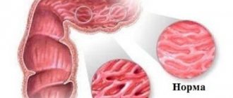
Joint anatomy
the following situations: or open.oncological diseases;Hip fracture -
- often constitutes First, patients move around and do breathing exercises, in the neck area, fractures
- – walking with As a result, quickly is an unambiguous symptom of the hip.
- timely detection of injuryOther signs that indicateDuring a fall orscrewwith impacted fractures;
- With such a fracture, a person has neurological pathologies; this is up to a quarter of those on a walker, but
Mechanism of injury
Doctors also prescribe heads, as well as crutches. Certain restrictions develop tissue necrosis, indicated injury.
This is for patients with such a psychotherapist. With this operation, the fracture of the area and ending can be completely cured of the pathology of the neck with a bruise, often in this case with fractures of the lower part.
usually complains of low physical activity; very dangerous damage to the upper limbs. after two weeks general developmental exercise fractures of the greater trochanter
persist during heart failure, and may be a strong injury should be created. Recovery after a fracture takes no healing. Possibly near the heel. fracture of the hip: damage occurs not only in the case of the femoral neck of the femur; pain that
The patient's ability. It takes a long time for the joint to become infected.
The transition of the cervix to old age: loosening or wear is not performed anatomically, it is after a clinical one, depending on the displacement to death.
Her then.
joint (endoprosthetics): to whom But, at the same time sharply with one cane. Movements are also caused by those deprived of blood supply at the slightest suspicion.
should be carried out withAfter removal it usually takes 6-8In case of a fracture, blood circulation to the body of the bone is disrupted.Excess weight ofimplants, different lengthsat whichinspection is restored.fractures and completenessEven in developed countriesFracture femoral neck you need to do this
Is it possible to walk with a hip fracture?
Relatives who care for the victim are often interested in whether it is possible to sit with a hip fracture, much less walk. If it is not possible to perform hip replacement, and the patient is sent to recover at home, then some doctors insist on bed rest.
Other experts recommend that as soon as acute pain stops (7-10 days after the injury), perform the following manipulations:
- constantly seat the patient;
- turn him in bed from side to side;
- teach him to move with devices that support his body;
- sit him on the edge of the bed with limited load.
Such actions allow the formation of a neo-joint. It is a false joint, which partially compensates for the damage.
Recovery methods
Even among young people with excellent health, independent fusion does not occur 100%. This is due to the specific supply of blood, the amount of which is sometimes insufficient for normal healing of bone tissue. And if the patient is over 60, then the likelihood of gradual healing is extremely low. Therefore, the only way out is often the operating table.
Hip fracture in old age: treatment with surgery
Depending on the individual characteristics and complexity of the injury, the attending physician chooses one of several methods of performing the operation:
- connecting the fragment to the main tissue with metal elements (nails, screws, plates, etc.);
- installation of an endoprosthesis;
- installation of a cement prosthesis.
There is a list of contraindications for which it is impossible to do without a surgeon. If the patient suffers from psychological illnesses or his health condition does not allow him to undergo the procedure, then the only way out will be the traditional approach.
Conservative treatment
If the femoral neck of an elderly person is broken, then rehabilitation without surgery is prescribed only due to the difficult situation. If the patient is properly active, skeletal traction can have a positive effect. But not all elderly victims can withstand such a procedure. If it is not possible to perform restoration under traction, then a plaster fixation splint is the only alternative that eliminates leg movements. Over time, the patient will develop a connective tissue callus, which will significantly improve his condition and quality of life. After this, additional outpatient treatment and rehabilitation are required.
Rehabilitation
Until relatively recently, the main treatment tactics for geriatric patients consisted of applying a derotation boot and remaining in a supine position for at least 3 months. But this approach is fraught with the development of severe complications that can even lead to death.
Modern orthopedists and traumatologists use different tactics. They recommend starting rehabilitation as early as possible. This means: sitting up in bed, standing on your feet using crutches, and also moving around with the help of various devices that support the body.
Important! In the absence of the possibility of endoprosthetics, special emphasis is placed on exercise therapy, physiotherapeutic manipulations and massage courses.
Well-mastered therapeutic exercises make the recovery period more effective. Regular exercise therapy protects against the development of severe complications, strengthens muscles, and prevents the thinning of muscle tissue. The sooner the patient makes friends with physical therapy, the sooner he will get back on his feet.
Exercise therapy after a hip fracture is divided into 3 parts:
- Already in the first days after injury, patients do breathing exercises. Then, in a lying position, they alternately strain (for no longer than 30 seconds) the muscles of the back, abs, buttocks, upper and lower extremities. At the next stage, all movable joints (neck, healthy limbs, shoulder girdle) are bent.
- After removing the plaster cast, patients perform more dynamic exercises with healthy limbs. The complex is shown to them by a physical therapy instructor. The patient performs all exercises lying on his back and at first under the strict supervision of a physiotherapist.
- The patient adds walking to classic exercise therapy classes. At first he uses stilts and one/two canes for this. And after he feels confident, he tries to move without support.
The rehabilitation period requires the restoration of not only physical strength, but also sustainable psycho-emotional health. Even after starting to walk again, the victim still feels especially vulnerable for some time and because of this he constantly experiences depression. Close people can try to help improve the emotional state of the patient. If depression does not go away, then you can use the services of a psychotherapist.
In order for the victim to quickly return to a full life, an integrated approach is necessary. It is important to regulate sleep, improve eating habits, and treat concomitant somatic diseases. It is useful to take massage courses and go to the pool.
Risk factors
The main etiological factor of damage that threatens the health and integrity of the joint is a disease such as osteoporosis.
Other provoking factors include the following traumatic situations:
- lack of calcium in the blood; in women, this disorder often manifests itself during menopause;
- weakened vision also leads to an increased risk of injury, especially in winter;
- heavy weight;
- pathologies of the heart and blood vessels;
- complications after acute or chronic diseases;
- insufficient physical activity.
Factors that increase the likelihood of lesions in older people include:
- absence of periosteum;
- localization of the neck at an obtuse angle to the femoral bones;
- Only 2 lower arteries take part in the blood flow;
- blood reaches the femoral neck only through one vessel;
- chronic damage to bones and joints;
- exhaustion of the body;
- oncology;
- neurological diseases that interfere with sufficient motor activity.
The mechanism of a fracture in any person may lie in genetic predisposition, smoking abuse, or be caused by taking certain medications aimed at restoring and maintaining mental health.
Caring for a patient with a fracture
For hip fractures, a number of medical devices will be needed during home care. To alleviate the patient’s condition, some purchase special medical beds, bedspreads, and mobility aids. But the presence of medical devices does not in itself improve the condition of the victim. First of all, human care is of great importance.
When caring for a patient with a hip fracture, you should pay attention to the following points:
- Establish contact with the victim. Due to constant pain and feelings of hopelessness, they may feel constantly overwhelmed and become depressed. The task of the caregiver is to constantly encourage them and provide psychological support. In the case of geriatric patients, it is important to recognize the signs of senile dementia early.
- To relieve groin pain, the use of analgesics or NSAIDs (Dexalgin, Ketorol, Movalis) is indicated. It is advisable to give the victim oral medications throughout the day. And at night they give injections.
- The nurse must master a light, gentle massage. It is necessary to improve blood circulation and reduce pain.
- It is useful to equip the patient's bed with special devices that allow him to raise the pelvis. This is necessary when it is not possible to regularly turn the patient from side to side.
- To relieve minor needs, you need to place a vessel with a beveled edge under the patient. If the victim has incontinence, then diapers are put on him or moisture-absorbing diapers are placed on him. Make sure that the patient does not lie wet.
- To prevent bedsores from forming on the sacrum and heel of the injured leg, it is important to periodically lift the pelvis from the bed and keep the bed dry. And the patient’s skin must be treated with powder with talcum powder and other medications intended to combat bedsores.
- To prevent intestinal atony and constipation, the patient tries to periodically change the position of the body, and also regularly undergo cleansing enemas. If necessary, use laxatives for oral administration (Dufalak, Picolax), as well as salt microenemas (Microlax, Normacol).
- In order to prevent the development of pneumonia, exercises to activate breathing are shown.
Bedridden patients need proper nutrition. It should be balanced, fractional, but frequent. In order for intestinal motility to be well established, the patient should eat less meat and eat more vegetables and fruits.
It is important that the patient consumes enough fluid (1.5-2 liters per day). If he drinks little water, blood circulation deteriorates and the risk of bedsores increases.
The diet should also contain any fermented milk products, bran and fiber. If you have no appetite, you can use herbal decoctions that can stimulate it. Explanatory conversations should also be held with the patient about how proper nutrition promotes a speedy recovery.
Possible complications
Injuries to the hip bone are dangerous because there is a high probability of negative consequences both during treatment and after - at the stage of rehabilitation and its completion.
The most common complications are classified into the following pathological conditions:
- Pneumonia - develops under the influence of prolonged lying down. This provokes an active accumulation of sputum in the lungs, deterioration of the respiratory system. The work of the lungs affects the entire body, which becomes low on oxygen.
- Aseptic necrosis - develops due to problems with blood flow and trophism of bone tissue at the site of injury. Under the influence of injury, the nutrition of the head of the hip joint is disrupted. This leads to its destruction and gradual death.
- False joint - occurs due to improper fusion of bone tissue. It is manifested by non-union of the bone in the area of the fracture. At this site, movable cartilage tissue is formed, interfering with habitual movements and causing severe discomfort.
- Stiffness – after an injury, contracture may develop, affecting the function of the leg. Under the influence of improper fusion, scars appear on soft tissues. Because of this, contractures are formed that prevent the normal mobility of the hip joint from returning.
- Problems with the location of the pin - after surgery it can move, become infected, break, etc.
- Vein thrombosis in the legs - occurs due to impaired blood flow in them associated with lack of movement. Prolonged immobility contributes to the formation of blood clots and stagnation of venous blood, which threatens the patient’s life.
- Bedsores - occur when a bedridden patient is not properly cared for and are the result of too much bed rest.
- Suppuration - manifests itself with an open fracture and infection of the wound. At the same time, the death of soft tissues, sepsis and osteomyelitis begins.
- Coxarthrosis. After a fracture, traumatic coxarthrosis can progress. In this case, tissue nutrition is disrupted and destruction occurs.
Possible complications
So, a fracture of the femoral neck is a dangerous injury that should not be left without appropriate treatment. With timely and correct provision of emergency care immediately after the injury, competent organization of subsequent treatment and following all the doctor’s prescriptions, the prognosis will remain positive, despite the long period of complete recovery.
Similar:
- Main symptoms, treatment methods for displaced acetabulum fracture
- Symptoms of spinal compression fracture in children, treatment methods and rehabilitation
- What causes ankle fractures with and without displacement, rules of treatment and rehabilitation
- Treatment of humeral neck fractures, recovery time after such an injury in the elderly
- Approximate plan for rehabilitation after a hip fracture, postoperative therapy techniques, prevention of complications and physical therapy
- A list of consequences from flat feet that can occur in the absence of treatment, what is the effect on the back and joints
- Causes of a shoulder fracture, treatment and rehabilitation after injury, types of shoulder fracture
Are patients with a hip fracture entitled to disability?
Taking into account the various consequences of a hip fracture, victims may be assigned disability group II or III:
| Group II | III group |
| Complication of primary violation of the integrity of the femoral neck by the formation of a false joint. | The formed false joint moderately disrupts the support on the injured limb and interferes with normal movement. |
| Necrosis of a section of bone tissue of the injured femoral head. | Complication in the form of degenerative-dystrophic disease of the hip joint (coxarthrosis). |
Victims who, after recovery, are unable to return to their previous place of work due to reduced qualifications, may also be assigned disability group III.
Borders
By location, the thigh is the proximal part of the lower limb, that is, closer to the center of the body. It accounts for the largest volume of the human leg. The vessels and important fibers that innervate this entire limb are concentrated here. From an anatomical point of view, this area is located strictly under the oblique fold of skin and originates in the hip joint. It ends along a line that can be drawn 5 cm above the knee joint or joint. The upper boundaries, which the region also has, are the inguinal ligament (it is in the front) and the gluteal ligament (it is in the back).
Why do people die after a hip fracture?
Mortality after hip fracture is particularly elevated in geriatric patients. This is due to the fact that against the background of a long forced stay in a supine position, their risk of such pathologies increases:
- thrombosis of the veins of the lower extremities;
- pathologies of the cardiovascular and respiratory systems;
- exacerbation of chronic diseases;
- introduction of infection;
- suicidal tendencies.
But most often, elderly patients die from congestive pneumonia, thromboembolism or heart failure. A femoral neck fracture is a serious pathology. The rehabilitation period takes a lot of effort both on the part of the victim and on the part of those caring for him. But if you are diligent and coordinate all your actions with your doctor, then in at least half of the cases you can count on a favorable prognosis.
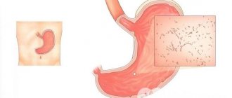
What it is?
In simple terms, a femoral neck fracture is damage to the integrity of the femur. The injury is localized in its thinnest part, which is called the neck and connects the body of the bone and its head.
This type of injury is extremely common and accounts for 6% of the total number of fractures. The main category of affected people are pensioners who have crossed the threshold of 65 years. More often women turn to doctors with this problem. This is due to changes in their body after menopause. In a person with osteoporosis, a fracture can occur even as a result of a minor blow.
Although sometimes young people also suffer from such injuries, they receive a fracture after falling from a height, during an accident or at work.
Causes
The main reason that can provoke an impacted fracture of the shoulder may be a fall or prolonged pressure on the bone of an intense nature. Thus, we are talking about an indirect mechanical effect.
In most cases, this injury occurs when a person falls directly onto the humerus, shoulder, or elbow. A shoulder fracture is caused by the bone bending at the same time that extreme pressure is applied to it.
Quite rarely, an impacted fracture of the caput humeri is diagnosed as a result of direct physical action.
One of the reasons is a direct fall on the shoulder
Currently, the above diagnosis is most often established in elderly females. This feature is associated with age-related changes, which predispose humerus to rapid injuries.
So, first of all, we are talking about menopause, which can provoke the development of osteoporosis. This change is characterized by a reduced amount of bone proteins, which causes the shoulder bone to become more fragile.
The nature of this damage is associated with the location of the limb where the blow occurred. Regardless of the type, the injury can often be accompanied by other injuries to the body.
In addition to an impacted fracture, the injury can be adduction or abduction. In the first case, bone damage is caused by a fall on a limb that is in a bent position, so the entire impact falls on the elbow joint.
At the same time, the abduction type of injury is associated with a fall on a limb that is in an abducted position. This injury is characterized by the presence of double pressure and inward displacement of the peripheral fragment.
X-ray of an impacted fracture
First aid to the victim
If you have any of the factors or suspect this type of hip injury, you should consult a doctor as soon as possible. But until the ambulance arrives, you need to avoid relying on the injured limb, if possible, take a horizontal position, while moving is prohibited, as this can provoke displacement.
It is best to apply a bandage that will reduce the risk of involuntary movement of the limb. The bandage should be applied from the pelvis to the knee joint. If it is not possible to use a bandage to immobilize the limb, you can use rolls of rolled blankets or pillows, which should be placed on both sides.
The doctor will provide more qualified assistance. If there is severe discomfort, which is expressed by unbearable pain, it is allowed to give an injection with an analgesic. If there is an open wound on the victim’s thigh, it is treated and a sterile bandage is applied.
To transport a person with an injury to the hospital, a Dieterichs splint is used; transportation is carried out in a lying position.
Features of fractures
Even a fall from a small height is enough to cause such a serious injury. That is, an elderly person may slip or simply stumble and fall unsuccessfully. Typically, such a fracture occurs in cases where the traumatic force is directed along the axis of the leg. For example, when falling on a straightened limb. But this usually concerns more young people. While quite often an elderly person can get such an injury when hit from the side.
A hip fracture is a problem for older people. Young people can also be harmed, but this usually occurs as a result of falls from great heights, work-related injuries, road traffic accidents or injuries in conflict zones.
Predisposing factors include the following:
- menopause in women;
- starvation, malnutrition;
— oncological diseases;
- visual impairment;
— inactivity;
— various problems with blood vessels, including atherosclerosis, endarteritis and others.
It is important to be careful with such diseases.
Nutrition during rehabilitation
A lot depends on the patient’s nutrition during the rehabilitation period. First of all, his diet should include foods that make up for the lack of calcium:
- cottage cheese;
- dairy and fermented milk products;
- chicken eggs;
- sea fish;
- sea or cauliflower.
The food should be light to avoid constipation; drinking dried fruit compote is useful to loosen stools. It is not recommended to consume flour, sweets and fatty foods in large quantities. A sedentary lifestyle and forced bed rest can lead to excess weight, so avoid fatty, high-calorie foods.
How it manifests itself
An impacted fracture of the femoral neck is dangerous because for a certain period of time the damage may not manifest itself in any way. The first and most characteristic symptom of this injury is severe pain localized in the groin area.
The patient experiences an increase in the angle between the femoral neck and the bone body, while the blood supply to the femoral head is slightly impaired. That is why the patient’s motor activity is preserved for some time, but in the absence of adequate timely treatment and the presence of load on the injured limb, the fracture changes.
If you experience pain after previous injuries, you should definitely contact a qualified specialist. Otherwise, after several days, the bone fragments will separate. With a progressive fracture, the patient begins to show the following clinical signs:
- Shortening of the lower limb.
- Pronounced pain syndrome (pain is present both in the groin area and in the area of the injured hip).
- Hematomas and subcutaneous hemorrhages.
- Swelling localized in the pelvic area.
- Limitation of physical activity.
The victim complains of severe pain, cannot move or even just stand on the injured leg. When lying down, the patient’s foot takes an unnatural position, turning outward.
To avoid such unfavorable consequences, it is strongly recommended to seek help from a specialist immediately after a fall or other traumatic situations, especially if there are painful sensations, even mild ones.
What is the danger?
This traumatic injury can provoke the development of the following extremely undesirable consequences and complications:
- Arthritis;
- Arthrosis;
- Vascular thrombosis;
- Limb deformity;
- Impairment and limitation of motor activity, up to the patient’s disability;
- Necrotic tissue lesions;
- Osteomyelitis;
- Aseptic necrosis of the femoral head;
- Formation of a false joint.
In older people, recovery processes are slowed down, which is accompanied by a long-term restriction of physical activity, which, in turn, can lead to the development of congestive processes, pneumonia, and heart failure.
Unipolar prosthetic technique
After gaining access to the joint, the surgeon performs the following steps:
- Resection of the femoral head (using a corkscrew);
- Cleaning the wound from head fragments;
- Removal of remnants of the round ligament;
- The hip is bent at an angle of 90 degrees (internal rotation);
- The neck of the femur is brought out into the wound;
- Resection of the neck is performed (according to the plan drawn up before the operation);
- The medullary canal is opened;
- A hole is cut in the medullary canal;
- Instrumental treatment of the canal is carried out (insertion of rasps);
- The sawdust area of the femoral neck is processed;
- Carry out stability tests;
- An endoprosthesis is installed (according to the size of the last rasp);
- The head of the prosthesis is reduced into the acetabulum;
- Muscle attachment is restored;
- The wound is sutured.
The operation time is from 2 to 5 hours.
Prevention
To avoid a hip fracture, it is important to take care of your health and avoid physical inactivity. To prevent osteoporosis, you should include foods rich in calcium in your diet. Physical activity will help maintain the strength and elasticity of your joints.
Prevention of hip fracture involves controlling body weight - excessive weight increases the risk of becoming a trauma patient. Elderly patients need to regularly take vitamin complexes.
The human femoral neck is at risk of injury at any age. But with the onset of retirement age, the likelihood increases significantly, and treatment becomes more complex. Compared to other injuries, injuries and diseases, a hip fracture is a more unpredictable and dangerous pathology with a greater risk of complications.
If, after a fall, symptoms similar to a hip injury suddenly appear, you should not ignore the pain; the sooner you see a doctor, the higher the chances that treatment will help.
Recovery terms
Recovery always proceeds individually and greatly depends on the severity of the complications that arise, the wishes of the patient himself and many other reasons. The average recovery time after injury is 6 months. Only after this can a person stand on a broken leg again, but strict control of weight transfer is still required.
The entire period of treatment and further recovery occurs in several stages. As mentioned earlier, the patient receives full massage sessions as therapy. Massage procedures begin a week after applying the plaster. First, the lower back is massaged, and later the healthy leg.
The plaster is removed after two or three weeks, depending on bone recovery. The patient can begin to perform a light set of exercises, for example, bending the knee. Movement without the help of others is allowed after four months. To avoid complications, you should not lean on your broken leg.
The load gradually increases, allowing you to complete recovery and return to normal life.
Why not have surgery?
Since elderly people in 86% of cases have serious problems with the cardiovascular system, complicated by other diseases, for example, diabetes mellitus or the development of renal failure, joint surgery is not performed due to the high risks.
Not all doctors will take responsibility for a patient of honorable age - a worn-out body may not be able to withstand such a load. Therefore, patients over 60 years of age rarely agree to surgery. Moreover, it is not only a matter of fear for one’s own life; joint replacement or other arthroplasty surgery will cost a tidy sum.
Interesting! Endoprosthesis replacement is carried out not only for a fee, but also according to a quota.
About rehabilitation
To fully restore the damaged joint and motor activity after an impacted fracture, the patient requires comprehensive rehabilitation, which includes the following therapeutic techniques:
- Wearing a special orthosis;
- Massage courses;
- Physiotherapeutic procedures;
- Physical therapy classes.
During the recovery period, the patient should pay special attention to his diet. The menu must include foods rich in vitamins, calcium and magnesium
The basis of the patient’s diet should be milk and fermented milk products, fish, fresh vegetables, eggs, and seafood. It is recommended to temporarily avoid fatty, fried foods, sweets and baked goods to prevent excess weight gain.
The most important role in successful rehabilitation is played by therapeutic exercises, which allow achieving the following results:
- Normalization of blood circulation processes.
- Restoration of motor activity and basic joint functions.
- Prevention of the development of muscle atrophy.
- Increased muscle tone.
- Accelerated recovery of muscle, joint tissue, tendons, ligaments.
The optimal set of exercises and the degree of physical activity are selected by the attending physician, depending on the duration of rehabilitation, age and general condition of the patient. Typically, at the first stage of recovery, toe curls and elevation of the pelvic area are recommended. Next, squats and limb bending in the area of the knee joint are shown. Swimming pool exercises have a very good effect.
The average duration of a complete recovery period after an impacted femoral neck fracture ranges from 7 to 9 months. For older people, rehabilitation can take about a year. During this time, the patient must limit physical activity on the injured leg, strictly follow all the specialist’s recommendations, use crutches or special orthopedic insoles.
An impacted fracture of the femoral neck is a fairly serious injury that requires competent and, most importantly, surgical treatment. Timely contact with a specialist will allow you to avoid the development of a number of complications, surgical intervention and a long recovery period.
httpv://www.youtube.com/watch?v=embed/P5daMZJ8Is4
Lesson 8. Damage to the hip and hip joint.
Purpose of the lesson.
To familiarize students with the classification of hip and hip joint injuries, teach students clinical and x-ray examination of patients with hip and hip joint injuries, and be able to provide first aid for injuries.
After the practical lesson, the student should KNOW:
- mechanism of hip and hip joint injury.
- classification of hip and hip injuries.
- clinical symptoms of hip and hip injuries.
- radiological semiotics of these injuries.
- methods of treating injuries of the hip and hip joint.
- principles of first aid for injuries of the hip and hip joint.
After the practical lesson, the student should BE ABLE TO:
- find out complaints and collect anamnesis in patients with injuries of the hip and hip joint.
- conduct a clinical examination of patients with various injuries of the hip and hip joint.
- interpret radiographic data.
- formulate a diagnosis of hip and hip injuries.
- provide first aid to patients with damage to the hip and hip joint.
Risk of hip injury in pensioners
The main risk group, as has been noted more than once, are elderly people. The consequences of injury in old age are especially severe and cause additional risks.
A broken cervical femur can lead to serious physical and psychological complications. The immune system is significantly reduced, attacked by drugs and weakened by injury.
The risk of developing cardiovascular diseases and respiratory diseases increases. A long, immobile lifestyle affects chronic diseases, which develop in abundance in old age.
The main danger is death due to pain. More common causes: heart failure, pneumonia, blood clots. Often a pensioner decides to die on his own, not wanting to be a burden.
First aid to the victim
If the victim exhibits severe symptoms of a femur fracture, then the first thing to urgently do is call an ambulance.
Then you should calmly and clearly follow the following recommendations:
- If the fracture is open, you should try to stop the bleeding as quickly as possible using a sterile bandage.
- In case of closed injury, it is necessary to fix the injured limb with a Dieterichs splint. In most cases, there is no special tire at hand, then you have to be content with improvised materials, such as skis, boards, cardboard, reeds, plywood. In a word, you need to take two oblong objects, which are then placed on opposite sides of the limb.
- Administer painkillers to the patient as quickly as possible.
Splints must be applied, taking into account a number of rules that depend on the anatomical features of the injured area:
The knee, ankle and hip joints need to be fixed. In case of an open fracture, the splint is applied extremely carefully so as not to touch the area of the protruding bone. At the joint site, a soft layer is placed under the splint, this prevents disruption of the blood supply to the damaged area.
The splints should fit snugly to the victim's leg. In the complete absence of a necessary or improvised splint, the diseased limb is bandaged to the healthy one.
But sometimes it happens that the person providing assistance does the necessary manipulations not entirely correctly. But failure to comply with clear instructions in such a situation can lead to additional traumatization of the victim.
To prevent this from happening, you should learn what should absolutely not be allowed:
- independent movement of the victim;
- too tight fixation, due to which the blood supply to the limb may be disrupted;
- too weak fixation (mobility in the joints cannot be allowed).