Author: A. Olesya Valerievna, candidate of medical sciences, practicing physician, teacher at a medical university
Cerebrovascular diseases constitute one of the most significant problems of modern medicine. Mortality from vascular accidents of the brain occupies a leading position among other diseases, and the frequency of disability is extremely high.
Carotid artery stenosis causes ischemic necrosis in the brain in about a third of all stroke cases. When the lumen of the internal carotid artery is closed by more than 70%, cerebral infarction occurs in almost half of the patients within the first year after a significant disruption of blood flow. Early diagnosis and timely elimination of the problem can help avoid such a dangerous consequence. Modern surgical treatment methods are safe, and with early detection of pathology, minimally invasive treatment is possible, which does not require large incisions and general anesthesia.
The carotid arteries depart from the aorta, go in the tissues of the anterolateral surface of the neck to the head, where they are divided into external and internal branches, carrying blood further to the vessels of the brain and tissues of the head. Stenosis can appear in any of the areas, but most likely in places of narrowing (ostia, division into branches).
The main volume of blood flows to the brain precisely through these large arterial trunks, so any disturbances in them lead to hypoxia and require immediate examination and treatment. If in the USA the number of surgical corrections of stenosis reaches 100 thousand per year, then in Russia only about 5 thousand are performed. Such a low figure does not allow covering all those in need of treatment, and this is one of the significant problems of the healthcare system.
Another problem is the late detection of pathology or the patient’s reluctance to “go under the surgeon’s knife,” however, all patients with critical stenoses should be aware that surgery is the only way to avoid a stroke and save life.
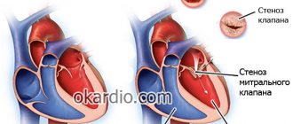
What is carotid artery stenosis
Impaired blood flow through the carotid artery due to narrowing of its lumen is called stenosis. This disease is found in every second patient with cerebral ischemia and in a third of patients with stroke. The disease, as a rule, occurs against the background of the formation of an atherosclerotic plaque inside the vessel. Depending on the degree of damage, the following options are distinguished:
- limited stenosis up to 1.5 cm, widespread more than 1.5 cm;
- segmental - up to a quarter of the circle, concentric - more than half the lumen;
- with complications (hemorrhage, blood clot formation) and without them.
We recommend reading the article about atherosclerosis of the neck vessels. From it you will learn about the pathology and symptoms of its development, causes, diagnosis and treatment.
And here is more information about examination for atherosclerosis.
Diagnostics
If signs of stenosis occur, you should seek first aid. At the same time, the attending physician will not be able to immediately make a diagnosis based on the symptoms, which, as already mentioned, are not specific. To do this, a series of studies are carried out, based on the results of which a diagnosis is made and treatment is prescribed.
Diagnostic methods:
- KTA;
- ECG of the heart;
- TANK;
- General blood and urine analysis;
- Ultrasound of the carotid arteries.
Diagnosis and CT angiography of cerebral vessels gives the most comprehensive results.
The technique of inserting an arterial catheter is carried out under local anesthesia and provides the ability to collect blood pressure data and free access to frequent blood sampling for subsequent laboratory testing.
Before diagnosis, you are not allowed to consume food and drinks in minimal quantities for 10 hours. Water procedures and preparation of the groin area for surgery (shaving) are also recommended. The images and results will provide the necessary information for therapy.
Risk factors
An atherosclerotic plaque forms on the wall of a vessel and contains cholesterol, triglycerides and platelets. Risk factors for this process are nutritional disorders (abundance of animal fats and simple carbohydrates in the diet), obesity, type 1 and 2 diabetes, smoking.
As it increases, it can independently block the lumen of the vessel or a blood clot breaks off from it, which moves with the blood into the internal arteries of the brain, causing their blockage. The plaque itself can also break off and, in the form of an embolus, disrupt the nutrition of deeper cerebral structures.
In addition to atherosclerosis, carotid artery stenosis is caused by:
- connective tissue diseases (fibromuscular dysplasia);
- autoimmune arteritis;
- traumatic injuries;
- blood diseases - increased platelet count, sickle cell anemia;
- thrombus formation in heart defects, endocarditis, atrial fibrillation, tumor processes;
- abnormal structure, including kinking (increased tortuosity).
Folk remedies
- Decoction of plantain and golden mustache.
- Hawthorn berry jam. Its use can significantly reduce the load on the heart of a sick person. To prepare, pour boiling water over the hawthorn berries and leave the mixture overnight. In the morning they need to be taken out of the water and ground in a bowl. After this, sprinkle the berries generously with sugar and make the jam using the traditional approach. You should take this jam once a day for a week, on an empty stomach, by dissolving a tablespoon of jam in a glass of water.
- Garlic tincture with honey and lemon.
- A mixture of honey and onion juice.
Before using these remedies, you need to make sure that the patient is not allergic to herbs. The cost of using such methods is noticeably lower than professional treatment, but you need to remember how to treat the disease without risk: only in combination with professional medicine methods and only with the permission of a doctor!
Also in treatment and prevention, household treatment methods are used, which serve to generally strengthen the body, for example, contrast showers. Used to quickly relieve vascular spasms. Every morning and evening you should pour your head first with hot and then with cold water. Such a shower can be taken for the whole body - this is a generally accepted method of hardening.
Carotid artery stenosis is a dangerous consequence of atherosclerosis and a number of other diseases, which, in turn, has quite frightening symptoms and development prognosis. Fortunately, modern medicine makes it possible to treat it, and a timely response to the disease will allow the patient to save his life and get rid of most of the serious consequences.
Symptoms of the onset of pathology development
If the lumen of the carotid artery is blocked gradually, then bypass pathways for blood supply begin to form in the brain, cells adapt to oxygen starvation and lack of nutrition, therefore, with unilateral and local stenosis, there may be an asymptomatic course and a relatively favorable prognosis. Sudden obstruction leads to an acute cessation of blood flow, that is, a stroke.
The first signs of obstruction of blood flow to the brain are called transient (temporary) cerebral attacks. Their main features:
- weakness (paresis) in one arm or upper and lower limb on the opposite side of the blocked artery;
- numbness of half the face and fingers;
- visual impairment on the side of the stenosis - darkening in the eyes, flickering dots or spots;
- difficulty pronouncing words;
- headache;
- fainting;
- difficulty swallowing;
- local convulsive muscle twitching.
Possible complications
The most serious complication of insufficient blood supply to the cerebral cortex is stroke. Doctors are considering various options for the development of a critical condition with atherosclerosis:
- Impaired blood supply to the brain. The free lumen of the arteries can be so narrowed that normal blood supply to the brain becomes impossible.
- Rupture of cholesterol plaque. Part of the growth may break off and enter smaller vessels with blood, blocking them.
- Blockage of blood flow by a clot. Some plaques rupture, and the body then sends platelets to the damaged area. Platelets form a blood clot, which can block or slow blood flow.
A stroke can lead to serious consequences for the normal functioning of the brain. In some cases, the critical condition can be fatal.
Degrees of carotid artery stenosis
To determine treatment tactics and prognosis of the disease, a criterion such as the degree of stenosis is used. It is calculated in relation to the reference size of the vessels:
- internal carotid artery superior to the bulb;
- common carotid artery before branching at 1 cm and 3 cm.
To calculate, measure the minimum lumen of the artery and divide it by the proper size. The data obtained can be as follows: moderate (above 50%), hemodynamically significant (from 70%), critical (up to 99%), complete occlusion. Moreover, narrowings of more than 75 percent are indications for surgical treatment.
Choice of carotid endarterectomy
Opinions differ regarding the advisability of endarterectomy. There is evidence of an increase in recurrent thrombosis and a significant risk of acute bleeding. Proponents of this intervention insist on observing precise indications and contraindications.
Surgery is recommended for patients with neurological symptoms when:
- narrowing more than 70%;
- acute thrombosis in the internal carotid artery;
- against the background of a stroke clinic;
- if the narrowing is embologenic and has stenosis from 30 to 69%;
- with simultaneous coronary artery bypass grafting;
- with existing acute aortic dissection;
- if while taking aspirin, the stenosis is less than 30% of the diameter.
Surgery is contraindicated for this group of patients if, without taking aspirin, the stenosis is less than 30% and has a chronic course.
For asymptomatic patients, the following indication is suggested: stenosis over 60%, while the prognosis of complications should not exceed 6%.
The following are considered obvious contraindications:
- narrowing less than 60%;
- the degree of stenosis is above 60%, but the risk of complications exceeds 6%;
- chronic form of occlusion;
- signs of carotid artery dissection.
The principle of the algorithm for choosing surgical treatment of stenosis should take into account that the frequency of complications cannot exceed the natural risk of the disease.
After discharge from the hospital, the patient must:
- constantly take a maintenance dose of antithrombotic drugs;
- give up smoking, alcohol, overeating, saunas and steam baths;
- undergo regular re-examinations.
What are the special types of pathology?
Depending on the effect of the degree of narrowing on the blood circulation of the brain, several clinical forms of the disease are distinguished.
Hemodynamically significant
Due to the blocking of more than 70% of the lumen of the artery, before the narrowing there is an increase in blood pressure, and after it the blood flow drops significantly, which causes a deficiency of nutrients and oxygen in the area of the branches of the middle cerebral artery and adjacent areas.
Clinical manifestations occur in the form of severe ischemic attacks, which may still have a reversible course. There are paresis of the limbs, decreased reflexes and sensitivity, visual and speech disturbances. With timely drug therapy, partial or almost complete restoration of functions occurs. 5% of patients develop a stroke within a year.
Critical stenosis
If the lumen is narrowed by 70 to 99 percent, then this is considered dangerous for the occurrence of extensive ischemic strokes. At this stage, atherosclerotic changes lead to massive damage to the arteries, which makes it difficult for the formation of collateral pathways to supply the brain.
Blood clots and emboli are formed that block small branches of blood vessels, so the course of the disease is resistant to conservative treatment methods, and complete rehabilitation of patients is difficult to achieve.
Watch the video about carotid artery stenosis:
https://youtu.be/49KdUldVbE8
Clinical forms
Atherosclerotic lesions of the carotid arteries most often differ according to the localization of the main process:
- Atherosclerosis of the common carotid arteries - rarely contributes to ischemic stroke; even with complete blockage of the common carotid artery, blood flow through the internal carotid artery can be preserved. However, there may be general cerebral disorders - poor memory, general weakness, headaches.
- Atherosclerosis of the bifurcation of the common carotid artery - atherosclerotic plaques narrow the entrance to the internal carotid artery and can completely block it. This is the most common form of damage. With this localization, transient ischemic attacks and ischemic strokes most often develop.
- Atherosclerosis of the intracerebral sections of the carotid artery - plaques are located in the carotid arteries and their main branches. Ischemic stroke often develops due to thrombosis of narrowed arteries.
Ultrasound identifies two main types of atherosclerotic plaques:
- Stable plaque - the narrowing of the artery has smooth contours, without undermined edges, with a slight acceleration of blood flow on the plaque.
- An unstable plaque is often a calcified plaque with jagged contours; moving elements and significant turbulence of blood flow during Doppler sonography may be observed.
Based on the degree of narrowing of the carotid artery, there are:
- Hemodynamically insignificant stenosis (narrowing of the artery by less than 70%, without local acceleration of blood flow)
- Hemodynamically significant stenosis (narrowing of the artery by more than 70%, with accelerated blood flow)
- Occlusion - complete blockage of the carotid artery
Diagnosis of the right and left arteries
In addition to the neurological status of the patient, instrumental methods are used to determine the diagnosis of stenosis:
- Ultrasound of the vessels of the neck: accelerated reverse flow through the external branches; when blocked, blood flows through them to the ophthalmic artery. When the temporal artery is compressed, the blood velocity decreases.
- Angiography of cerebral vessels is used only for preoperative examination. It is being replaced by magnetic resonance imaging.
- MRI with vascular contrast is the most valuable method. Ischemic foci in the brain and sites of vascular embolism are determined.
The difficulty in diagnosing a stroke is that in about a third of patients on the first day of its development there are no visual manifestations in the brain tissue.
How is the disease prevented?
By controlling your weight, a person will prevent such a dangerous disease.
It is much easier to prevent a disease than to deal with its consequences. Doing exercises, monitoring your diet, and providing the body with necessary vitamins and microelements is required from an early age. In young people, the body is much more susceptible and can cope with the problem without serious complications. To prevent the disease, you should follow medical recommendations, which include:
- giving up destructive addictions;
- control of weight fluctuations;
- proper diet;
- eradication of psycho-emotional situations;
- introduction of physical exercises;
- timely medical examination.
Treatment as a chance for life
In order to completely restore blood supply to the brain during stenosis, only surgical treatment methods can be used. But if the operation is performed when a stroke has already occurred, then the mortality rate for such patients is about 40 percent. Therefore, radical therapy is recommended for the following indications:
- suffered a transient cerebrovascular accident;
- cured stroke with minor neurological abnormalities;
- blockage of more than 70% of the artery lumen (even in the absence of manifestations);
- presence of sources of thromboembolism;
- decreased cerebral blood flow.
Operation options
When the lumen of the vessels is completely closed, an anastomosis is created between the external and internal cerebral arteries to bypass the area of stenosis. To do this, connect the subclavian and common carotid or external carotid arteries.
If there is partial occlusion, then surgical treatment options may include:
- carotid endarterectomy or similar reconstructive methods;
- stent installation;
- shunting.
The first method has a large evidence base for its effectiveness in preventing strokes for 3 to 5 years. To carry it out, the artery is dissected at the location of the plaque, which is separated from the vascular wall. Then a Teflon shunt is installed into the lumen of the vessel, which is removed after the plastic surgery.
The eversion method involves isolating the carotid artery, inverting it like a stocking, and layer-by-layer separation of the plaque. After this, the original tightness of the vessel is restored.
Carotid endarterectomy
Recovery after
For the first two days, the patient is in the intensive care unit, where basic vital functions and cerebral hemodynamics are monitored. Particular attention is paid to blood pressure and blood electrolyte balance. Before discharge, duplex scanning, CT and Doppler ultrasound are performed. As a rule, the patient goes home after a week of hospital stay.
Complications of the operation may include heart attack, stroke, bleeding, vascular thrombosis, and nerve damage. In the long term, a relapse of the disease may occur.
To prevent such conditions, preventive treatment is prescribed:
- Plavix,
- Warfarin,
- Acetisalicylic acid,
- Chimes.
Anticoagulant therapy is carried out under the supervision of blood clotting tests.
Carotid artery stenosis is a condition in which there is narrowing (stenosis) or complete closure (occlusion) of the carotid artery. A person has two carotid arteries located in the neck (right and left). These blood vessels carry blood to the brain and face. Most often, the patency of arteries suffers as a result of the development of atherosclerotic plaques in their walls, narrowing the lumen.
Symptoms of carotid artery stenosis
Most people with carotid artery lesions have no symptoms. If symptoms are present, the risk of ischemic stroke increases several times. The most common symptoms are transient (i.e., temporary) ischemic attacks, which are sometimes called minor strokes.
During an ischemic attack, blood supply to certain areas of the brain is reduced. This may cause temporary dizziness, blurred vision, numbness and tingling of the skin of the extremities, and weakness in an arm or leg, which usually lasts no more than 30 minutes. The risk of stroke is very high in people who have had a transient ischemic attack.
A stroke occurs when there is a sharp decrease in blood supply to a vessel supplying the brain or when it is occluded. Depending on the area of the brain affected, a stroke manifests itself as paralysis of an arm and/or leg, visual and speech disturbances, and behavioral changes. The larger the area of the brain affected, the greater the risk to life. Cerebrovascular accidents are one of the main causes of death in Russia. Ischemic stroke confidently ranks first among diseases leading to permanent disability. Only 10 - 20% of patients who have suffered a stroke recover their ability to work. The rest become disabled and have persistent neurological defects. Along with this, the risk of developing a recurrent stroke remains, since the root cause (atherosclerotic narrowing of the carotid artery) has not been eliminated.
Risk factors for stroke:
- Atherosclerosis
- Diabetes
- High blood pressure
- Smoking
- Eating fatty foods
- Excess weight
- Increased thrombus formation
Diagnosis of carotid artery stenosis
To determine whether you have stenotic lesions of the carotid arteries or not, your doctor will examine you. Even if you have no symptoms, your doctor may hear a murmur over your carotid arteries caused by blood flowing through the stenotic area. If necessary, Doppler ultrasound of the main arteries of the head (USDG MAG) will be prescribed first. It allows you to determine the localization of the narrowing, its degree and significance.
For a more detailed assessment of the condition of the carotid arteries, your doctor may recommend angiography (x-ray examination of blood vessels). This study is performed by catheterization, usually of the femoral or wrist artery, under local anesthesia in a special operating room equipped with an angiography unit.
Angiography of the carotid arteries
During this test, a very thin catheter is inserted into an artery in the leg or wrist and advanced toward the neck. A contrast agent is then injected through the catheter, making the carotid arteries and other arteries in the neck visible under X-rays.
On the eve of the examination, you should shave the skin in the groin area. You should also refrain from eating and drinking in the evening (except for medications). In the operating room, you will be covered with sterile sheets, which you cannot touch so as not to compromise sterility. During the test, your doctor will monitor your electrocardiogram (ECG) and blood pressure (BP). The catheter insertion site will be treated with an antiseptic and numbed. After this, the doctor will puncture your artery, through which he will pass a catheter to your neck. You won't feel it, but you will be able to see the catheter on the monitor.
You must follow all doctor's instructions. Sometimes you will need to hold your breath and not move. From time to time, from the administration of a contrast agent, you may feel warmth or a rush of heat in your head. The doctor will take pictures of the artery. If you have stenosis or occlusion of the carotid arteries, they will be detected.
Depending on the results obtained, you may be recommended a treatment procedure or scheduled for re-examination at a later date.
Treatment of carotid artery stenosis
There are two methods of surgical treatment. The first is an open endarterectomy performed by vascular surgeons. The second is a modern, minimally invasive, x-ray surgery - stenting. Both methods have their indications and contraindications. Therefore, the question of choosing one of them is always decided individually.
Carotid endarterectomy is a surgical operation aimed at removing the inner wall of the carotid artery affected by atherosclerotic plaque. The surgical technique is as follows: under anesthesia, an incision is made in the neck in the projection of the carotid artery. The artery is isolated and opened at the site of narrowing. The inner part of the artery wall is removed, along with the atherosclerotic plaque. Then plastic surgery of the artery is performed and the wound is sutured layer by layer.
Stenting is the installation of a stent, which is a metal tube consisting of cells, into a narrowed part of an artery. Expanding, the stent pushes the narrowed walls of the artery apart from the inside and constantly maintains them in a straightened state. Thanks to this, the internal lumen of the artery is restored and thereby the blood supply to the brain is improved. The following goals are pursued with stenting: elimination of chronic cerebral ischemia and prevention of ischemic stroke (or prevention of its recurrence).
The first stages of carotid artery stenting are carried out in the same way as an angiographic study: preparation, local anesthesia, puncture of the artery, insertion of a catheter and administration of a contrast agent. Before the operation, the patient is connected to special monitoring equipment that monitors parameters such as blood pressure and heart rate. The entire operation takes no more than an hour. From time to time you may feel hot flashes in your head.
Stenting of the carotid arteries is performed with protection against microembolism of cerebral vessels during surgery. Today, many of the world's leading experts prefer so-called filters. The filter is a metal frame on which a membrane is located (vaguely reminiscent of an umbrella). The filter retains microemboli without interfering with blood flow through the vessel: blood flows through micropores in the membrane that do not allow emboli to pass through. In some situations, according to indications, other microembolic protection devices are used.
After puncture of the artery, having installed a guide catheter into the carotid artery affected by atherosclerosis, the doctor passes the guide with a filter above the narrowing of the artery. Then a stent is installed along the guidewire into the area of stenosis. On the monitor, the doctor can see and evaluate the result. In some cases, it may be necessary to inflate the installed stent with a special balloon catheter. At this point, you may feel some discomfort in the neck and a change in heart rate. This is normal and should not be a concern. All manipulations are monitored using X-rays using a special high-tech angiographic device. The radiation dose is minimal and absolutely safe. Many such operations are performed around the world every year.
At the end of the intervention, the filter, balloon (if used) and guiding catheter are removed. The stent remains in the artery permanently, keeping it open. The doctor will apply pressure to the puncture site of the femoral artery for several minutes until the bleeding stops completely. The patient may be transferred to the intensive care unit for several hours to monitor vital signs. Strict bed rest should be observed for 24 hours after stenting. After returning to the room, you can eat and drink as usual.
The length of hospital stay depends mainly on the patient's well-being. After returning home, it is important to strictly follow all doctor’s instructions and regularly take prescribed medications. The further success of the operation depends on this. You should undergo regular examinations by a neurologist. If new complaints appear, you should immediately consult a doctor. For postoperative dynamic monitoring, Doppler ultrasound of the carotid arteries is very informative.
Following the doctor's recommendations after surgery improves treatment results and prognosis of the disease.
Prevention
The earlier stenosis of the artery carrying blood to the brain is detected, the easier it is to prevent the development of ischemic stroke and prevent disability of the patient. Therefore, at the first symptoms you should immediately consult a doctor.
Indications for intervention:
As has been described in detail above, candidates for carotid artery stenting are patients with hemodynamically significant stenoses of the internal carotid arteries. If the patient has already suffered an ischemic stroke or transient ischemic attack (TIA) in the territory of a stenotic artery, then the latter is considered symptomatic (that is, with a high degree of probability, it is the cause of the cerebral catastrophe that occurred). In such cases, stenosis of the internal carotid arteries (ICA) of more than 50% in diameter is subject to surgical treatment. If the patient has not suffered an ischemic stroke or TIA, but an ultrasound examination reveals ICA stenoses of more than 70% in diameter, the patient is also subject to surgical treatment. In cases where patients meet the indications described above, surgical treatment of targeted ICA stenoses has been shown to reduce the risk of ischemic stroke.
Clinical examples:
To illustrate clinical examples of performed stenting of the carotid arteries, there is no need to describe in detail the condition of the patients before and after the intervention, because the patient's well-being may not change significantly. The main goal of the intervention is stroke prevention.
Below are some illustrative examples of stenting:
Now patients have the opportunity to undergo examination and treatment at Medservice LLC by specialists with extensive experience in this area using the most modern angiographic complex Philips Allura Xper FD20 (Netherlands). This device with a new digital image processing system has a unique technology for suppressing noise and artifacts, which makes it possible to significantly increase image clarity without increasing radiation exposure and to see the thinnest vascular structures and stents. The latest generation X-ray tube minimizes radiation exposure to the patient.
You can ask questions related to angiographic and X-ray endovascular interventions at Medservice LLC:
Tel., email mail
Ivanov Andrey Gennadievich (head of the department of x-ray surgical methods of diagnosis and treatment, doctor of the highest category in the specialty “endovascular diagnostics and treatment”)
Expert advice: lifestyle changes
In addition to drug and surgical treatment, lifestyle correction plays an important role. I always recommend that my patients stop smoking, exercise regularly, and make changes to their diet (eat more vegetables, fruits and fish). Patients with diabetes need to periodically donate blood for glycated hemoglobin, follow a low-carbohydrate diet, visit an endocrinologist and take prescribed medications to lower blood sugar.
Favorite spots for plaques
Most often, atherosclerotic plaques are located in the following places:
- in the area of the bifurcation of the carotid artery, i.e., where the common carotid is divided into external and internal;
- at the mouth of the ICA – the initial segment of its branch from the ECA;
- at the mouth of the vertebral arteries;
- in the ICA siphon (the place of bend at its entry into the skull).
This is explained by the fact that in these places, due to the unevenness of the vessel, a turbulent (vortex) blood flow is created, which increases the likelihood of endothelial damage.
Main vessels of the neck: location and functions
In humans, there are two main types of main arteries of the neck - carotid and vertebral (vertebral).
Both are paired. The right one arises from the brachiocephalic trunk, the left one from the aortic arch. Further, at the level of the upper edge of the thyroid cartilage, they are divided into the external and internal carotid arteries (ECA and ICA, respectively). The common carotid artery supplies blood to the scalp, tongue, pharyngeal muscles, the internal artery supplies the organ of vision, the cerebral hemispheres and the diencephalon (thalamus and hypothalamus).
The vertebral arteries depart from the subclavian and through the openings in the vertebrae of the cervical spine are directed to the brain, where they form the so-called vertebrobasilar basin, which supplies blood to the spinal and medulla oblongata, as well as the cerebellum. The ICA and vertebral arteries, as the main sources of nutrition to the brain, are combined under the name “brachiocephalic” or “extracranial”.
The main problem is atherosclerosis of the internal carotid arteries.
The most popular recipes from the people
The statement that such complex and diverse pathologies as aortic stenosis, blockage of the lumen of great vessels with blood clots or cholesterol formations are not subject to effective treatment only with herbs does not need confirmation. At the same time, modern doctors recommend including simple, accessible recipes for various types of stenosis in the treatment course:
- Serious improvement in the condition and reduction of pathological signs in stenosis of cerebral vessels allows the use of a fairly simple medicine. In a glass container, mix 1 bottle of pharmaceutical tinctures - peony, hawthorn, motherwort, valerian and Corvalol. Take 1 teaspoon per 25 ml of water twice a day before lunch.
- Stenosis of the ducts of the digestive glands will partially eliminate the infusion of coltsfoot. To prepare it, take 1 tsp. mixture of flowers and leaves is poured with 1 glass of boiling water, after 30 - 40 minutes, the infusion is filtered and drunk 100 ml before meals.
- The atherosclerotic process will help slow down the use of garlic tincture. To prepare, take 80 g of crushed garlic cloves, pour in 1 glass of vodka, and leave for 10 days in an opaque glass container. Take 10 drops 3 times. / d. 90 - 95 days.
- Atherosclerosis of the cerebral vascular basin helps to significantly slow down the rowan bark. The decoction is prepared from 200 g of ground bark and ½ liter of boiling water, cooked in the stew mode for at least 2 - 2.5 hours. Take 3 tbsp. l. before meals three times a day, a course of 30 days is repeated three times a year.
- A good effect can be obtained by consuming a decoction of golden mustache. To prepare it, mix plantain juice with golden mustache juice (20/1), and honey in the same volume, bring to a boil in a steam bath, drain and store in a dark, cool place. Take exclusively after one daily meal.
- Manifestations of narrowing of the aortic lumen affect the activity of the entire cardiovascular system. To reduce the load on the heart, traditional healers recommend medicinal jam from hawthorn berries. The fruits, pre-steamed for 10 - 12 hours in hot water, are strained, pounded, then boiled for 5 - 10 minutes with sugar in a ratio of 1 to 2 or 1 to 3. Take 1 tbsp for no more than a week. a spoonful of jam dissolved in 200 ml of water before meals once a day. Hawthorn reduces hypertension, which simplifies the work of the heart muscle.
- Blockage of blood vessels by blood clots is also very relevant for traditional medicine. Here you can find a recipe for infusion of white onion juice with May honey in equal parts. The resulting mixture must be infused for 20 days - in the room, then in a cooler place. It is recommended to take 1 tbsp. l. three times a day before meals, course 5 - 8 weeks.
- In certain cases of stenosis of the great vessels, the effect of consuming gruel from raw grated potatoes 20-30 minutes before eating is to grate along with the peel.
- If not eliminated completely, then a decoction of motherwort herb will help solve problems of the cardiovascular system of various types in a certain way. To prepare, you need to take 4 tablespoons of dry crushed stems and leaves of motherwort, pour ½ liter of boiling water and simmer for no more than 5 minutes. Take 50–60 ml before meals, for a course of 25–30 days.
- The use of pharmaceutical herbal mixtures No. 1, 2, 3, 4 based on meadowsweet, mint, motherwort, birch leaf, nettle, lemon balm, yarrow, cudweed, elderberry and other equally useful gifts of nature will significantly complement traditional medical treatment methods and improve them efficiency.
Traditional recipes as an alternative type of therapy should not be perceived as a panacea; they are a small part of helping the body. The choice of treatment method based on the diagnosis lies solely in the competence of the doctor.
How to treat the disease
- Dizziness.
- Pain syndrome. In some cases, stenosis may cause neck pain. Headaches similar to a migraine attack also indicate this pathology. It is impossible to relieve symptoms with conventional analgesics.
- Impaired brain activity - problems with concentration, fainting, sudden falls and restoration of vision for no apparent reason.
- Ultrasound scanning of blood vessels - allows you to see the structure of blood vessels and blood circulation in real time. Stenosis is diagnosed when the blood flow after the site of the lesion decreases by 50% or more. The disadvantage of the method is that the result largely depends on the qualifications of the specialist. Determining the degree of stenosis using vascular ultrasound should be carried out exclusively by a doctor with extensive experience. The results are not absolute; if in doubt, additional studies are prescribed.
- Angiography – shows the most detailed and complete picture of the condition of the vessels. It is carried out using contrast. It helps to see not only the structure of the vessels of the neck on the right and left sides, but also to determine the causes of pathological changes. The need to use a contrast agent somewhat limits the application of the diagnostic method.
- MRI - shows changes in soft tissues. Magnetic resonance imaging is mainly prescribed when malignant vascular occlusion is suspected. MRI helps to see the structure of the cholesterol plaque and simultaneously diagnose the neck vessels on the left and right sides.
https://youtu.be/_k7a_pBe0IY
Living with this disease
- Bad habits - you will have to reconsider your attitude towards smoking, excessive drinking, etc. Particularly dangerous is the habit of drinking a cup of coffee in the morning and smoking a cigarette at the same time. To prolong your life and improve your well-being, you need to quit smoking once and for all.
- Dietary habits – you will need to refrain from fatty meats, flour and other foods high in cholesterol. Therapeutic fasting can bring results, but its use is limited for certain diagnoses (diabetes).
- The use of non-traditional methods of therapy. Traditional medicine contains a large number of recipes that help normalize vascular tone and stop the development of the disease. Decoctions and tinctures help improve blood circulation in damaged arteries. Folk remedies include the use of hirudotherapy, acupuncture and massage.
- Alternative Therapy – Homeopathy may help in the early stages. The essence of the homeopathic method comes down to prescribing narrowly targeted drugs and eliminating the very cause of stenosis.
Atherosclerotic lesions of other vessels of the neck: signs, diagnosis and treatment
Atherosclerosis of the vertebral arteries is less common and develops at a later age than the carotid arteries. However, it is responsible for 20% of ischemic strokes in the occipital region of the brain.
Clinical symptoms are as follows:
- vision problems - double vision, flashes, lightning, blurred or loss of visual fields;
- hearing loss, tinnitus;
- imbalance, body instability when walking or standing;
- dizziness, sometimes so severe that it causes nausea and vomiting.
A very characteristic sign of damage to the vertebral arteries are drop attacks. These are sudden falls without loss of consciousness that occur during sudden turns or throwing back the head. With severe stenosis, the acute conditions described above (TIA and stroke) may develop. Diagnosis is carried out using the same methods as for atherosclerosis of the carotid arteries - Doppler ultrasound and duplex scanning. CT angiography is sometimes used.
Drug therapy remains unchanged, but the indications for surgery are somewhat different.
If the patient does not have any symptoms of cerebrovascular accident, then surgery is not performed, even despite the severe degree of stenosis.
If clinical signs are present and stenosis is more than 50%, surgical intervention is performed. Preference is given to open endarterectomy, since endovascular stenting in such patients has shown very low effectiveness.
Classification by reason of origin
Some vascular pathologies are considered to be the immediate causes of SA stricture:
- obliterating endarteritis - an autoimmune-allergic disease that leads to a gradual narrowing of the lumen of the vessel until it is completely blocked due to the accumulation of immune complexes in its wall;
- atherosclerosis is the most common cause of narrowing, occurs against the background of impaired lipid metabolism and leads to the appearance of plaques that clog the vessel;
- nonspecific aortoarteritis (Takayasu syndrome) is an autoimmune disease mainly of the aorta, which leads to increased thrombus formation;
- congenital developmental anomalies;
- fibromuscular dysplasia is a systemic lesion of the connective tissue of blood vessels, which is manifested by a lack of elastic fibers and increased proliferation of fibrous and muscle fibers.
According to the mechanism of development of ischemic changes, three main forms are distinguished:
Hemodynamic - a decrease in blood flow due to vessel narrowing by more than 75%. It is the main cause of dyscirculatory encephalopathy.
- Microembolic – separation of microthrombi from the altered vascular wall. Usually small vessels are blocked, causing transient attacks.
- Thrombotic - the formation of large blood clots that completely block the lumen and lead to extensive strokes. The predominant localization is the place of bifurcation (bifurcation) of the common carotid artery into internal and external. It is considered the most dangerous form and often ends in death.
Causes of plaque formation
The manifestation of atherosclerosis of the carotid arteries is the final stage of damage to the body by cholesterol. Factors that may negatively affect the development of the disease:
- Bad habits . Give up bad habits, eliminate smoking and alcohol from your life.
- Health problems . Monitor blood pressure readings and the functioning of internal organs, in particular the gastrointestinal tract. If any deviations from the norm are detected, consult a doctor.
- Excess weight . Monitor your weight and do not exceed it. Don't overeat, eat right. If you have already been prescribed a diet, follow it strictly.
- Nervous overload . Try to avoid nervous overload.
- Slow metabolism . Play sports, as it helps speed up the metabolism in the body.
The reasons for the development of atherosclerosis can be:
- genetic predisposition;
- diabetes;
- some infectious diseases;
- dysfunction of the immune system;
- elderly age.
The risk of the onset and progression of the disease depends on the ratio of “good” and “bad” cholesterol in the human body. Up to 80% of “good” cholesterol is formed by liver cells. Without it, the human body will not be able to function normally.
The so-called “bad” cholesterol, which enters the human body with food, has a great influence on the development of the disease. It is this that promotes the intensive formation of plaques. This process starts when the amount of such cholesterol begins to regularly exceed 20% of the total amount.
An important role is played by dyslipidemia, that is, a disorder of lipid metabolism. In the body of a healthy person, all processes are balanced. Cholesterol produced by the liver is transported by lipoproteins. They also help in the construction of cells. If there is a metabolic disorder, the amount of lipoproteins may become excessive and a person will develop hyperlipoproteinemia, causing pathologies of the cardiovascular system.
Surgical intervention
If there is a large narrowing of the free lumen of blood vessels, surgical treatment may be prescribed. Surgery for plaque on the carotid artery allows you to remove growths and restore the diameter of the vessel. Patients undergo carotid endarterectomy (through a small incision, the surgeon removes a growth that is blocking the flow of blood) or angioplasty and stenting (the surgeon artificially widens the artery by installing a special stent; this operation is recommended if the patient has concomitant diseases or the problem areas are in a hard-to-reach place where it is dangerous perform a standard operation). Angioplasty and stenting are a new method, so the results of the operation in the long term have not yet been studied much.