What is invasive breast cancer and the reasons for its formation
Often when breast cancer is not diagnosed at an early stage, many patients are diagnosed with invasive carcinoma.
This type of cancerous tumor (BC) rapidly progresses, and once it enters the lymphatic system, it spreads to all internal organs. This is a nonspecific type of invasive cancer. There is also a non-invasive type of carcinoma, which is characterized by the growth of cancer cells into the organ where it formed. Metastases with this type of tumor appear later than with the invasive type. If there are metastases, such carcinoma is called metastatic. https://youtu.be/uiBSKKo0yBU
Symptoms of Invasive Cancer
Depending on the stage of the disease, invasive cancer manifests itself differently. Before cancer cells spread beyond the boundaries of the structure, many patients do not feel anything; some complain only of discomfort and pain when feeling the mammary glands. Morphological signs of early invasive cancer are practically absent. Only with further development of the tumor do the following symptoms begin to appear:
- nipple pain;
- change in breast shape;
- bloody discharge from the nipples;
- a “bump” or compaction appears without precise boundaries;
- The skin of the breast in some areas becomes red, pale or wrinkled.
Leading clinics in Israel
Invasive cancer occurs more often in people with a history of the following diseases and conditions:
- first pregnancy terminated by abortion. During pregnancy, changes occur not only in the genitals, but also in the mammary glands. An abrupt interruption of such a process is a prerequisite for the development of such cancer;
- mastopathy. Cavities in which fluid is present (cysts) and fibrosis (foci of connective tissue) appear due to hormonal imbalance. Being a collection of altered cells, they seem to be a convenient focus for the formation of cancerous tissue;
- fibroadenoma. The presence of elastic nodules of connective tissue, which appear due to hormonal imbalance in the body, can contribute to the development of cancer. To prevent such malignancy from occurring, fibroadenoma must be treated in a timely manner, without giving it the opportunity to grow and transform;
- refusal to breastfeed. In women who do not breastfeed, various lumps may develop in the breasts, which can subsequently develop into invasive ones.
The following types of factors may also contribute to the development of invasive cancer:
- Chronic pathologies of the female reproductive system, as a rule, are those that have led to complete or partial infertility;
- Long absence or irregularity of sexual activity;
- The presence of a similar disease in close relatives.
Signs and symptoms of CC
It is impossible to accurately predict how the disease will manifest itself - in medicine, both practically asymptomatic cases and serious deterioration in the condition of patients against the background of pathological processes have been recorded.
First of all, the intensity of symptoms is associated with the degree of spread of the malignant tumor formation. For example, non-invasive changes do not manifest themselves in any way, since the cancerous tumor is localized exclusively in the epithelial layer and does not extend beyond it. Most often, the disease is detected during a thorough gynecological diagnosis.
With invasive cervical cancer, signs of pathological changes are most often present. First of all, vaginal bleeding that occurs after sexual intercourse, physical activity or thermal procedures can be alarming. If cancer has damaged the smallest vessels of the lymphatic system, then watery exudate may appear from the genitals.
As the disease progresses, cells degenerate in increasingly deeper layers of tissue, and the tumor invades areas outside the uterine cervix. If this happens, the symptoms become more pronounced:
- Bleeding often occurs between periods;
- the nature of the discharge changes, it loses its transparency and acquires a foul odor;
- bothered by painful sensations in the lower part of the peritoneum and in the sacral area;
- defecation is difficult;
- urination is impaired.
In severe forms of cancer, general disturbances appear that resemble intoxication syndrome. Patients may complain of increasing weakness, increased fatigue, significant weight loss, close to exhaustion, pale skin, attacks of nausea, vomiting, and a slight constant increase in temperature.
Any of the alarming symptoms deserves attention. The sooner a woman seeks medical help, the sooner the disease will be diagnosed, and, accordingly, the higher the chances of a complete cure.
Oncology pays special attention to cancer diseases of the female reproductive system; for this purpose, additional examination rooms are being commissioned at clinics. Experts urge all women to be more attentive to their own well-being and not to neglect scheduled visits to the gynecological office. Any precancerous condition is reversible, and even non-invasive cancer is curable in 90% of cases, which cannot be said about its invasive form.
Leading clinics in Israel
Ductal carcinoma in situ is often called precancer, so being treated by an incompetent doctor who is unaware of the high risk of recurrence of the disease and believes that the tumor can simply be removed and forgotten about is deadly. In Israel, at the Top Ichilov MC, doctors exclusively practice complex therapy aimed at completely removing all cancer cells and reducing the risk of tumor re-development in the future. Complex treatment consists of the following procedures:
- Surgical excision of the tumor - lumpectomy. The clinic’s surgeons adhere to international standards for performing this operation: the edges of the tumor removed from the gland must be “clean,” that is, free of cancer cells. Thus, by capturing a certain amount of healthy tissue, doctors prevent cancer cells from “hiding” in the gland and starting to develop.
- Radiation therapy. According to Israeli doctors, postoperative radiation therapy must be carried out without fail, because irradiation of the body allows you to destroy atypical cells if they suddenly remain after the operation.
- Hormonal therapy. It is carried out if DCIS is a hormonal-dependent tumor.
This approach to the treatment of patients with ductal breast cancer is considered the most optimal today and is practiced in medically developed countries such as Israel, Germany, and the USA.
Classification
In medical practice, there is a generally accepted classification of forms and stages of the disease according to the TNM system.
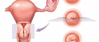
The exophytic form of cervical cancer is diagnosed in situations where the vaginal lumen is completely filled with pathological tissues. This type of tumor has a favorable prognosis for recovery, provided that the woman regularly undergoes preventive gynecological examinations. It is not difficult to identify this form of cervical cancer even at an early stage of development, therefore the effectiveness of the therapeutic effect on the body is quite high.
Invasion in cervical cancer. Source: myshared.ru
The endophytic form of cervical cancer is not as common as the previous type. A neoplasm grows in the inner part of the isthmus, which connects the uterus and vagina. If we talk about the presence of external visible changes, they will be noted on the exocervix, but this happens only in advanced stages of the development of the pathology. In this situation, the cervix or part of it will have a concave and uneven surface.
Stages
- Stage 1 (degree) of invasive breast carcinoma – when the tumor is no more than 2 cm, does not have metastases and does not penetrate nearby structures;
- Stage 2 invasive streaming breast cancer has a neoplasm - 2-5 cm, tumor cells are localized in one or several nodes in the axillary fossa, but they are not fused with each other and with nearby tissues, there is no metastasis;
- Stage 3 of invasive unspecified cancer - at this stage, the tumor does not have clear boundaries in a lobular or ductal neoplasm, the lymph nodes have cancer cells that are “glued” together, there are no distant metastases yet;
- Stage 4 - with this carcinoma, the lymph nodes are already affected and metastases are present in distant organs.
Identification of the disease
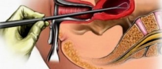
Diagnosis of invasive cervical cancer is carried out in a comprehensive manner, including laboratory and instrumental examination methods. The main research method is biopsy and cytology. The doctor collects the affected cells, which are then studied in the laboratory.
The result of such an examination is accurate; it helps to determine whether the neoplasm is malignant and to thoroughly study the structure of cancer cells.
Colposcopy is also prescribed. During this process, the affected tissues are enlarged several times, which makes it possible to examine the cells. If the doctor identifies suspicious areas, an extended colposcopy is performed using a special solution.
To obtain more detailed information about the condition of the uterus, as well as other abdominal organs, ultrasound, CT, and radiography are prescribed.
Diagnostics
Diagnosis of ductal breast cancer in Israel is carried out using highly informative and at the same time painless and atraumatic methods for the patient. At the Top Ichilov Clinic, the following diagnostic program is used to confirm the diagnosis of “ductal carcinoma in situ”:
- Mammography, in which, in the case of ductal breast cancer, the doctor identifies microcalcifications on the mammogram.
- Fine needle aspiration biopsy. A small amount of tissue is “pumped out” from the thickness of the mammary gland using a syringe with a thin needle. After such an intervention, the patient does not even have a small scar.
- Core needle biopsy. A larger diameter needle is inserted through a small incision in the skin of the gland and more tissue is taken. The small scar after the needle is inserted disappears in just a couple of weeks.
Breast mammography
It is almost impossible to detect the initial stage of the disease without special equipment and tests. Symptoms such as seals in the areola or nipple discharge are observed in only 1% of patients.
A biopsy is usually sufficient to make an initial diagnosis. If suspicions are confirmed, studies are carried out that should confirm or refute the initially obtained results.
To determine the extent of the lesion and confirm the diagnosis, mammography, ultrasound, and also:
- MRI, light scanning;
- CT scan;
- ducto- and thermography.
Carrying out various studies is caused by the need to accurately determine the stage of the disease and establish a diagnosis by excluding other forms of breast cancer.
Lumpectomy
When collecting anamnesis, the doctor may inquire about the presence of dizziness, nervousness, chronic fatigue, as well as rapid weight loss and a number of other signs characteristic of certain stages of oncology. A survey is required about the time of occurrence of complaints and the methods used to combat the manifestations of the disease, as well as family history and existing allergic reactions.
The choice of methods and further prognosis for ductal carcinoma of the breast occurs after the doctor summarizes all the information received. This pathology can only be treated through surgery.
- For the non-invasive form, a lumpectomy or quadrantectomy is performed. In both cases, a certain part of the breast tissue is removed.
- Mastectomy is performed only in case of significant growth of the tumor and the transition of the cancer to stage 3-4.
Therapeutic actions are taken after prompt elimination of problem areas. Treatments include:
- Radiation and chemotherapy. They are aimed at destroying atypical cells remaining after surgery, stopping the development of metastases.
- Hormone therapy. They are used if the tumor is hormone-dependent and provokes hormonal imbalances in cells.
- Molecular targeted therapy. The destruction of cancer cells occurs at the gene level by blocking growth stimulation receptors.
To diagnose a breast cancer tumor and determine the exact size of the affected area, a woman is sent for an ultrasound examination of the breast. To confirm or deny developing carcinoma, the doctor prescribes a number of diagnostic measures. Diagnostics includes the procedure:
- Ductography is an X-ray examination of the chest area. The method involves the use of a contrasting element. The product fills the milk duct, which helps to examine in detail the clinical picture and characteristics of the compacted affected area.
- Puncture of the affected area of the breast and further biopsy - a sample of cellular material is sent for histological analysis to determine the form of the tumor.
- Immunohistochemical tests are intended to assess the sensitivity of pathological formations to female sex hormones. The final information obtained from the examination allows us to understand the likelihood of successful removal of a malignant lesion using hormone therapy.
To determine the stage of development of cancer, the patient is sent to undergo a CT scan of organs and structures susceptible to the development of metastases. If there is a suspicion of the existence of an oncological process in these systemic parts, it is recommended to conduct a histological examination. The method involves an early biopsy to collect a sample of tissue material. Oncology uses a system for determining cancer growth.
The Gleason classification allows you to understand the speed and spread of tumor formation to nearby structures and organs. To establish the listed characteristics of the patient, a biopsy of the tumor site is performed. Afterwards, the doctor analyzes the taken cellular sample of the material through a microscope. The calculation of undifferentiated cell chains helps to obtain a result marked with the G value. The marked indicator is deciphered as follows:
- G1 – well-differentiated carcinoma;
- G2 – moderately differentiated growth;
- G3 – low differentiation of malignant neoplasm;
- G4 – tumor is highly malignant;
- Gx – the study does not allow establishing the level of differentiation.
A low degree of differentiation is characterized by difficulties in the fight against oncological pathology. Some cases require the use of a group combination of treatment techniques.
From the anamnestic data, much attention is paid to risk factors that affect the hormonal status of a woman. Clinical examination includes palpation of the lymph nodes and mammary gland in the supine, sitting, frontal and dorsal positions, bimanually. Reliable results of palpation of the breast are obtained in the middle of the monthly cycle. Instrumental research methods include:
- Mammography – allows you to detail the nature of the tumor and perform a targeted biopsy. The method is informative for preclinical cancer and diffuse forms of the disease.
- Ductography is an X-ray method of contrasting the milk ducts to detect intraductal pathology.
- Ultrasound of the mammary glands is informative and harmless, helps in diagnosing formations not detected by x-ray. A tumor biopsy is performed under ultrasound guidance.
- Computed tomography and the use of nuclear magnetic resonance to examine the breast, regional lymph nodes, liver, bones, and lungs.
- Cytological, histological and immunohistochemical studies are required.
To determine the degree of malignancy of the tumor and select treatment tactics, histological classification of breast cancer is used. The degree of differentiation of the primary tumor or G histopathological classification are taken into account. It provides the following gradations:
- Gx – degree of differentiation cannot be assessed.
- G1 – high degree of differentiation.
- G2 – medium degree of differentiation.
- G3 – low degree of differentiation.
- G4 – undifferentiated carcinoma.
Diagnosis of the disease
Diagnosis always begins with a gynecological examination and visual examination of the affected area using a microscope. Then the following is carried out: smear collection for cytological examination, colposcopy, screening, biopsy, DNA test for HPV (human papillomavirus).
Colposcopy is used to identify changes in the cervical epithelium
Next, hardware diagnostics are prescribed, which includes various tomographies (this can be magnetic resonance, computer and positron emission).
At advanced stages of oncology, to determine the condition of other organs, radiography and ultrasound may be prescribed (but these methods are additional and are not always used).
General tests are required, and laboratory blood tests are performed to determine the content of tumor markers.
In medicine, there are many ways to examine a woman for malignant tumors in the cervical area, but to make an accurate and final diagnosis it is necessary to carry out a whole range of examinations, consisting of laboratory tests and diagnostic procedures.
The optimal set of measures includes colposcopy, histology, and tomography of various organs. Let's look at each method in more detail.
What is invasive cervical cancer and the factors behind its occurrence?
Malignant formation in the cervix is in second place in the frequency of diagnosis after breast cancer. Depending on the stage of formation, it can be non-invasive or invasive. The transition from one type to another can take a long period.
Invasive uterine cancer usually occurs in females after 40 years of age, with the peak of this disease occurring between the ages of 48-55 years. Before the age of 30, the chance of developing this disease is quite low - 7%, and the risk of getting invasive uterine cancer after crossing the 70-year mark is also low (16%).
The development of the disease can be influenced by many factors. Among them is infection with the HPV virus (human papillomavirus). But even its presence in a woman’s body does not always indicate the necessarily onset of the cancer process. Among the factors influencing the development of invasive cancer, the following should be noted:
- sexually transmitted diseases, also HIV;
- a fickle partner who has a large number of sexual relationships with different partners;
- promiscuous sex life;
- sexual activity started at an early age;
- a large number of births;
- use of hormonal drugs;
- previous oncological disease of the genitourinary system;
- active and passive smoking.
The risk also increases if you have the following diseases:
- cervical erosion;
- dysplasia;
- leukoplakia.
Timely detection of such diseases will help to begin treatment on time and prevent it from developing into cancer.
It is also customary to divide ongoing transformations in the cervix into the following types:
- Cervical dysplasia (this includes pathologies such as polyps, pseudo-erosions, leukoplakia, condylomas)
- The precancerous process (this is cervical dysplasia in different stages) is considered a reversible process;
- Pre-invasive (or non-invasive). This stage is characterized by the completion of epithelial changes and the completion of infiltrative growth;
- Invasive cancer. Cancer cells spread.
The most common signs of invasive cervical cancer are usually bleeding during sexual intercourse, unstable menstruation, watery discharge with an unpleasant odor, pain in the lower abdomen, and problems with urination. These signs usually indicate the presence of a tumor and are specific.
Although this disease is a clinically pronounced cancer, early stage invasion in oncology may not always have pronounced symptoms to give a prognosis for the disease. This disease can be detected by histology and can also be confirmed by biopsy.
Scheme of mastopathy in the chest
- Mastopathy - a disease of the mammary glands in frequent cases favors the appearance of pathological tumors. The development of mastopathy is based on a violation of hormone levels in women in the age category from 30 to 40 years. Signs of the pathological process appear in the form of impassable pain in the chest area and discharge from the nipples of fluids other than lactation fluids. A woman is faced with the formation of lumps in the mammary gland, which, in the absence of proper treatment, can deform and modify the tissue of the gland and develop into a malignant course of oncology.
- Fibroadenoma is a benign cancer formation formed from connective material. The disease is diagnosed in young girls as a result of hormonal imbalance or being in a constant stressful situation that affects the nervous system. If the fibrous tumor is not eliminated from the mammary gland, it becomes malignant.
- Abortions – they provoke a severe disruption of the normal level of hormones in the female body. The situation favors the development of inflammatory foci in the uterus or ovaries. Abortion in frequent situations precedes and causes infertility. During pregnancy, the glandular structure of the breast prepares for breastfeeding the unborn child. Abortion triggers the opposite effect, leading to disastrous consequences. As a result, the woman’s body becomes overgrown with various compactions, and tubular carcinoma occurs.
- Refusal of breastfeeding - the breasts of a pregnant girl and after the birth of a child undergo major changes in structure. The functioning of the mammary glands is adjusted to breastfeeding. When a woman does not want to breastfeed, a disruption occurs in the normal processes in the body. Drastic changes can subsequently cause triple-negative breast cancer.
Clinical picture
The clinical picture of the disease is quite broad. The first stage of development of the pathology can be hidden, there are no symptoms. In this case, the disease can be identified by a specialist during an examination of the patient and additional diagnostic methods.
Symptoms of invasive cervical cancer are severe. Experts urge women to pay special attention to the following warning signs:
- contact bleeding that appears after sex;
- watery discharge from the genital tract;
- bleeding between periods;
- vaginal discharge that has a strong, foul odor. Their color may change, they become cloudy;
- pain, which is localized mainly in the lower abdomen, in the sacral area;
- problems with urination;
- constipation
Along with the occurrence of these symptoms, the woman begins to complain of increased fatigue, she feels weak when performing usual activities. Many people notice a sharp weight loss up to critical levels. Nausea may occur periodically, the skin may become pale, and this is accompanied by an increase in body temperature.
The combination of these signs should be the basis for immediately contacting a doctor and undergoing a full range of diagnostic measures. Ignoring the problem will aggravate the health condition in the future, and the tumor may be detected too late.
Diagnosis
In order to detect invasive breast cancer in a timely manner, it is recommended that once a year (you should start such regular examinations after 20 years of age) you undergo screening tests - X-ray mammography or ultrasound scanning.
If such a study confirms the presence of a tumor, then a more accurate examination is prescribed, which consists of:
- MRI of the mammary glands;
- Positron emission tomography;
- Ductography (x-ray of glands using contrasting X-rays with a substance).
But an accurate diagnosis is possible only after examining the cells obtained by puncture; the discharge from the nipples is also examined. Immunohistochemical tests are performed on the resulting cells to determine sensitivity to sex hormones in order to select hormone therapy.
To accurately determine the stage of invasive cancer, tomography of regional lymph nodes, bones, and lungs is performed. If tumor cells are found there, they are also studied by biopsy.
To determine the rate of tumor growth, a Gleason classification is performed, which is based on studying the area of malignancy obtained by biopsy. During the study, undifferentiated chains of cells are counted, and based on the counting results they are assigned to one of the categories:
- G1 – well differentiated cancer;
- G2 – moderately differentiated cancer;
- G3 – low differentiation carcinoma (if this cancer is not a lobular type, but a ductal one, it has the maximum ability to penetrate structures that differ from its own);
- G4 – extremely malignant, undifferentiated;
- Gx – the degree of differentiation cannot be established.
If histological analysis confirms the presence of a tumor, then the following studies are prescribed:
- Pyelography (a test to detect problems in the urinary system);
- Chest X-ray;
- Cystoscopy;
- Sigmoidoscopy.
The main reasons for the development of cervical cancer
Malignant tumors of the cervix arise from the epithelium of the organ. It is important to remember that a tumor is not able to develop from healthy tissue. The impetus for pathological degeneration of cells is often a change in the mucous membrane caused by some disease.
The following borderline precancerous conditions are considered predisposing to the development of cancer:
- erythroplakia – pathological atrophy of the exocervix mucosa;
- endometrial adenomatosis - is a common atypical form of precancerous condition, in which dishormonal hyperplasia of the endometrium develops;
- Leukoplakia is a pathology in which the integumentary epithelium lining the uterine cervix thickens and becomes keratinized.
With any of the above diseases, the risk of developing cancer increases several times.
Experts also identify indirect reasons due to which degeneration of cervical cells occurs:
- infection of a woman with HPV;
- smoking;
- inflammatory diseases of the genitourinary system in chronic forms;
- gaining early sexual experience;
- promiscuous sexual relations, and this applies not only to the woman, but also to her sexual partner, who has numerous sexual contacts with other, often casual, partners;
- infection with sexually transmitted diseases, human immunodeficiency virus, AIDS.
Risk factors that increase susceptibility to cancer cannot be ignored. These include:
- too frequent pregnancies and childbirths;
- treatment with hormonal drugs, including long-term use of combined oral contraceptives;
- deficiency of vitamin components;
- cancer of the urinary and/or reproductive system, previously suffered;
- immunodeficiency condition resulting from chemical therapy.
In almost all of these conditions, it is possible to reduce existing risks and prevent the occurrence of malignant tumors.
Prognosis and prevention
Basically, the prognosis for invasive breast cancer depends on the results of the therapy. Invasive lesions of the mammary glands are characterized by a high percentage of deaths. Breast cancer stage 1 or 2 ends in recovery in 90% of cases. Positive dynamics are possible only with proper treatment.
To treat invasive cancer, both local (radiation therapy, tumor removal) and systemic (chemotherapy, biological and hormonal therapy) methods are used. Combinations of several methods are often used. The choice of treatment is based on:
- Tumor size;
- Location of the neoplasm;
- Stages of the disease;
- Sensitivity of the tumor to estrogen;
- Menopause (age of the patient).
The usual treatment regimen looks like this:
- Hormone therapy is prescribed to reduce the size of the tumor and its fusion with neighboring structures;
- For invasive breast cancer, the tumor itself is surgically removed. Mastectomy (complete removal of the gland) or lumpectomy (removal of the tumor, axillary lymph nodes and part of the healthy tissue around the perimeter of the tumor) is used;
- For invasive cervical cancer, the cervix and body of the uterus are removed (the ovaries and tubes are also removed) or the uterus is removed, including part of the vagina and nearby lymph nodes;
- Treatment is carried out with chemotherapy and radiotherapy to avoid relapse of the disease.
The prognosis for this type of cancer depends on several points:
- At what stage was the disease discovered? Started treatment at stage 1 gives a 90% recovery rate, at stage 2 - 66%, stage 3 - only 41%, at stage 4 survival is less than 10%;
- Location of carcinoma (if it is located within the gland tissue on the outside, the prognosis is more favorable);
- Tumor diameter (five-year survival rate for tumors up to 2 cm - 93%, 2-5 cm - 50-70%);
- Degree of tumor differentiation;
- The presence of estrogen and progesterone receptors;
- The presence of other foci of cancer and lymphedema of the breast and arm.
After treatment for cervical cancer, the patient should be observed by a doctor every three months for two years, and later once every six months.
Invasive breast cancer of a nonspecific type is considered an oncological pathology that establishes a prognosis based on the stage of diagnosis. If carcinoma is detected in the early stages of development, five-year survival is observed in 90-95% of patients. When breast cancer is detected at stage 2, the positive predicted result is reduced to 70%.
Correct and favorable disease prevention includes following the rules:
- Every month check the body condition for the presence of atypical manifestations.
- Be examined by a mammologist once a year.
- Stick to a healthy weight limit.
- After giving birth, do not give up breastfeeding for at least six months.
- Stop using alcohol and tobacco products.
- Check the amount of hormones in the body.
- Refuse abortion. If you do not want to have a child, you should use contraceptives.
- Use hormonal medications exactly as prescribed by your doctor, strictly following the prescribed dosage.
If invasive G2 breast carcinoma is detected, the prognosis will depend on the degree of spread of the tumor to neighboring tissues and organs. When prescribing treatment, the oncologist takes into account various factors: the degree of differentiation, stage of development, type and location of the tumor, the patient’s health status and diseases associated with cancer.
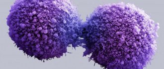
Poorly differentiated breast cancer is an aggressive form of cancer that requires aggressive treatment. The tumor grows rapidly, affects the lymph nodes, and invades neighboring tissues. These types of malignant tumors are difficult to treat and have a negative prognosis when detected in late stages.
Forecast
With the development of non-invasive neoplasms, the prognosis is quite favorable; about 90% of women successfully cope with the disease and fully recover from it. A woman with invasive cervical cancer that has spread to nearby tissues has a significantly reduced chance of cure. With timely diagnosis and adequate treatment methods, with a 60% probability, the patient will be able to get rid of the second stage of cancer.
At the third stage, the chance of recovery decreases to 30%. At the last, stage 4, it is much more difficult to stabilize the patient’s condition and the activity of metastases. Only 10% of women with stage four can get rid of the disease.
Cervical cancer is a dangerous disease because malignant cells spread quickly, affecting many organs and systems. The early stages are often asymptomatic, so the woman may not be aware of the presence of pathology. Only regular examinations by a gynecologist will help to timely detect any disorder and begin the necessary therapy.
Diagnosis of breast cancer
A malignant formation of this type does not have the characteristic features of this type of tumor; all cells of the neoplasm are similar. G2 nonspecific invasive breast cancer is an intermediate-grade breast tumor, a type of cancer that has spread beyond the primary tumor, with difficult definition of the type of disease.
Invasive unspecified G2 breast cancer can be hormone-dependent or hormone-negative. Hormone-dependent cancer is diagnosed most often during the postmenopausal period. Hormone-negative cancer affects premenopausal women. A grade 2 tumor is a malignant neoplasm that is treated conservatively or with surgery.
The degree of tumor differentiation is designated by the letter “G” and is described in five options:
- Gx – the degree of differentiation is not determined.
- G1 – highly differentiated malignant tumor.
- G2 – average degree of tumor differentiation.
- G3 – poorly differentiated malignant neoplasm.
- G4 – undifferentiated malignant process.
Histology
There is another classification of cervical cancer based on histological changes. Here the type of tissue from which the tumor is formed is taken into account. The following types of disease are distinguished:
- adenocarcinoma – columnar epithelial cells with glandular components undergo malignancy;
- squamous cell carcinoma - a tumor develops in stratified squamous epithelium, which is reflected in the name of the disease.
The study also evaluates the degree of cell restructuring. A malignant tumor formation can be highly, moderately or poorly differentiated. With the latter option, we are talking about the most malignant process, when cells degenerate very quickly and their growth is uncontrollable.
Survival prognosis
Poorly differentiated ductal breast cancer forms inside the wall of the breast duct. As the tumor develops, it begins to grow into neighboring tissues and affects the breast tissue; metastases affect the lymph nodes and spread throughout the body. Invasive ductal breast cancer is characterized by the appearance of a dense neoplasm adherent to adjacent tissues.
The tumor causes the nipple to be pulled inward along with the isola. Poorly differentiated ductal carcinoma is characterized by a high degree of aggressiveness, a high rate of cell division and spread. Rapid onset of symptoms is common in aggressive G3 invasive breast cancers.
Oncologists at the Yusupov Hospital use their experience to diagnose the early stages of cancer, determine the type of tumor and prescribe effective treatment. Depending on the state of health, the woman receives treatment using innovative methods that are used by progressive oncology clinics around the world.
In the hospital, the patient is under the supervision of various specialists - a mammologist, a gynecologist, an oncologist. The Yusupov Hospital includes a 24-hour hospital, a rehabilitation clinic, and a diagnostic center. If symptoms of trouble or discomfort appear, you should seek advice from a mammologist or hospital oncologist.
Head of the Oncology Department, oncologist, chemotherapist, Ph.D.
Ductal breast cancer
This pathology received its name from the initial stage of its development, since the pre-invasive form of the neoplasm is formed from the tissue cells lining the breast ducts. Depending on the spread of the lesion, the clinical form of the disease can be non-invasive and invasive:
- In the first case, spread to other tissues does not occur, and growth occurs in the lumen of the duct. This is the initial stage. This phenomenon is called ductal carcinoma in situ. Its degree of danger is low, medium and high.
- In the second case, as the tumor grows, all surrounding tissues are affected. Infiltrative ductal carcinoma is quite aggressive. In 80% of cases, it rapidly progresses, forming secondary lesions in distant organs and lymph nodes.
The concept of carcinoma indicates that the development of atypical cells occurs in epithelial tissue. In this case, the formation comes from the inner walls of the milk canals and does not affect the petals of the gland. The term in situ indicates the stage of the pre-invasive form.
Inverted nipple, swelling - signs of cancer
Mammary glands are a distinctive feature of all mammals. Accordingly, this type of cancer occurs not only in women, but also in men. The structure of their breasts is completely identical to women's, differing only in the degree of development. The symptoms of breast pathology are also identical:
- the appearance of tumors and swelling;
- discharge of fluid or blood from the nipple;
- atypical appearance of the areola or inverted nipple;
- peeling and redness of the breast, the appearance of ulcers;
- lymph nodes in the armpits are enlarged.
With the invasive form, lumps in the breast tissue are already clearly palpable.
Symptoms
The clinical picture of the pathology is divided into two groups: general and specific. The first involves a deterioration in the woman’s general condition due to the development of the disease. These include signs such as:
- Fast fatiguability.
- General weakness.
- Attacks of dizziness.
- Lack of desire to eat.
- Dramatic weight loss.
- Fever.
Specific symptoms appear due to damage to the reproductive organ itself. They occur already at a late stage of tumor development. Manifestations of oncology include:
- Discharge containing bloody impurities.
- Bleeding after sex.
- Increasing the duration of menstruation.
- Pain in the lower abdomen.
- Specific smell of discharge.
- Swelling of the lower extremities.
- Problems with urination.
When metastases spread at the last stage of the disease, the functioning of many internal organs is disrupted in patients. At this stage, it is impossible to save the woman’s life.
Bibliography
- ICD-10 (International Classification of Diseases)
- Yusupov Hospital
- Cherenkov V. G. Clinical oncology. — 3rd ed. - M.: Medical book, 2010. - 434 p. — ISBN 978-5-91894-002-0.
- Shirokorad V.I., Makhson A.N., Yadykov O.A. The state of oncourological care in Moscow // Oncourology. - 2013. - No. 4. - P. 10-13.
- Volosyanko M.I. Traditional and natural methods of preventing and treating cancer, Aquarium, 1994
- John Niederhuber, James Armitage, James Doroshow, Michael Kastan, Joel Tepper Abeloff's Clinical Oncology - 5th Edition, eMEDICAL BOOKS, 2013
Treatment
The choice of therapeutic tactics depends on several indicators, including the size of the tumor formation, the stage of the oncological process, the location of the tumor, and the age category of the patient.
On this topic
- Oncogynecology
The first signs and symptoms of ovarian cancer
- Natalya Gennadievna Butsyk
- December 4, 2020
To reduce the volume of the tumor, treatment with hormonal medications is prescribed.
When invasive breast cancer is diagnosed, the tumor is removed in several ways:
- mastectopia – complete excision of the affected breast;
- lumpectomy - removal of the tumor, lymph nodes located in the armpits, and part of the normal tissue surrounding the tumor.
If cancer is detected in the cervix of the uterine body, then the tumor is removed along with the cervix, tubes and ovaries. Surgery can also be performed, which involves complete excision of the uterus, part of the vagina and lymph nodes located nearby.
Chemotherapy
The essence of this treatment method is that the patient is prescribed to take potent chemical medications that have a destructive effect on cancer cells, contribute to the cessation of growth and prevent the recurrence of the disease.
Chemotherapy treatment can be carried out as an independent method or in addition to surgery, both before and after it. Concomitant use with radiation therapy may also be prescribed.
The peculiarity of this procedure is that thanks to it it is possible to obtain more effective treatment results. It also significantly reduces the likelihood of further metastasis. Among the drugs, Bleomycin, Etoposide, Cispltin and Hydroxycarbamide are the most popular.
Radiation therapy
The lesions are exposed to high-frequency radiation. The advantage of this procedure is that the beam is directed directly to the location of the malignant neoplasm, which prevents damage to healthy tissue.
Radiotherapy can be used as an independent method or in combination with surgery and chemotherapy. Also used for extensive metastasis.
Complications
Since invasive cancer at an early stage can occur without the manifestation of specific symptoms, it is often diagnosed at a later stage. This, in turn, leads to the fact that the malignant tumor begins to metastasize and spread to nearby and distant anatomical structures. As a result, when vital organs are damaged, their functioning is impaired, which can lead to even more serious problems.
In addition, complications include infertility, which results in the removal of the reproductive organs along with the tumor.
Also, consequences may occur during chemotherapy, which is manifested by nausea, vomiting, brittle nails and hair loss, as well as other side effects.
consultations
Link to the official website of GKOD>
The questions are answered by the WINNER OF THE NOMINATION “BEST ONCOLOGIST” SKVORTSOV VITALY ALEXANDROVICH, doctor of the highest category, kmn, mammologist, plastic surgeon, oncologist of the State Clinical Oncology Department
Presentation of the “We Will Live” award in the category “Best Oncologist” to Dr. Skvortsov>link>
Non-invasive breast cancer in situ.
12.07.2017
QUESTION: Doctor, according to the results of histology, I have non-invasive breast cancer in situ. I am 37 years old. What treatment would you prescribe in this case? And how does non-invasive cancer differ from invasive cancer? Thank you in advance. Love. ANSWER: Hello! I would prescribe you radiation therapy, determine the expression of estrogen and progesterone receptors, if they express, then add Tamoxifen and regularly see your doctor. Non-invasive cancer means it does not metastasize distantly, while invasive cancer means it metastasizes to other parts of the body.
Mammary cancer
26.12.2017
QUESTION: Vitaly Alexandrovich, sometimes ductal carcinoma in situ (DCIS) is not removed because one of its types is low-grade and never develops into invasive cancer. Or is this a time bomb? After all, even fibroadenoma can degenerate into cancer. ANSWER: Hello! in situ is also cancer, studies have shown that it is enough to irradiate it and it does not metastasize, does not grow into the wall of the basement membrane! So it is also impossible to argue that fibroadenoma can degenerate! What if it doesn’t get reborn?! Therefore, each pathology has its own operation! But you shouldn’t look into the future! If you protect yourself from breast cancer, another one will appear, for example, in the stomach!
Oncology
12.04.2018
QUESTION: Hello St:0T:is N0M0 intraductal cancer non-infiltrating NOS morphological diagnosis proliferative lobular and ductal fibroademotosis IHC Her2neu negative Est Pr negative negative, Ki -67-60-80% of cancer cells, amputation of the breast was performed. Tell me, please, what other herbs should I drink? Thank you. ANSWER: Hello! In this case, breast cancer is treated only with radiation therapy, but since you had a mastectomy, it is not indicated for you. In your case, only observation by an oncologist and that’s it!
Cancer in situ
24.03.2019
QUESTION: Vitaly Alexandrovich, my oncologist told me that cancer in situ is not considered cancer. As far as I understand, this is stage zero and the cancer is non-invasive, that is, it has not grown into the surrounding tissues. Does in situ cancer always become invasive? ANSWER: Hello! Cancer in situ does not always become invasive; or rather, one cannot say that it can become invasive; most likely, it has already been invasive. Cancer in situ is a rather dangerous form, in which invasion is actually sometimes observed.
carcinoma in situ
24.04.2019
QUESTION: Vitaly Alexandrovich, hello. I went to the doctor with pain in my right breast in November 2020. They did a puncture and found nothing. Come back in three months, don’t worry, the vessel is inflamed, this also happens. At the beginning of March I had an ultrasound and mammogram. Let's run to the doctor, everything is not okay. They took a puncture - suspicion of cancer. 03/27/19 sectoral resection. Urgent histological examination - sector 6×4×2 cm, on the section there is a node with an unclear contour 3.5×1×2.5 cm. Micro: Fragments of breast tissue with multiple structures of carcinoma in situ. NO SIGNS OF INVASIVE GROWTH. Conclusion: Urgent examination of 1 frozen section revealed ductal carcinoma in situ. To exclude an invasive component, it is necessary to study the entire volume of material. The answer from the protocol for studying the biopsy material is fragments of breast tissue with a large number of complexes of ductal breast cancer in situ grade 3, without signs of invasive growth. They didn’t do IHC, they say it’s not necessary for DCISN0M0. It's right? Next LT. Thank you. ANSWER: Hello! IHC must be performed! Determine whether he is hormone dependent or not. It is also necessary to irradiate the chest.
Cancer in situ and timing of irradiation
10.06.2019
QUESTION: Sorry to bother you, but I also wanted to clarify the timing of the radiation. I read that the timing of radiation for cancer in situ is within 8 weeks after surgery, and 6 months after surgery if chemotherapy is done. Those. If with in situ RT they will do it after 4.5 months (as planned), then its meaning is lost? Thanks for the answer. ANSWER: Hello! The guidelines state that it is advisable to do it within 8 weeks after surgery, up to a maximum of 6 months, this makes sense, and radiologists determine the meaning and effectiveness of radiation therapy after surgery!
Microinvasive carcinoma, in the background and with predominance of ductal carcinoma in situ
19.06.2019
QUESTION: Hello, Vitaly Alexandrovich. I wrote to you on April 24, 2019. The hospital never did IHC for me. While I was doing RT in 16 fractions of 2.67 Gy, I took the blocks and, according to compulsory medical insurance, they did it in 1 onc. center on Baumanka. Microdescription: in the studied histological preparations stained with hematoxylin and eosin, fragments of breast tissue are presented, with the presence of dilated ducts, with the proliferation of large antipic cells in them, with large hyperchromic nuclei and a wide rim of eosinophilic cytoplasm. Tumor cells are located in several layers, completely filling the lumen of the ducts and the presence of focal comedonecrosis. On the periphery of the site there are foci of microinfestation. The stroma is fibrous, with periductal lymphocytic infiltration. This is how I understand the revision of histology. IHC was performed using antibodies to: Smooth Muscle Aktin (SMA), CD10, ER, PgR, Ki-67. ER by D.C. Allred: PS-2 IS-ITS=3 weak expression. PgR: Ps-0 IS-0TS=0 negative expression. Ki67 staining of 10% of nuclei. SMA AND CD10 were not able to evaluate express. I will continue, not everything fit from 06/19/19. . Results of IHC studies (non-invasive, intraductal component). CONCLUSION: in the scope of the research material, the picture corresponds to microinvasive carcinoma, against the background and with a predominance of ductal carcinoma in situ, G3 (Duktal Carcinoma in situ (DCIS) of high nuclear grade, ISD-0 code 8500/2. Reading this, I have microinvasion, and not in situ with stage 0???? , somewhere else to review the blocks? And should I take hormones?. I’m really looking forward to your answer. Thank you very much for your answers. Good health to you. ANSWER: Hello! There are areas of microinvasion and this is stage 1 - the very initial one. There is no need for a review and you are advised to take tamoxifen 20 mg. It is very difficult to make a diagnosis with such a histological examination. An unusual case. In principle, it was necessary to irradiate the nodes, but it is better for you to consult a radiologist!
Cancer in situ and ovestine
02.07.2019
QUESTION: Please tell me, for breast cancer in situ, a radical mastectomy was performed on both sides. Due to the presence of the BRSA1 mutation gene, the ovaries were removed. No treatment was prescribed, only observation. The question is this: can I take Ovestin with all my indicators? ANSWER: Hello! If you have hormone-dependent breast cancer, then you definitely cannot take this drug! But there are other mechanisms for the occurrence of breast cancer, but they are not hormone dependent! And yet there is evidence that estrogen should not be taken for any molecular subtype of cancer!
cancer in situ
12.08.2019
QUESTION: Good afternoon, please tell me what the conclusion means: no amplification of the HER2 gene was detected by fluorescence in situ hybridization (label index i=1.75)? ANSWER: Hello! This means that this is cancer in situ and there is no point in defining HER2NEU, since this indicator is not relevant for this form of cancer. This issue can be discussed with an oncologist.
Cancer in situ and lymphorrhea
02.09.2019
QUESTION: Vitaly Aleksandrovich, after the operation, resection of the left breast and removal of 5 lymph nodes under the arm, almost a month has passed, but every other day 8 syringes of lymph are pumped out, after 10 days radiation is prescribed. Is it possible to do it in such a situation!? And is such a large outflow of lymph normal? According to histology results, there is no cancer in the lymph nodes. IDO conclusion: p63, alpha-active, calponin-negative in the ducts. Coll. IV-bright preserved basement membrane around the streams. Repeat: ER, Pr-positive nuclear uneven reaction. Conclusion: intraductal cancer, G3, receptor-positive, HER 2 has not been studied in such tumors. Please explain whether the decision to leave the breast was justified, although the tumor was close to the nipple? Thank you! ANSWER: Hello! The accumulation of lymph in this case is quite normal and this volume is not at all large and radiation therapy can be carried out. You have breast cancer O st. (in situ). There is no need to remove the breast with this histology, even if it is close to the nipple. It is sufficient to irradiate this cancer after sectoral resection and prescribe endocrine therapy.
Is it possible to install the system on the side of the operated arm?
26.09.2019
QUESTION: Hello, Vitaly Alexandrovich! Thanks for the previous answer. I have one more question. Is it possible to install the system on the side of the operated arm? There was a sectoral resection (cancer without removal of axillary lymph nodes, cancer in situ). It turns out that you need to put 5 systems in this hand. And is it possible to install a system with pentaxiphylline? Thanks for the answer. ANSWER: Hello! If the axillary lymph nodes have not been removed, then you can definitely do intravenous infusions on the arm on the affected side. Even if there was an operation on this side, then according to vital indications, the system can still be installed.
Why were Tamoxifen and radiation therapy prescribed for cancer in situ?
08.10.2019
QUESTION: Hello! I have carcinoma in situ of the mammary gland, a sectoral resection was performed, no tumor cells were found in the deep edge, the lymph nodes were clean, ER 99% PR 99%, Ki-67 0.8%, Tamoxifen and radiation therapy were prescribed. Why, if the tumor was removed, histology confirmed in situ G1. ANSWER: Hello! This is an international standard that says that even if you have an sinus infection, you must undergo treatment! And this is mandatory - radiation therapy and taking antiestrogens according to the regimen.
Tamoxifen 20 mg was prescribed for 3 years
31.10.2019
QUESTION: Good afternoon, at the beginning of October of this year, a resection of the permanent breast was performed (removal of fibroadenoma); based on the results of hypoxia, it was written that there was a suspicion of cancer in situ in the nearby tissues. IHC exp. estrogen (100%), progesterone (100%), ki67 - 3%. They wrote lobular carcinoma in situ. Tamoxifen 20 mg was prescribed for 3 years. Is the doctor's prescription correct? ANSWER: Hello! The prescription is correct, but I would also schedule a consultation with a radiologist regarding irradiation of the chest; usually in such cases radiation therapy is also prescribed.
Cancer in situ
24.11.2019
QUESTION: Hello, Vitaly Alexandrovich! Help decipher the results of the trephine biopsy: Is the expression of estrogen determined in the ductal streams (5PS+3IS points according to Allred)? expression of progesterone receptors (1PS+3IS points according to Allred) The index of Ki-67 labeled nuclei of tumor cells was 40%. HER2/neu status according to ASCO/cap 18 is negative (+1), HER2/neu overexposure was not detected. In the basal regions, nuclear expression of p63 and membrane cytokeratin 5/6 is detected. G3 intraductal carcinoma in situ. ANSWER: Based on this conclusion, we can say that this is cancer in situ with sensitive receptors to estrogen and progesterone. In this case, you need to contact an oncologist to determine a treatment regimen.
Treatment of cancer in situ
09.12.2019
QUESTION: Vitaly Alexandrovich, continuing about my treatment - I’m 42 years old. Cancer of the right breast pTisN0M0 (grade 0). Intraductal carcinoma in situ, 2 l/u without tumor growth. The doctor said that I did not need either chemistry or radiation. What to do? Go to another doctor? The operation took place 1 month ago. ANSWER: Hello! After mastetomy for this form of cancer in situ, you do not need any other treatment, only taking Tamoxifen, since you have cancer in situ. With this form, preventive mastectomy of the opposite breast is definitely not necessary.
Cancer in situ
13.12.2019
QUESTION: 54 years old diagnosed with C50 DCIS pTis cNO R1MO right breast? there was a sectoral resection of the mammary gland, multiple, merging growth foci of high grade DCIS are determined? The loss of Er and Pr expression is determined by reaction with Her2+++/ IHC: KI67 - 50%. What should I do? Do I undergo chemotherapy or remove the breasts, then the hormonal ones? Two clinics, two opinions. ANSWER: Hello! This is non-invasive breast cancer and if there is no cancer in situ in the area, then simply radiation therapy. Surgery and chemotherapy are not indicated for you, because this is a non-invasive cancer, and it does not metastasize to distant organs, but the fact that HER2 NEU 3+ is often the case with this form of cancer and this does not mean that chemotherapy is necessary. If the edges still contain cancer in situ, then the decision is at the discretion of the surgeon: you can excise the edges wider, or if the breast is small, then sometimes you have to remove the entire breast, but cancer in situ can be irradiated very well by a radiologist without surgery! In any case, discuss this issue with your doctor.
Taking tamoxifen
06.03.2020
QUESTION: Hello, doctor. I am 47 years old. 3 weeks ago I had a mastectomy for intraductal carcinoma of the breast. pTis(m)NOMO, 0 tbsp. The oncologist surgeon in the hospital prescribed tamoxifen for 5 years and told me to take it right away. I came to the clinic to register, and the oncologist told me it was not cancer, but pre-cancer. For now, do not take the drug, but bring an examination from a gynecologist and endocrinologist, saying that it’s okay if you don’t take it for 1.5 months. I have fibroids, endometriosis and a nodule in the thyroid gland, I’m undergoing an examination, there are queues everywhere, then I have to wait another week for the results. Time passes, I haven’t started treatment, it’s starting to worry me. What is your opinion: should I start taking tamoxifen now or wait for the results, which is another two weeks? ANSWER: Hello! Have you had immunohistochemistry performed? What is the IHC result? If it is positive for estrogen and progesterone receptors, then you need to take it, if there are no these receptors, then you don’t need to take it! Discuss this issue with a hospital doctor, not with a clinic doctor!
Tamoxifen and cancer in situ
12.04.2020
QUESTION: Hello, Vitaly Alexandrovich. My name is Irina. I also decided to contact you. In February of this year, I underwent a sectoral resection of the left breast. Histology showed fibrolobular filtroadenomatosis of the mammary gland with the presence of mixed fibroadenoma, popillary cysts with areas of atypical ductal hyperplasia and a microfocus “Cancer in situ” T1NOMO. Particular emphasis was placed on the microfocus, although everything sounds scary to me. As I understand correctly, I have stage 1 cancer, which was successfully removed. The surgeon prescribed Tamoxifen 20 mg for a year, but the oncologist says that I need to take it for at least 5 years. What is your opinion about the duration of taking the drug? Can Tamoxifen Hexal cause a dry cough as a side effect? Thanks in advance for your answer. Vitaly Alexandrovich, I forgot to ask. After a course of treatment with Tamoxifen, cancer may appear, for example, in another mammary gland, or some other organ? Or does Tamoxifen completely cure and there are no relapses? And one more thing: if the thyroid gland is enlarged, can Tamoxifen bring the thyroid gland back to normal, or does it only act as an antitumor agent and has nothing to do with the thyroid gland? Thank you. ANSWER: Hello! You clearly have cancer in situ, which means this is not stage 1, but stage O, and now it is practically classified as benign tumors. You have stage 0 cancer. Tamoxifen should be taken if you have positive estrogen and progesterone receptors. Only in this case. You shouldn’t just take it without this immunohistochemical analysis. It should have been done for you. Tamoxifen has no effect on the appearance of new cancer in the breast, and it inhibits the appearance of distant cancer metastases, but since you have stage 0, cancer in situ does not metastasize and does not progress and there are no relapses. Amoxifen has nothing to do with the thyroid gland It has. You can read about Breast Cancer in situ>
Ask a Question
CATEGORIES
| Make an appointment [10] |
| Sign up for a consultation online [9] |
| Reviews about the work [6] |
| Internship of Skvortsova V.A. in the USA, MD Anderson Cancer Center [2] |
| Quota for breast reconstruction [13] Reconstruction of the mammary gland (breast) after mastectomy according to quota |
| Breast cancer [1258] All about breast cancer: diagnosis, treatment, breast restoration and reconstruction. |
| Diagnosis of breast cancer [108] |
| Breast diseases [111] |
| Mammography [22] |
| Ultrasound of the mammary glands (breasts) [87] |
| Computed tomography [28] |
| Scintigraphy (osteoscintigraphy) [13] |
| Cytological examination of breast cancer [11] |
| Biopsy and histological examination of breast cancer [42] |
| Immunohistochemical study of breast cancer [210] Immunohistochemical study |
| Her2neu [39] |
| her 2 [11] |
| ER and PR [11] |
| ki67 [58] |
| Surgical treatment of breast cancer [52] |
| Mastectomy [63] |
| Organ-preserving operations [24] |
| Preventive mastectomy [7] Preventive mastectomy |
| Breast reconstruction according to quota [44] What is included in breast reconstruction according to quota, breast correction after reconstruction, lipofilling |
| Breast reconstruction consultation [25] |
| Breast reconstruction (breast surgery) [175] Breast reconstruction |
| Breast reconstruction [109] |
| Reconstruction of the nipple and areola [9] |
| Breast lipofilling [20] |
| Mammoplasty (breast augmentation) [1] |
| Breast removal [3] |
| Chemotherapy for breast cancer [158] |
| Side effects of chemotherapy [22] |
| Taxanes (paclitaxel, docetaxel) [18] Taxanes (paclitaxel, docetaxel) |
| Paclitaxel [6] |
| Zoledronic acid [5] |
| Targeted therapy [51] |
| Herceptin [14] |
| Antihormonal endocrine therapy [188] Treatment of hormone-dependent breast cancer |
| Tamoxifen [125] Tamoxifen |
| Radiation therapy [78] Radiation therapy |
| IORT [10] Intraoperative radiotherapy |
| Cancer in situ [18] |
| Treatment of breast cancer [136] |
| Luminal A breast cancer [49] |
| Luminal In breast cancer [38] |
| Luminal In her positive breast cancer [25] |
| Her2neu positive breast cancer [12] |
| Triple negative breast cancer [52] |
| Breast cancer prognosis [19] |
| Treatment of breast cancer [27] |
| Treatment of stage 1 breast cancer [28] |
| Treatment of stage 2 breast cancer [21] |
| Treatment of stage 3 breast cancer [26] |
| Treatment of stage 4 breast cancer [37] |
| Breast cancer recurrence [4] |
| Monitoring the effectiveness of breast cancer treatment [10] |
| Medicines [31] |
| Vitamins [7] |
| Lymphostasis and lymphorrhea [55] Lymphostasis and lymphorrhea |
| Immunomodulators for cancer [4] |
| Keloid scar [1] Keloid scar |
| Ovariectamia for breast cancer [3] Ovariectamia |
| BRCA1, BRCA2 [5] |
| Tumor markers [35] |
| Mastopathy [2] |
| Fibroadenoma [24] Fibroadenoma |
| Breast cyst [15] |
| Injuries to the mammary gland (chest) [2] |
| Follow-up after breast cancer treatment [16] |
| Life after breast cancer treatment [108] |
| Myths and truths about breast cancer [31] |
| After breast cancer surgery [16] |
| Rehabilitation after breast cancer [11] |
| Miscellaneous [53] |