If a woman has been diagnosed with endometriosis, then she must undergo additional studies aimed at identifying concomitant pathological processes.
CA 125 for endometriosis allows you to determine whether there are any other pathologies in the uterus and ovaries. Based on the results of this study, a further treatment strategy for the underlying disease is determined.
If after the examination it turns out that the level of CA 125 is moderately elevated, then there is nothing to worry about, since an increase in the indicator can occur due to the disease itself.
So, let's figure out what role tumor markers play in diagnosing endometrial disease, what results they can give, and decipher these indicators for each individual case.
What is CA 125?
Endometriosis markers ca 125 are high-molecular glycoproteins (substances of protein origin) that are found in the uterine fluid and healthy tissues of the female body. They are present in small quantities, which is considered normal.
The information content of this method is high. However, in some cases, the results show an increased level of it in the body for other reasons, including physiological ones (pregnancy, onset of menstruation), so the specialist must take this factor into account.
In addition, at the initial stage of development of endometriosis, as a rule, the test shows a negative result, which complicates diagnosis. Accurate indicators are guaranteed for moderate and severe degrees of pathology development.
Tumor markers are substances produced by the body in response to the development of tumor cells in it. It is worth keeping in mind that tumor markers can be produced not only in response to the development of tumors.
Detection of CA 125 allows you to determine the presence of a tumor in the body even before it can be diagnosed using ultrasound or other methods. Thanks to the determination of tumor markers, it is possible to perform highly effective operations that will help prevent relapse of the pathology.
CA 125 is a tumor marker that allows one to diagnose the presence of an ovarian tumor or cyst in a woman’s body. However, it is worth considering that CA 125 does not always increase in tumor diseases. It may also increase when:
- development of myomatous nodes in the uterus;
- for endometriosis and endometrial cancer;
- for breast cancer;
- with cancer of the bronchi.
Today, there are still no tumor markers that make it possible to determine with one hundred percent probability the presence of cancerous tumors in a particular organ. CA 125 also does not 100% indicate ovarian cancer or an ovarian cyst.
Why test CA-125 for endometriosis?
Several years ago, CA-125 was determined for all patients with suspected endometriosis. Today this practice is being abandoned. There are explanations for this:
- CA-125 is not a very specific indicator. It can increase in various diseases, including those not related to the reproductive sphere;
- The level of this tumor marker has no practical significance in endometriosis. Its concentration does not affect the choice of treatment tactics;
- The CA-125 indicator cannot be used as a screening for endometriosis or to assess the dynamics of the process - for example, after removal of an endometrioid cyst or lesions on the peritoneum.
Indications for tumor marker determination:
- Ovarian cyst. Ultrasound cannot always accurately determine the nature of the cyst. Sometimes the doctor cannot understand whether a benign growth has grown in the ovaries or is it cancer. Then a test for CA-125 is prescribed. If the indicator is normal, most likely we are dealing with a functional cyst or a true benign tumor. A slight increase in tumor markers speaks in favor of endometriosis. A sharp rise in tumor marker levels suggests ovarian cancer;
- Differential diagnosis of nodular adenomyosis and fibroids. According to ultrasound, these conditions are very similar. But CA-125 is extremely rarely elevated in uterine fibroids. If the tumor marker increases, adenomyosis can be assumed;
In differential diagnosis, a slight increase in the CA-125 tumor marker may indicate adenomyosis.
- Suspicion of a malignant tumor. Endometriosis is a risk factor for cancer. Against the background of heterotopias, foci of malignant degeneration often form. If the tumor marker level is increased two or more times, an oncological process must be excluded;
- Relapse of endometriosis. If symptoms of the pathology appear again after the operation, you need to undergo further examination. But it is not advisable to immediately perform diagnostic laparoscopy or hysteroscopy, and you can first determine the concentration of the tumor marker.
A blood test for CA-125 does not replace other examination methods and does not replace histological verification of endometriosis. If it is possible to take tissue for research, this should be done.
https://youtu.be/jbejchLXNwA
Reasons for exceeding the norm
Experts note that an increase in the norm of indicators can occur with such diseases as:
- Endometriosis is a common disease in gynecology. It is characterized by the proliferation of endometrial cells outside the uterus.
- ovarian cyst - is a specific formation on the walls of the ovaries, filled with fluid;
- cirrhosis of the liver;
- peritonitis - inflammation of the peritoneum accompanied by a serious condition of the body;
- chronic hepatitis;
- pancreatitis is an inflammatory process in the pancreas.
- pleurisy – inflammation of the pleura;
If the results of CA 125 are over 100 units/ml, this is an alarming factor that makes one suspect the presence of malignant tumors in the body and conduct additional examination.
If the tumor marker CA 125 is elevated, this indicates ovarian cancer in women. But this is not the only cancer that can be detected when the marker level is above the reference values. When decoding the data, cancer of the mammary glands, endometrium, uterus, rectum, stomach, lungs and liver can be detected.
According to statistics, among women who suffer from ovarian cancer, only eighty percent have a high level of this type of marker. This shows the importance of undergoing tests if malignant tumors are suspected in combination with other examinations.
In conclusion, it is worth noting that there are cases when the ovarian tumor marker CA 125 does not exceed the norm, but the woman is at risk. In this case, a test for HE 4 should be prescribed, which will allow timely detection of epithelial ovarian cancer and detection of relapses of the disease.
A specialist cannot make an oncological diagnosis based solely on CA 125 readings. All you need is a timely, comprehensive study, which will help identify cancer, determine its stage and increase the chances of successful treatment.
Preparing for the examination: how to donate blood for tumor markers
Rules for donating blood:
- The CA-125 blood test must be taken outside of menstruation. During menstruation, the level of tumor markers increases. It is better to undergo examination in the first phase of the cycle - before 12-14 days. During menopause, you can donate blood any day,
- Testing is carried out on an empty stomach - 8-12 hours after the last meal. It's better to donate blood in the morning
- The day before the examination, you need to exclude physical and psycho-emotional stress, do not drink alcohol,
- You should not smoke half an hour before donating blood.
It is advisable to take a blood test in the morning on an empty stomach, having eliminated all kinds of stress and alcohol consumption the day before.
If these conditions are not met, the analysis must be retaken.
Cyst symptoms and how they differ from cancer symptoms
According to the classification, there are three main types of these benign formations.
The first is functional cysts, which go away on their own within a couple of months. They represent the accumulation of fluid in the follicle or its growth if ovulation has not occurred.
The second type, an endometroid cyst, is a cavity in the tissues of an organ filled with blood.
The third (or true tumor) is cystic, developing from its tissues. Rarer types of cysts are hemorrhagic or ordinary cysts of the corpus luteum, retention, mucinous, dermoid, paraovarian cysts, etc.
Regardless of their origin, all cysts present with similar symptoms:
- pulling pain in the lower abdomen, on the right or left, sometimes intense (up to nausea, vomiting, spreading throughout the pelvis and radiating to the rectum);
- disruption of the menstrual cycle - intermenstrual bleeding or delays;
- with a significant size of the cystic tumor, the abdomen enlarges.
If the norm of the tumor marker CA 125 in women after 50 years of age is exceeded two or more times, a suspicion of ovarian cancer arises. Risk factors should also be taken into account: most often, adenocarcinoma develops in women who have not given birth or have a family history of cancer. Although scientists and doctors cannot yet name the exact reasons for the degeneration of cells.
How are the symptoms of cancer different from the signs of a cyst? Both pathologies are often virtually asymptomatic at the very beginning. While the tumor is localized in the organ itself, it can manifest itself as pain only when the leg is torsed (sudden and sharp pain).
As it grows, symptoms characteristic of cancer of any location appear in the form of low-grade fever, weakness, loss of appetite, weight loss, menstrual irregularities, and the development of anemia.
Already at the second stage of adenocarcinoma, the abdomen enlarges, which also happens with a cyst. But the difference is that in the case of cancer it is an accumulation of fluid (ascites), and in the case of a cyst it is overgrown tissue. You can distinguish a benign tumor from a cancerous one by the resumption of uterine bleeding during cancer.
With the formation of metastases, problems with the gastrointestinal tract, heart and respiratory failure appear. At this stage of the disease, the prognosis is already unfavorable, so you need to be examined as early as possible, even with minor symptoms of the disease.
Normal values
The value of CA 125 is shown in the table
| CA value 125 | IU/ml |
| Average value in healthy women | 8 |
| Permissible upper limit | 35 |
| Normal for women of reproductive age | 11-13 |
| Normal for menopausal women | 20 |
| Normal for men | 10 |
As can be seen from the table, the norm for women of reproductive age is in the range of 8-13 IU/ml, but in some cases it is possible to increase the rate to 35 IU/ml, and this is not a pathology.
The change in tumor marker in endometriosis ranges from 25 to 37 IU/ml, but in some cases the marker may increase by 2-3 times. If CA 125 values exceed 100 IU/l, oncological pathology should be excluded:
- ovarian or uterine cancer;
- cancer of the gastrointestinal tract - liver, intestines, pancreas, stomach;
- lung tumor:
- breast tumors.
Types of tumor markers
Currently, scientists have identified more than 200 types of tumor markers, because all neoplasms secrete their antigens.
The most commonly used tumor markers in diagnostics include:
- Alpha fetoprotein (AFP) is determined to detect liver carcinoma, the formation of metastases of oncological pathologies in other organs and monitor the effectiveness of therapy;
- Carcinoembryonic antigen (CEA) is a protein secreted by embryonic cells; its detection in an adult allows one to detect colorectal cancer with more than 50% accuracy, monitor postoperative relapse, and determine the stage of cancer;
- Human chorionic gonadotropin (hCG) is a marker that increases during pregnancy (it is the increase in the hormone that confirms its presence). If the indicator increases in men and non-pregnant women, testicular or ovarian cancer can be suspected, respectively;
- Prostate specific antigen (PSA) is a polypeptide, a high level of which allows one to suspect a benign prostate tumor or prostate cancer in a patient;
- CA 15-3 (breast tumor marker) is a highly specific marker that allows you to diagnose breast cancer pathology in the initial stages, evaluate the effectiveness of therapy and identify relapses and metastases in the earliest period;
- CA 19-9 (tumor marker of the pancreas) is a glycoprotein that does not have sufficient specificity, suitable for studying the dynamics of tumor development and differential diagnosis with other formations of the pancreas;
- CA-125 is a specific high-molecular glycoprotein, a tumor marker used in the diagnosis of both ovarian cancer and its metastases;
- HE 4 (epididymal secretory protein) is a glycoprotein, increased production of HE4 has been detected in ovarian and endometrial cancer, and rarely in lung adenocarcinoma; has greater sensitivity than CA-125 and is used together with it to confirm the presence of ovarian cancer.
Diagnostic value
https://youtu.be/w0TWDVCxIbw
Determining the level of tumor markers is an important diagnostic criterion used in oncology practice when conducting screening, identifying relapses of malignant tumors after the end of a course of radical treatment, to assess the effectiveness of treatment, and determining prognostic indicators.
An increase in the level of tumor markers is observed in non-oncological diseases.
The diagnostic value is not the level of content of one or another marker, but the multiplicity of excess values of normal indicators.
All known tumor markers have a certain degree of specificity in relation to pathological processes of various localizations. Today, studies of the level of tumor markers are widely used to detect tumors of the ovaries, breast, stomach, liver, prostate, and lungs.
The marker of ovarian tumor process most often determined is CA125.
What best helps with women's diseases?
The downside of most drugs, including those described in this article, is side effects. Often medications greatly harm the body, subsequently causing complications in the functioning of the kidneys and liver.
To prevent the side effects of such drugs, we would like to pay attention to special phytotampons BEAUTIFUL LIFE.
They contain natural medicinal herbs - this gives amazing effects of cleansing the body and restoring women's health.
Read more about how this drug has helped other women here in our article about phytotampons.
We wish you good health!
Testing for tumor markers is recommended annually after 40 years of age, since the risk of malignant tumors increases with age. In women after menopause, the reproductive system is especially vulnerable, so an analysis for the specific “female” antigen CA 125 is mandatory for them.
This tumor marker is a glycoprotein produced by epithelial cells of the digestive tract, kidneys, and pancreas. But the maximum concentration is found in the ovaries, which allows the antigen to be used as a marker of diseases of the female reproductive system. The maximum permissible value is 35 U/ml; venous blood is taken for research.
Concentration indicators also increase in benign formations - ovarian cysts, fibroids. A physiological increase in the norm is typical for the first trimester of pregnancy and menstruation.
To diagnose ovarian cancer in women of fertile age, CA 125 is rarely used because it is characterized by low sensitivity. In addition, in the first stages, when there are no metastases yet, the amount of glycoprotein increases slightly.
To suggest adenocarcinoma, additional tests for antigens HE4, CA 15-3, CA 72-4, CEA and others are required. The tumor marker is of practical value at the stages of monitoring treatment and after it, for predicting relapses.
The information content of the analysis increases in the postmenopausal period, in women over 50. It is for this age category that CA 125 is recommended as the main tumor marker for ovarian cancer. In addition, specific values make it possible to judge the presence of certain diseases.
The CA 125 marker is present in absolutely every female body. It is always present in small quantities and can penetrate into the bloodstream if it is disturbed. During the menstrual cycle, the test results for the CA 125 tumor marker may increase slightly; this is normal. The upper limit of normal can also be detected in the first trimester of pregnancy.
If there is no pathology, then tests should show no more than 15 units/ml. If the readings are more than 15 units/ml, but do not reach a value of 35, the specialist should prescribe additional examinations for the presence of benign neoplasms and inflammatory diseases of the ovaries.
Also, the CA 125 tumor marker may show a high result due to the woman’s age. Because during menopause all processes in the body occur especially, this is what affects the composition of the blood. There is a slight increase in indicators during ARVI.
Why does CA-125 increase in endometriosis?
Endometriosis is one of the main reasons for elevated tumor markers. It is this pathology that is most often detected during a targeted examination. CA-125 may begin to rise before the first symptoms of the disease appear. Sometimes there are no changes on ultrasound yet, but the marker is already increasing.
It is not known for certain why the rate of this tumor marker increases in endometriosis. Several theories have been put forward:
- The phenomenon of micromenstruation. Endometriotic lesions are a collection of cells similar to the cells of the mucous layer of the uterus. They are sensitive to the influence of sex hormones and transform monthly. At the end of the next cycle they increase in size, at the beginning of the next they bleed. With the bloodstream, specific proteins are carried throughout the body, and CA-125 increases;
- Chronic inflammatory process. Micromenstruation leads to tissue damage. Blood in closed cavities does not find a way out, and the inflammatory process is activated. An increase in tumor markers is observed with any inflammation, regardless of the location of the process.
An increase in tumor marker is observed in endometriosis of any localization:
- Internal endometriosis (adenomyosis) – damage to the uterus;
- External endometriosis - the process involves the ovaries, fallopian tubes, peritoneum, vagina;
- Extragenital endometriosis - lesions are detected outside the reproductive organs: intestines, bladder, skin.
Foci of endometriosis spread.
In endometriosis, CA-125 rarely increases more than twice. Usually its level remains at the upper limit of normal or reaches 40-50 U/ml. The concentration of the marker does not depend on the localization of the process. There is evidence that the marker increases more often in endometrioid ovarian cysts and adenomyosis, but there is no reliable evidence of this.
Clinical trials have shown that CA-125 is more likely to increase with significant spread of the process and/or deep tissue damage. This often correlates with the severity of pain. The stronger the pain, the higher the tumor marker rises. But here it is important to remember: the pain threshold varies. Where one woman takes pills and cannot leave the house during menstruation, another leads a normal lifestyle and does without analgesics.
CA 125 and pregnancy
Since menstruation is present during premenopause, although its rhythm is disturbed and ovulation is unstable, this cannot exclude the occurrence of pregnancy at this time. CA 125 levels are elevated to eighty-five U/ml before three months of pregnancy.
The presence of this protein during pregnancy is concentrated in the amniotic fluid, blood serum and milk of a woman. A slight increase in antigen is also observed during menstruation. Gynecology does not practice prescribing this test to women under these circumstances.
Significant increase in marker level
Definitely, the highest level of CA-125 from 110 to 1200 U/ml is observed in serous ovarian tumors. However, despite scientific advances in medicine, early diagnosis of ovarian cancer is very difficult because at the first stage of the disease the level of tumor marker can remain normal, only with further progression of the disease the level of protein in the blood begins to increase significantly.
The danger of such delay is that this tumor develops quickly, and it ranks first in terms of deaths in the structure of gynecological tumors.
SA-125 analysis indicators
CA 125 is a tumor antigen (tumor marker). This substance is a glycoprotein that is synthesized by derivatives of coelomic epithelium. This tumor marker is specific for malignant neoplasms of the female reproductive system and, in particular, for ovarian pathology.
It is worth noting that such a marker is not found in the blood in all cases when a woman has endometriosis. Deviations from normal values are most often detected only in cases where the pathological process reaches great severity.
Such an analysis for CA 125 will not harm absolutely any representative of the fair half of humanity. At the same time, there are a number of conditions in which it is recommended to undergo such an analysis without fail. The main ones among them are the following:
- Menstrual irregularities.
- Inability to conceive a child.
- Periodic or constant pain in the lower abdomen.
- Bloody discharge from the external genitalia between menses.
Even if the general condition of women is normal, they are still recommended to undergo such a study. It may be especially useful for those who have a family history of gynecological cancer and endometriosis.
There are several options for how to undergo such an examination. To begin with, it is recommended to contact a gynecologist and express a desire to undergo an appropriate examination. If you wish, you can also contact a private medical center directly, pay for and take such a test.
In the future, to interpret the examination results, you will still have to consult a gynecologist. If an elevated level of this tumor marker is detected, the doctor prescribes all the necessary diagnostic measures.
If, as a result of such additional examination, no pathology is detected, then the woman is recommended to repeat the set of studies after a certain time. This will make it possible to identify severe pathology at the earliest stages of its formation.
- First of all, this test must be taken by every woman who monitors her health. For screening purposes, the analysis is carried out for early detection and most effective treatment of cancer. The sooner an increase in tumor marker levels is detected, the greater the chances of successful treatment of the disease.
- It is important for those women whose relatives have been diagnosed with cancer to undergo the test. For this purpose, it is recommended to carry out the analysis once a year.
- If a woman has previously been diagnosed with benign neoplasms, such as leiomyoma, fibromyoma, functional ovarian cysts, neoplastic lesions, the doctor may prescribe a blood test for tumor markers to diagnose and differentiate tumors.
- It is mandatory for women who have symptoms of a malignant neoplasm to undergo the test. However, do not forget that a positive test for tumor markers is not specific and 100% confirmation of cancer, so additional instrumental research methods (ultrasound, tumor tissue biopsy, MRI) will be prescribed.
- After diagnosing a malignant tumor and carrying out conservative (chemotherapeutic, radiotherapeutic) and surgical (radical removal) treatments, the doctor prescribes repeated blood tests for CA-125 tumor markers. This is done to evaluate the effectiveness of the therapy.
- Subsequent analysis is carried out to identify metastases in distant organs, as well as for the early detection of tumor recurrence. To do this, the test is taken monthly in the first year after treatment, then once every 2 months during the second year, and once every 3 months in the third year. In the absence of relapses and metastases, the test is performed 1-2 times a year until the end of the woman’s life.
To obtain the most accurate result of a tumor marker test, you need to prepare and adhere to simple but very important rules:
- Blood from a vein must be taken on an empty stomach; the patient must not eat 8 hours before taking blood for analysis. Only water is allowed among drinks to prevent distortion of results.
- It is better to take the test in the morning, between 8 and 11 o'clock.
- A woman should give up alcohol and smoking at least three days before taking the test.
- Immediately before the test, you need to calm down, because... Nervous strain, along with nicotine and alcohol, can affect the final result.
- For a certain number of days before the analysis, you should not engage in intense physical activity.
- It is necessary to exclude medical procedures (physiotherapy, massages, ultrasound examinations) 3-4 days before the test.
- It is very important to monitor your diet during the week before the test: avoid fatty, fried and spicy foods.
- Consult your doctor about taking medications before the test, because some of them may affect the result of the analysis.
- If a woman has any inflammatory diseases, the test should be postponed and taken after they are completely eliminated.
- The test cannot be performed during menstruation, because... how this period can be accompanied by a physiological increase in the level of tumor marker in the blood.
If all these rules are followed, the analysis will be clearly and correctly interpreted by the doctor. The result can be expected within 1-2 days after delivery.
After blood is drawn, it is sent to a laboratory where the tumor marker level is determined. After receiving certain numbers, a very important and responsible stage begins - deciphering the results. It requires a high level of professionalism for accurate verification of the diagnosis, and, accordingly, correctly selected treatment for the patient.
Results:
- Threshold values for protein in laboratory diagnostics are up to 35 U/ml.
- Under normal conditions, in the absence of pathology, the tumor marker level fluctuates between 10-15 U/ml.
- An increase in its level to 35 U/ml is observed in women during menstruation, as well as in the first trimester of pregnancy.
- If a woman’s screening examination revealed an increase in the level of the CA-125 tumor marker above 35 U/ml, you should not make hasty conclusions and think about the worst prognosis.
An increase in the CA-125 antigen in the blood to 100 U/ml can result in various non-tumor processes in a woman’s body:
- inflammatory changes in the abdominal cavity (chronic hepatitis and cirrhosis of the liver, chronic pancreatitis, peritonitis),
- pelvis (pelvioperitonitis),
- cystic ovarian abnormalities,
- endometriosis,
- adnexitis,
- other gynecological infections, pleurisy, autoimmune diseases.
The thing is that the forms of endometriosis that cause serious complaints, in their clinical signs, can proceed very similarly to a malignant neoplasm of the female genital organs, including ovarian pathology with regional metastases.
During initial treatment, it is indicated in case of various uterine bleeding, in order to exclude possible neoplasm, which is hidden under the mask of endometriosis. After an ultrasound, in the case of an unclear picture and the presence of cysts in the ovaries that are suspicious of tumor growth, CA-125 is also prescribed.
Gynecological oncologists use this marker in two cases: firstly, to assess the quality of treatment in the dynamics of ovarian cancer, and secondly, to determine the possible risk or fact of metastasis in the absence of clinical symptoms.
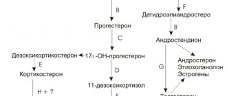
In some cases, an increase in CA 125, the rate of which depends on the level of estrogen, indicates the progression of endometriosis and malignancy of lesions.
Other methods are used to diagnose the disease - colposcopy, hysteroscopy, pelvic ultrasound. The gynecologist performs an examination using a speculum and determines the size of the uterus and its appendages. A mass formation in the area of the gonads, adherent to the body of the uterus, may indicate an endometrioid ovarian cyst, which is also accompanied by high CA 125 numbers.
Treatment is carried out with hormonal drugs, after which the level of the tumor marker usually decreases.
The test is carried out in the first phase of the monthly cycle, the optimal period is 2-3 days after the end of bleeding. Donate blood on an empty stomach, in a calm state. If the woman has previously undergone an ultrasound or x-ray, the doctor should be informed about this. The results may be distorted when taking hormonal drugs.
Let's summarize:
- The test result is positive during menstruation and pregnancy.
- An increase in CA 125 occurs in other non-tumor diseases.
- The study must be repeated to clarify the diagnosis.
- An increase in the marker indicates the progression of endometriosis.
Tactics when increasing CA-125
The blood test results are interpreted by a gynecologist. If the CA-125 level exceeds the norm, he prescribes additional testing:
- ROMA index. Includes determination of CA-125 + HE The latter is a more accurate indicator. Its growth is more common in malignant neoplasms. HE4 may be elevated even with normal CA-125. The growth of HE4 is observed in the preclinical stage of cancer development - when there are no obvious symptoms of the disease. In endometriosis, HE4 remains within normal limits;
- Ultrasonography. It is necessary to assess the condition of the uterus, ovaries and fallopian tubes, according to indications - skin scars, intestines, bladder. Endometriosis is not always clearly visible on ultrasound, but there are indirect signs that allow one to suspect the disease;
- Magnetic resonance imaging. MRI makes it possible to identify small foci of endometriosis – those that are not visible on ultrasound;
- Diagnostic laparoscopy is the gold standard for diagnosing external endometriosis. Allows you to identify lesions on the pelvic organs and take material for histological examination;
- Hysteroscopy – examination of the uterine cavity. It is carried out for adenomyosis, including for differential diagnosis with uterine fibroids and endometrial polyps;
- Histological verification makes it possible to make a final diagnosis.
Further tactics will depend on the diagnosis. If endometriosis is detected, various options are possible:
- Observation. Treatment is not prescribed, but you need to regularly visit a doctor - undergo an examination and ultrasound, monitor the growth of lesions. Allowed for minor forms of endometriosis and minor spread of lesions;
- Conservative therapy. Hormonal drugs are prescribed to suppress the growth of heterotopias. The course of therapy lasts 3-9 months. Can be combined with surgical treatment;
- Surgical intervention. Using laparoscopic access, all accessible lesions are removed, cysts and adhesions are excised.
After therapy, a control determination of the tumor marker is often prescribed. If its level increases, it is worth looking for another cause of the pathology or assuming a relapse.
Quite often, when diagnosing diseases, as well as to monitor the effectiveness of treatment, doctors prescribe a blood test to determine the level of tumor markers. In gynecology, one of these tumor markers is CA 125.
Exceeding the norm for the tumor marker CA 125 may indicate the development of endometriosis. This disease is the third most common among women of reproductive age. This marker, in conjunction with HE 4, is used in the verification of ovarian tumors.
Endometriosis is said to occur when endometrial cells, normally localized in the inner layer of the uterus, are found in other tissues and organs. In this case, irritation and inflammation of tissues occurs, and persistent pain syndrome develops due to changes in endometrioid cells according to the phases of the cycle.
Endometriosis can have two main forms, which are classified depending on the organ affected by the disease.
- Genital, characterized by the spread of endometrioid cells in the form of foci to the organs of the reproductive system. In the genital form, lesions are found in the uterus, vagina, cervix, ovaries, and tubes.
- The extragenital variety of the disease is the involvement of the lungs, intestines, conjunctiva of the eyes, and bladder in the pathological process.
Features of the procedure
Markers of endometriosis, as a rule, are taken repeatedly, and several times for an accurate diagnosis. The test is performed on an empty stomach in the first phase of the female menstrual cycle. Venous blood is taken for this.
Endometriosis is a benign pathological growth of tissue, identical in morpho-functional composition to the endometrium, outside the uterine mucosa. Some women are not even aware of the disease, considering the clinical manifestations of endometriosis to be normal.
Symptoms of endometriosis, which can aggravate the disease and lead to severe forms:
- heavy, prolonged menstruation;
- intermenstrual bleeding;
- pain in the lower abdomen, especially during menstruation;
- pain during sexual intercourse;
- pain during bowel movements (when lesions spread to the rectum);
- enlarged uterus (detected by palpation or ultrasound);
- infertility or miscarriage.
Many scientists determine that each of the modern types of treatment for endometriosis is not always fully able to compensate for the problem completely, and in this regard, the possibility of relapses of the disease cannot be ruled out.
Surgical treatment of endometriosis is one of the difficult tasks of our specialists. Considering the duration of treatment for endometriosis, the final stage of the pathology is surgery.
Surgical action is the maximum mechanical effect on the foci of endometriosis along the border of unchanged tissues. The extent of surgical manipulation depends largely on the age and site of the lesion.
The extent and severity of the disease, the spread of the pathological process to neighboring organs and tissues, as well as the volume and method of surgery depend on the patient’s age and reproductive function. Treatment with these methods is carried out exclusively in a hospital.
Surgical treatment is indicated in the following cases:
- If there are contraindications to drug treatment, hormonal therapy is not suitable for the patient. (Chronic diseases of the gastrointestinal tract, immune system, hormonal imbalance, blood diseases). It is also worth considering allergic reactions to medications or their components. Since the use of medications takes a long time.
- In the absence of the expected effect from drug therapy, for more than six months, when it is not possible to stop the symptoms and progression of the disease, reduce the risk of relapse.
- If there are lesions larger than 2 cm or the process is neglected up to the third and fourth stage.
- When the disease develops into endometrioid cysts or extensive adhesions.
- In complex and severe forms of the disease involving other organs and disruption of their functioning.
- And also, there is a certain type of endometriosis disease - retrocervical endometriosis, in which drug treatment does not produce results at all.
Surgical treatment of uterine endometriosis can be carried out in several options. One of the treatment options is conservative surgical treatment; organ-preserving operations are performed to excise foci of destruction.
This operation is performed only in cases where: the course of endometriosis is moderate or severe, when a woman is planning a pregnancy, to solve problems with infertility. For the woman, all foci of destruction are excised and endometriosis cysts are removed.
Implants larger than 2 cm are also excised, which leads to prolonged pain and discomfort. The purpose of treatment of this method is excision of the focus of endometriosis in various locations of the organ, endometrioid cysts, dissection of the adhesive process, and restoration of body functions, in particular childbearing. Restore the anatomy of the organ as much as possible.
The operation can be performed laparoscopically, however, in case of extensive infiltrative lesions, with resection of adjacent organs, the abdominal approach should be considered safer. Laparoscopy is considered a minimally traumatic operation, which is carried out in a surgical, gynecological hospital.
In this operation, the surgeon makes a minimal incision near the navel and in the lower abdomen above the pubic bone. The device used to perform the operation is called a laparoscope - a thin, optical one. It is inserted into the incision near the navel in order to see and differentiate the abdominal organs and pelvic organs, and through another hole, instruments are inserted to excise the focus of heterotopia or endometrioid cyst, as well as to cut the adhesive process.
Since the operation is small, after the procedure only small scars in the form of dots remain on the skin, and the woman herself recovers much faster. This operation reduces the risk of developing adhesions in the future.
The second type of surgery used to treat endometriosis is the radical method. The operation is carried out not only with the removal of the entire organ, but also with its appendages or adjacent parts (ovaries, fallopian tubes).