Recurrence of cervical cancer
– resumption of the oncological process in the area of the primary lesion after completion of radical treatment and subsequent well-being. It manifests itself as nagging pain in the lower back, perineum and sacral area, watery or sanguineous discharge, urination disorders, swelling, weakness, apathy, exhaustion and appetite disturbances. Sometimes it is asymptomatic and is discovered during a routine examination. The diagnosis is made taking into account the medical history, complaints, gynecological examination, angiography, lymphography, cytological examination, biopsy and other studies. Treatment is surgery, radiotherapy and chemotherapy.
General information
Recurrence of cervical cancer is the re-development of a malignant tumor some time after radical treatment of the tumor. Relapses are understood only as cancer lesions that occur after a period of well-being lasting six months or more. In the absence of such a period, they speak of the progression of the oncological process. The likelihood of recurrence of cervical cancer after combined treatment (surgery and radiotherapy) is approximately 30%, and most tumors are diagnosed within 2 years after completion of therapy. Recurrent neoplasms are characterized by a more aggressive course. Treatment is carried out by specialists in the field of oncology and gynecology.
https://youtu.be/zBdP-QH3tb8
Functionality of the cervix
The cervix undergoes significant changes during nine months of intrauterine development.
In the normal state, the uterine organ is closed and has a dense structure; at the exit, the canal is blocked by a mucus plug. Thus, the cervix protects the fetus by preventing infections from entering the uterine cavity. Additionally, the successful course of pregnancy depends on the condition of the organ; in the presence of abnormalities, the likelihood of childbirth before the due date increases. Ultrasound of the cervix during pregnancy is used both as part of the necessary studies and separately if there is a possibility of abnormalities. During the procedure, the length of the uterus is determined by week; for women with their first child, the indicator is within 3-4 cm. It is these dimensions of the cervical canal that allow the fetus to be held in the uterine cavity. Closer to childbirth, at 36-38 weeks of gestation, the cervical canal softens and shortens, which becomes one of the signs of impending labor.
Classification and causes of cervical cancer recurrence
A.I. Serebrov distinguishes two types of relapses: local and metastatic. According to the classification of E.V. Trushinkova there are four types of relapses:
- Local – damage to the vaginal stump.
- Parametric – an oncological process in nearby tissue.
- Combined - a combination of local and parametric processes.
- Metastatic - involvement of lymph nodes and other organs.
In 70% of cases, recurrence of cervical cancer occurs in the pelvic area. Most often the lymph nodes and ligaments of the uterus are affected. Local tumors are diagnosed in only 6-12% of cases and are usually detected in patients who suffered from endophytic forms of cancer. The cause of the development of the tumor is malignant cells remaining in the pelvic cavity after surgery and radiotherapy due to the rapid growth of the tumor or too non-radical treatment due to underestimation of the severity and rate of progression of the disease.
What happens to her after the operation?
After the operation, the adaptation period begins. Rehabilitation can be:
- Early. The patient must be in a hospital setting under the strict supervision of the attending physician. Lasts from 7 to 12 days depending on the type of operation chosen. During this period, the stump is just healing, so there is a high probability of bleeding and inflammatory processes. Due to the removal of the uterus, complications from various internal organs may occur. This condition may be accompanied by severe pain in the area of the surgical sutures, which the woman must inform the doctor about.
- Late. This is a long period of time during which the body gets used to the changes, but only after discharge from the hospital. However, a woman can be discharged from the hospital only after confirmation that everything is normal with the stump and there is no inflammatory process in the area. This may require an ultrasound.
During the postoperative period, the woman will have to wear a bandage to support her abdominal muscles. Any physical labor is contraindicated, as this can lead to rupture of the seams. After discharge from the hospital, a woman should not be sexually active for 2-3 months after surgery. Swimming in open waters is prohibited.
During the normal course of the recovery period, scar and fibrous tissue forms at the suture site. They separate the vagina from the cervix and prevent the connection of the genitals with the abdominal cavity. Due to this, it is impossible for sperm to enter here (with improper tissue healing, there is a high risk of ectopic pregnancy). If the sutures were applied incorrectly, benign cysts may form on them, which are a cosmetic defect.
https://youtu.be/GTN3U1aaYX0
Symptoms of cervical cancer recurrence
Recognizing recurrent lesions is often associated with significant difficulties, especially at the initial stage. The reasons for the difficulties are the asymptomatic or low-symptomatic course, as well as difficulties in interpreting the manifestations of the oncological process against the background of postoperative scars and sclerotic changes caused by previous radiotherapy. The first symptoms of relapse of cervical cancer are usually apathy, unmotivated fatigue, appetite disturbances and dyspeptic disorders.
After some time, pain appears in the abdomen, perineum, sacrum and lower back. The intensity of the pain syndrome can vary significantly. The pain is usually nagging and worsens at night. While the patency of the cervical canal is preserved, sanguineous, watery or purulent leucorrhoea is observed. When the canal is closed, there is no leucorrhoea, fluid accumulates, and the uterus enlarges. Possible swelling and urinary disorders. Some patients with recurrent cervical cancer develop hydronephrosis. With distant metastasis, the functions of the affected organs are disrupted.
During a gynecological examination, an ulcer with hardened edges is discovered in the cervical area. As the tumor grows, the neck expands and becomes lumpy. When the canal or upper parts of the vagina are closed, an elastic formation is palpated above the cervix. As the recurrence of cervical cancer progresses, the general signs of cancer lesions become more pronounced. The patient suffers from disability, fatigue and depressive disorder. Exhaustion and hyperthermia are detected.
Measuring the cervix during ultrasound during pregnancy: subject of study
Ultrasound results allow the doctor to give a detailed description of the cervix. All the nuances must be deciphered. During the ultrasound procedure, the specialist evaluates the following organ parameters:
- tone is one of the most important indicators. Hypertonicity indicates a high probability of premature birth;
- shape and length - these characteristics change as the fetus develops. Non-compliance with the norm indicates abnormal development of pregnancy;
- consistency;
- density;
- the presence or absence of dilation - normally the cervix is securely closed from both the uterus and the vagina. The presence of holes requires the application of tightening sutures or the installation of an obstetric pessary;
- changes in the state of the internal pharynx;
- lengthening, expansion and patency of the cervical canal;
- is there ectopia of the cervix;
- whether there are polyps or cysts in the cervix;
- the likelihood of cervical rupture and scarring. This complication threatens women who have had a cesarean section in the past.
Highly sensitive ultrasound technology allows you to obtain accurate and objective information about the course of pregnancy. The procedure does not harm the development of the fetus in any way.
Diagnosis of cervical cancer recurrence
The diagnosis is made on the basis of anamnesis, complaints, gynecological examination data and additional studies. A fairly effective way to early diagnose relapses is to determine the level of tumor marker for squamous cell carcinoma SCC. An increase in the level of a tumor marker at the preclinical stage is observed in 60-70% of patients and can serve as a basis for an extended examination. When examining patients with clinical forms of recurrent cervical cancer, an ulcer is detected in the affected area. During bimanual examination, infiltrates may be palpable in the surrounding tissue. To identify renal dysfunction, excretory urography is performed.
To detect vascular networks in the tumor growth zone, percutaneous transfemoral angiography is performed, indicating the presence of new randomly located vessels with characteristic “brooms” at the end. To confirm recurrence of cervical cancer with metastases to regional lymph nodes, direct lymphography is prescribed. The affected nodes are enlarged, with uneven contours, the contrast passage is slow. During the examination, ultrasound of the female genital organs, CT and MRI of the abdominal cavity are also used. If metastatic damage to distant organs is suspected, CT and MRI of the brain, ultrasound of the liver, scintigraphy of skeletal bones and other studies are prescribed. The final diagnosis is established taking into account the data of a cervical biopsy or cytological examination of a cervical scraping.
Treatment of cervical cancer recurrence
Radical surgical intervention is possible in the absence of hematogenous metastases and extensive infiltrates. Patients undergo panhysterectomy - removal of the uterus (hysterectomy) with adnexectomy. For single lymphatic metastases, lymphadenectomy is performed. After surgery, radiotherapy and chemotherapy are performed. The best option is considered to be a combination of intracavitary and remote gamma therapy. Sometimes transvaginal radiotherapy and close-range intravaginal radiotherapy are additionally prescribed.
In case of relapse of cervical cancer with spread to the pelvic tissue and multiple lymphatic metastases, radiotherapy and drug therapy are used. For relapses in the vagina, surgery is usually not indicated. Patients undergo combined radiation therapy. With single nodes in the liver and brain in young, somatically safe patients, surgical removal of metastatic tumors is possible. For multiple distant metastases, chemotherapy, radiotherapy and symptomatic therapy are prescribed.
Forecast and prevention of cervical cancer recurrence
The prognosis in most cases is unfavorable. The best results are observed with local relapses that do not spread beyond the uterus and vaginal vault. The average five-year survival rate after surgery combined with radiotherapy and chemotherapy in such cases is 27.4%. In the presence of lymphogenous and distant metastases, 10-15% of patients manage to survive a year from the moment of diagnosis.
The importance of early detection of recurrent cervical cancer necessitates thoughtful preventive measures. During the first year, examinations are carried out once every 4 months, over the next two years - once every 6 months. The examination includes a speculum examination, rectovaginal examination, general and biochemical blood tests, cytological examination of vaginal fluid, excretory urography, chest radiography, ultrasound of the female genital organs, CT scan of the abdominal organs and dynamic renal scintigraphy (if appropriate equipment is available). In doubtful cases, a puncture biopsy of the cervix is performed.
Ultrasound cost
An ultrasound is performed in a antenatal clinic on the direction of a gynecologist. The procedure is free if you have a medical insurance policy.
You can get tested for a fee in private clinics and reproductive health centers.
| City | Cost, rubles |
| Moscow | 500-1200 |
| Saint Petersburg | 450-1100 |
| Ekaterinburg | 500-1000 |
| Novosibirsk | 400-1000 |
| average cost | 462-1075 |
Ultrasound of the cervix allows you to determine a woman’s readiness for childbirth and indications for cesarean section. During pregnancy, the procedure helps to promptly identify the threat of miscarriage.
Leave comments on the article and share your own experience. Share useful information with your friends on social networks.
Ultrasound during pregnancy is included in the examination program for expectant mothers. A woman undergoes ultrasound scanning several times throughout her pregnancy. The main goal of a screening study is to monitor fetal development and monitor the condition of the expectant mother. The doctor will be able to detect possible anomalies and complications of pregnancy (post-term pregnancy, premature birth, miscarriage) in the early stages and take appropriate measures.
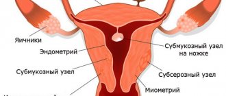
Operations to remove the uterus: types and indications
Removing the uterus is a very risky step for a woman, since such an intervention completely eliminates future childbearing. That is why to perform an operation there must be strict indications, which can be divided into absolute and relative.
Absolute indications for surgery are a malignant tumor of the female genital organs, uterine prolapse, incessant postpartum bleeding (this is an emergency indication). Absolute indications imply that it is impossible to cure the disease in any way other than surgery. Such operations are performed on women (sometimes teenage girls) regardless of age and number of children born, since this is the only way to save the patient’s health and life.
Relative indications are large or multiple fibroids that progress rapidly, endometriosis, adenometriosis, inflammatory diseases of the female genital organs, frequent and heavy menstrual bleeding. In all these cases, we talk about removal only when conservative treatment methods are ineffective. In some cases, removal of the uterus for the listed conditions is performed on postmenopausal women or those who already have two children (at the patient’s request), even if the woman’s condition is not so critical.
There are several main types of hysterectomy. The choice of a specific type remains with the doctor. The two main types are amputation (the neck of the tag remains in place) and extirpation (the uterus is removed along with the cervix and part of the vagina). Both of these types of operations may affect the appendages (fallopian tubes and ovaries), or may leave them intact.
Since during extirpation the cervix is completely removed, in the future we will only talk about amputation of the uterus. In terms of volume, this intervention can be of two types - subtotal and total amputation. The first type is the most sparing, while the vagina, cervix up to the internal os, fallopian tubes (sutured) and ovaries are preserved. In the second case, the uterus is completely removed along with its appendages, but the vagina and cervix remain in place.
According to the method of carrying out, uterine amputation can be hysteroscopic, laparoscopic and laparotomic. Hysteroscopic is the most gentle option, performed under local or subdural anesthesia. The instruments are inserted through the vagina, leaving no scars on the skin. Laparoscopy is a surgical procedure performed through a small puncture in the skin, if there is a need to perform a large volume. Transsection is used in severe cases, for example, postpartum hemorrhage.
In addition to indications, there are also contraindications to hysterectomy. These include the serious condition of a woman caused by decompensation of a chronic pathology - in this case, the operation is performed after stabilization of the patient’s condition. Acute infectious diseases are also a contraindication for intervention - they need to be cured before surgery. And the third absolute contraindication is cancer of any organ, including the uterus, stage 4.
How to prepare for the examination?
In practice, several methods are used to conduct an ultrasound of the cervix (as well as the scar on the uterus) when monitoring pregnancy:
- The transvaginal method
involves the use of a special vaginal sensor, which is placed in the vaginal cavity during examination. This procedure does not require special preparation. - Transabdominal
. Before such a procedure, it is recommended to refrain from eating food that contributes to excessive gas formation in the intestines for 2-3 days. - Transrectal method - examination of the cervix through the anus. Before such an ultrasound, it is necessary to do a cleansing enema. Pregnant women are rarely prescribed such an ultrasound; it is more acceptable for virgin girls.
- Ultrasound through the skin of the perineum. No preliminary preparation is required.
What is a cervical stump?
A stump is the remnant of an organ left after its removal. In the case of the cervix, this is a fragment from the internal os (sutured during surgery) to the external os (vaginal part). The vagina remains completely intact. The stump of the cervix is a consequence of a previous operation - amputation of the uterus (with or without appendages).
The absence of a uterus and the presence of a stump in no way affects the appearance (especially if the operation was performed using the hysteroscopic method) or the woman’s quality of life. Patients who have undergone uterine amputation continue to live a full sexual life, experiencing no less vivid sensations. However, after removal of the uterus, reproductive ability is completely lost, so in young women who do not have children, doctors are extremely reluctant to prescribe this operation if there are no vital indications.
There is a widespread belief that the consequences of uterine amputation have a significant impact on the female body - for example, that a woman ages much faster or has an increased likelihood of other gynecological diseases. Here it should be clarified that the changes that occur depend on the volume of the operation.
If a woman has only her uterus removed, but the ovaries and fallopian tubes remain, then her hormonal levels after the operation remain normal, she looks healthy and feels good. Therefore, this type of intervention is preferable in young women.
If a total amputation of the uterus and appendages was performed, then in the absence of female sex hormones, menopause actually occurs earlier. If a woman is postmenopausal, then these changes do not affect her, but if before the operation she had a stable menstrual cycle, then the symptoms of premature menopause are eliminated with the help of hormonal therapy.
As for the likelihood of developing gynecological diseases, in particular breast cancer, in this case the connection is not with the operation itself, but with the reasons that caused the disease. In particular, the likelihood of breast cancer is higher in women who have been diagnosed with fibroids (if conservative therapy is ineffective, this disease is an indication for uterine amputation).
The most noticeable change in a woman’s lifestyle after surgery is the disappearance of menstruation. Many patients consider this feature to be rather positive, especially if during the illness they suffered from heavy and painful periods. However, if the intervention is performed incorrectly or in insufficient volume, and the factor causing the disease is not completely removed, complications in the late postoperative period are possible - prolapse and cancer of the cervical stump.
Why do ultrasound of the cervix during pregnancy?
During pregnancy, the cervix gradually changes its structure, size and shape. This is a natural process that indicates the body’s preparation for the upcoming birth process. Detection of any deviations and disturbances in this process can be dangerous for bearing a fetus or pregnancy.
During gestation, the cervix performs the main function of holding the fetus. For most of pregnancy, it is in a closed position, protecting the fetus from pathogenic bacteria. From 37-38 weeks of gestation, the cervix begins to open. It moves towards the center, becomes shorter and more elastic. In this case, a single canal is formed that connects the external os of the uterus with its body. After opening the neck, the pharynx can reach 12 cm in width.
The muscle ring that holds the fetus is located in the internal pharynx. When its muscles lose tone and weaken, the cervix loses its ability to hold the fetus.
Another important indicator that is assessed during an ultrasound scan is the length of the cervix. Its shortening may be a sign of cervical insufficiency, which provokes a miscarriage. This is due to the physical inability of the organ to withstand the load of amniotic fluid and a growing child. As a result, the uterine cervix opens earlier than expected.
With the help of ultrasound diagnostics, the doctor can detect the slightest deviations and irregularities in the structure, location and shape of the cervix, and develop suitable treatment. This will stabilize the condition of the organ and prevent premature labor.
Prolapse of the cervical stump
This is a fairly rare condition. Most often, its reason is that before the operation there was uterine prolapse or prerequisites for it, which were not completely eliminated. The reasons for this condition are a history of traumatic childbirth, connective tissue diseases, and improperly performed amputation of the uterus.
Even more interesting:
Tongue with HIV infection photo
Yarina or Dimia
Prolapse of the cervical stump develops rather slowly and rarely tends to correct itself. Patients report a feeling of a foreign body, abdominal pain, which intensifies when coughing, sneezing, or lifting heavy objects. Urination and defecation are also impaired.
Treatment for this condition is fixation of the cervix using special materials. In this case, transsection is most often performed to provide the greatest scope for the surgeon’s actions. The cervix may be artificially attached to the uterosacral or other ligaments that support the uterus.
When to measure the length of the cervix using ultrasound during pregnancy
The ultrasound procedure is timed to coincide with certain periods of pregnancy:
- The first ultrasound will be scheduled for the newly minted expectant mother before the 13th - 16th week of gestation. The study will allow you to exclude an ectopic pregnancy, as well as find out whether the woman is at risk of miscarriage;
- the second time the procedure must be completed between the 17th and 24th weeks of pregnancy. Based on the results of the study, it will become clear whether the expectant mother should be afraid of bleeding and spontaneous abortion;
- The pregnant woman may be invited to the third ultrasound when she is 32-34 weeks old. But if there is no evidence, the procedure is not necessary.
During each screening, the doctor assesses the overall picture of the pregnancy, determines the length of the cervix and the degree of its maturity. An unscheduled ultrasound is prescribed in case of a high risk of premature birth or in the presence of abnormalities in the development of the fetus.
Cervical stump cancer
Cervical cancer is a common oncological pathology in gynecological practice. After amputation of the uterus, the likelihood of developing a malignant process in the cervix remains. Moreover, there are situations when, at the time of making a decision on intervention for fibroids or endometriosis, a woman already has a cancerous tumor at an early stage, but doctors mistake the symptoms of cancer for symptoms of uterine disease and ignore them. This leads to the fact that instead of extirpation, a more gentle intervention is performed, and the cancer remains and progresses.
Symptoms of the disease are vaginal discharge, which in the early stages is brownish, then becomes bloody, with an unpleasant odor, and its abundance can vary. The appearance of discharge should especially alert a woman who has had her uterus removed, because normally after this all discharge stops or becomes insignificant.
Later, abdominal pain, urination and defecation problems, and general toxic syndrome appear. A peculiarity of cervical cancer and its stump is the development of metastases to the pelvic lymph nodes. This leads to swelling of the lower extremities and external genitalia, a little later swelling of the fingers and hands occurs, swelling of the veins in the neck are symptoms of late stages with extensive metastases.
Treatment of stump cancer after removal of the uterus is carried out in several ways. Radiation therapy with a maximum dose in the affected area is considered preferable. Chemotherapy is rarely prescribed because it is not always effective. If radiation therapy is ineffective, they resort to surgical intervention - removal of the cervical stump.
Laparoscopy and Operative Gynecology
Uterine cerclage is performed on patients planning pregnancy with a history of ineffective surgical correction of isthmic-cervical insufficiency, as well as after surgical treatment for precancerous and cancerous diseases due to the absence or significant shortening of the cervix.
Isthmic-cervical insufficiency (ICI) is considered the most common cause of miscarriage in the second trimester, and its frequency in patients with recurrent miscarriage reaches 13–20%. Miscarriage is one of the pressing problems of modern obstetrics. This is a pathological condition characterized by incompetence of the cervix and/or isthmus of the uterus, leading to spontaneous abortion in the 2nd and 3rd trimesters of gestation.
| Cervical insufficiency | Functional form |
| Organic form |
Functional form of cervical insufficiency
Functional ICI develops with hyperandrogenism, connective tissue dysplasia, increased levels of relaxin in the blood (noted in multiple pregnancies, induction of ovulation by gonadotropins), as well as with increased load on the cervix (polyhydramnios, multiple pregnancies, large fetuses)
Ultrasound criteria for the progression of ICI during pregnancy: shortening of the closed part of the cervical canal, formation of a funnel-shaped internal os, prolapse of the amniotic sac, development of amnionitis, rupture of amniotic fluid, leading to late spontaneous miscarriage or premature birth.
The history of surgical correction of ICI goes back decades. Currently, vast experience has been accumulated in the use of various methods of surgical and non-surgical correction, and the effectiveness of the methods has been assessed. Vaginal cerclage was first proposed in 1950 by A.F. Lash. The most frequently performed manuals at present are the Shirodkar technique, proposed in 1955, as well as a modified and simplified version of it, proposed by McDonald in 1957. Both procedures are performed via vaginal access to correct ICI during pregnancy.
The effectiveness of sutures reaches 70%, but the question arises: what to do in other cases?
Organic form of cervical insufficiency.
This form of ICI develops as a consequence of previous cervical injuries:
- Damage to the cervix during childbirth [ruptures not repaired surgically, surgical delivery through the birth canal (use of obstetric forceps, delivery of a large fetus, a fetus in the breech presentation, fetal-destroying operations)
- Invasive methods of treating cervical pathology (conization, cervical amputation, radical trachelectomy)
The results of numerous worldwide statistical studies indicate a steady increase in the frequency of detection of dysplastic processes and cervical cancer in young women, with a particularly noticeable increase in incidence under the age of 29 years, many of whom were unable to realize their generative function.
Adequate treatment of dysplasia, preinvasive and microinvasive cervical carcinoma in women of reproductive age is conization and/or amputation of the cervix. Carrying a pregnancy to term in such patients is very difficult due to the high incidence of cervical stump failure and the development of organic ICI. The maximum number of complications is observed after knife amputation of the cervix for CIN II/III and the minimum after radiocoagulation for CIN I. The main complications of gestation in such cases are the threat of termination of pregnancy at different times, reaching 62%, early reproductive losses and premature birth in 64% of cases.
Radical trachelectomy is a unique operation that allows preserving reproductive function in patients with invasive cervical cancer stages Ia2-Ib1 of the disease. For the first time, radical vaginal trachelectomy with laparoscopic ileoobturator lymphadenectomy was performed by the French surgeon D. Dargent in December 1986, the total recurrence rate does not exceed 5%, which indicates the high oncological effectiveness of the method, not inferior to classical radical hysterectomy. However, pregnancy in these patients is very difficult due to the high frequency of functional failure of the uterovaginal anastomosis. At the same time, the frequency of premature births and perinatal losses in the 3rd trimester reaches 75%.
Thus, pregnancy after various cervical surgeries is a high-risk pregnancy. Considering the complete absence of the cervix or its vaginal portion, classical surgical procedures for the formation of isthmic-cervical insufficiency during pregnancy are impossible, which became the reason for the search for new technologies for preconceptional preparation of patients.
Uterine cerclage
All of the above has given rise to the search for new technologies aimed at reducing the risk of miscarriage among patients with a burdened obstetric and gynecological history. A necessary condition for the progression of pregnancy in such patients is the application of a circular synthetic prosthesis, which reduces the load on the distal parts of the cervical stump or uterine vaginal anastomosis.
In 1965, Benson proposed a technique for performing cerclage using an abdominal approach. A technique for transvaginal cervico-isthmic uterine cerclage has also been developed. In this case, the synthetic prosthesis is located at the level of the cardinal and uterosacral ligaments.
A comparison was made of the effectiveness of transvaginal cervico-isthmic and transabdominal uterine cerclage in patients who had previously encountered the ineffectiveness of classical cervical sutures during pregnancy, complicated by perinatal losses. The author shows that the abortion rate in the abdominal group is 6% versus 12.5% in the vaginal group, indicating a higher efficiency of the overlying prosthesis.
| Indications for uterine cerclage |
|
The application of “classic” sutures to the cervix during pregnancy in patients who have undergone surgical interventions is very difficult, and in many cases impossible due to the lack of a vaginal portion.
Thus, based on large cohort international studies, as well as as a result of their own research work, doctors have formulated indications for uterine cerclage.
According to research work conducted by Prior Clinic , Ph.D. Fedorov A.A. and Ph.D. Since 2011, Vrotskaya V.S., performing cerclage of the uterus in patients at the stage of pregnancy planning provides high rates of newborn survival, a history of recurrent miscarriage
Based on our experience, as well as data from foreign authors, it is recommended to perform cerclage of the uterus at the stage of pregnancy planning to minimize the risks of complications.
Prior Clinic surgeons perform cerclage of the uterus using laparoscopic access using a patented technology, which includes the following stages (Click VIDEO):
- mobilization of the bladder, uterine isthmus or uterovaginal anastomosis
- A synthetic prosthesis was placed around the selected area at the level of the internal os or proximal to the uterovaginal anastomosis, positioning it medial to the ascending branches of the uterine arteries and ureters on both sides.
- fixation of a synthetic prosthesis behind the uterus at the level of the uterosacral ligaments.
Ethicon SurgicalTM mersilene tape is used as a synthetic prosthesis
The optimal period for the operation, length of hospital stay and restrictions and recommendations in the postoperative period.
The optimal period for the operation is the first phase of the menstrual cycle, that is, immediately after the end of menstruation.
The operation is performed under general anesthesia (endotracheal anesthesia). The patient's stay in the hospital varies from 1 to 3 days.
The limitation period for pregnancy after cerclage of the uterus is 2 months.
Dangerous symptoms, or when to see a doctor?
Both of the above conditions significantly threaten the health and life of the patient, so it is important to recognize dangerous symptoms in time so that the treatment measures taken are effective. You should consult a doctor immediately if:
- Pain appeared in the lower abdomen;
- Any discharge from the genital tract has appeared, especially if the vagina is bleeding;
- Urination and defecation are impaired.
These symptoms occur with both prolapse and cancer of the cervical stump; it is impossible to distinguish these diseases at home without being a specialist. To make a diagnosis, it is necessary to undergo a series of procedures, starting with an examination in a gynecological chair. It should be remembered that one of the diseases does not exclude the other; both diagnoses can accompany each other.
Reviews from various sources
Kristina, 34 years old, Omsk
My second birth was difficult - there was bleeding, the doctor says that they could barely pump it out, although I myself don’t remember that day well at all. Due to bleeding, I had to have my uterus removed, but the cervix was left in to avoid any side effects. Three months have passed, the tears have healed safely, the baby is healthy. Everything is fine with my husband, too, although at first I felt strange. In general, I wouldn’t say that life has improved, but it hasn’t gotten any worse.
Anastasia, 48 years old, Kemerovo
14 years ago I was diagnosed with ovarian endometriosis. Two years of treatment, different medications, the result is quite weak. I wanted a third child, but the gynecologist said that with my illness I would not be able to get pregnant. But I couldn’t recover, it only got worse. Together with the doctor, we made the difficult decision to remove the uterus and appendages. After the operation I had to take hormones, but overall I felt good. I am seeing a gynecologist, and in the near future I will have to stop taking hormones in order for menopause to occur.
Daria, 46 years old, Novosibirsk