Scar on the uterus after cesarean section: what is it, what does it look like?
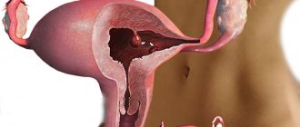
The scar on the uterus after cesarean section is the result of healing of the incision; it looks thin at first, then thickens. Consists of connective tissue fibers (scar) and muscle cells. Outside of pregnancy, it most often does not manifest itself, but when the uterine cavity is stretched and during childbirth, it can rupture with bleeding and disruption of labor.
Reasons for appearance
The main reasons for the appearance of a scar on the uterus are indicated in the table.
| Type of uterine injury | Circumstances of occurrence |
| C-section | Planned in case of pre-established impossibility of normal childbirth, at the request of the woman in labor, emergency in case of sudden complications |
| Operations | Removal of fibroids, fallopian tubes, ectopic pregnancy, plastic surgery for anatomical defects (for example, removal of a horn for a bicornuate uterus) |
| Birth rupture | Suturing the body and neck if it is possible to preserve the organ |
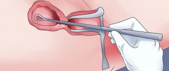
| Diagnostic procedures or mini operations | Abortion, curettage of the inner layer of the uterine cavity (endometrium), endoscopy, all of them are usually small |
| Injury | Penetrating injury in the pelvic area |
What does it look like
At first, the scar looks like a wide, swollen red stripe, then it darkens. During healing, a thin strip of compacted tissue with a ribbed surface is visible at the incision site, since connective tissue is the first to form in the wound. It is rigid, does not stretch well, and does not contract enough.
This period normally lasts up to six months. Then gradually blood vessels and nerve endings begin to grow in, and new muscle fibers are formed. Only with their appearance does the scar become both strong and extensible. Complete scarring can be expected by the end of 16-18 months.
Metroplasty in the broad sense of the word is a reconstructive operation on the uterus, which consists of eliminating certain congenital or acquired disorders of the anatomical structure of the organ in order to restore its reproductive function. We are interested in metroplasty for failure of the uterine scar after a cesarean section, which consists of excision of the old, incompetent scar outside of pregnancy and restoration of the integrity of the uterine wall.
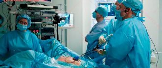
There are several ways to perform metropalectomy: transabdominal - through the anterior abdominal wall (laparotomy or laparoscopically) - and transvaginally (through the vagina). Each method has its own indications and contraindications.
Of course, laparoscopic metroplasty is the most common and least traumatic method. Excision of an incompetent scar is carried out under general anesthesia, through small (5 to 10 mm) punctures on the anterior abdominal wall at standard points. The postoperative period, as a rule, is 2-3 days, and if the patient’s condition is satisfactory, the patient is discharged home.
Of course, the task of any doctor is to preserve the health and life of the patient, and in the case of an obstetrician-gynecologist we are talking about two patients. Management of childbirth through the birth canal, in the absence of indications for cesarean section, is optimal for achieving the set goals. Correct diagnosis of uterine scar incompetence and timely correction is the right step towards a healthy pregnancy and happy motherhood.
Diagnostics
The presence of scars in the cervical canal is assumed based on the history of difficult childbirth and previous invasive manipulations.
Confirmation of suspicions is carried out using the following diagnostic methods:
- Gynecological examination - the procedure shows non-standard dilatation of the cervical canal, ruptures, signs of eversion of the cervical mucosa. A vaginal examination allows you to insert your fingers into the external os.
- Colposcopy - using a microscope, a specialist examines the nature of the changes that have occurred on the surface of the uterine cervix and in the cervical canal.
- Cytomorphological study - assessment of the collected material shows the state of cellular structures and allows us to identify the degeneration of structures.
- Laboratory research - determination of specific and non-specific counterparty is prescribed for inflammation. To identify pathogenic microflora, serology, PCR, and bacteriological culture are used.
Complications of the underlying disease require additional consultation with an oncologist or venereologist.
Advantages of laparoscopic reconstructive metroplasty
- Low-invasiveness - thanks to the use of video endoscopic equipment, only the scar is excised, the surrounding tissues are minimally affected
- No blood loss
- Reliable suturing - thanks to the author’s technique used, pregnancy and childbirth naturally become possible in the future
- The use of anti-adhesive barriers during surgery on the uterine scar reduces the risk of formation of adhesions
- Short rehabilitation period and minimal hospitalization period
- Fast recovery after
- Excellent cosmetic result - only three barely noticeable marks remain on the skin.
- Our Center for Operative Gynecology annually conducts about 40 master classes on laparoscopic operations; the clinic has more than 40 patents for bloodless interventions.
- Some specialists at our clinic have performed more than 3,700 low-traumatic surgeries on the uterus, including interventions to treat an incompetent scar on the uterus after cesarean section.
- Some gynecological interventions can be performed only in 2-3 Russian clinics, including ours.
- Thanks to the use of modern technologies, more than 600 of our patients were able to give birth to a child, despite previously diagnosed infertility.
- We perform about 120 operations per month, and treatment tactics for each patient are selected individually.
- We have accumulated vast experience in conducting simultaneous operations. If there are several diseases that require surgical treatment, it is possible to perform a simultaneous operation that allows you to get rid of 3-4 pathologies at once.
- During the operation, equipment, instruments and suture material of the latest generation produced by well-known companies are used, which allows surgical intervention to be carried out quickly, efficiently and reliably. The protocol for operations performed in our clinic also includes the use of anti-adhesive barriers, which reduce the risk of adhesions, which has a beneficial effect on a woman’s fertility in the future.
Treatment methods
It is impossible to cure deformation with medications. Folk remedies will also be powerless. The only effective way is surgery. His technique is selected in accordance with the individual characteristics of the patient and is:
- Conization;
- Reconstructive plastic surgery;
- Laser correction;
- Radio waves;
- Ultrasound surgery.
For minor damage to epithelial tissue, vaporization, cryodestruction or dermatocoagulation are used.
Deformation changes in the uterine cervix lead to physiological abnormalities in the cervical and vaginal canal. The pathological process is rare compared to erosions and leukoplakia. The disease can cause the development of cancerous tissue lesions, threatening the patient’s life.
Types of scars on the cervix and body of the uterus, their classification
Classification of scars on the body and cervix is carried out according to the following criteria:
- solvency (insolvent and wealthy);
- direction (longitudinal and transverse);
- homogeneity (heterogeneous and homogeneous structure).
Insolvent
An incompetent uterine scar means that pregnancy is dangerous for the woman and the fetus, since it can rupture when the uterus stretches.
Known reasons why a woman may have it even after 2 years include:
- suffered chronic inflammatory processes of any localization;
- endometritis after cesarean section (including asymptomatic);
- a long period after the water breaks before the onset of labor;
- dehiscence of seams on the skin (lifting weights, physical activity, sexual activity earlier than recommended);
- curettage of the uterine cavity (surgical abortion) before the end of the scarring period (especially dangerous before 1 year);
- new pregnancy within 2 years after cesarean section.
Incompetent scar on the uterus after cesarean section on ultrasound
If the scar is defective during pregnancy, there may be a threat of miscarriage, women report pain in the lower abdomen, and intrauterine development of the fetus is often disrupted.
Wealthy
A scar is considered healthy if it has a predominance of muscle tissue (85% or more) and a small amount of connective tissue fibers (up to 15%).
It is characterized by:
- smooth and clear contours;
- homogeneous structure;
- thickness is in the range of 3-5 mm;
- the location of the uterine cells is correct;
- there is sufficient blood supply.
With a healthy scar, pregnancy proceeds normally, since the uterus stretches well, and during childbirth its strength protects the organ from rupture. A fully formed scar appears no earlier than 16 months after cesarean section. In many ways, this period of time is determined by the woman’s general health, the presence of heart disease, and metabolic disease (for example, diabetes mellitus inhibits wound healing).
The transverse and longitudinal scar reflects the direction of the incision during cesarean section. The first is located in the lower third of the abdomen and is the best option for surgery.
It works in 95% of cases because:
- healing is faster and less likely to be accompanied by complications;
- the edges are neat and even, comparable in thickness;
- makes it possible to carry out the next natural birth through the vagina.
Longitudinal and transverse scar
The longitudinal scar after cesarean section runs along the body (corporal) or the middle and lower segment of the uterus (isthmic-corporal). It is longer than transverse, the recovery is long, there are risks of infection, but it is faster in terms of execution time. Therefore, this method is chosen when complications arise before or during childbirth that threaten the life of the mother or fetus.
The countdown starts in minutes when there is bleeding, the fetal heartbeat stops, or the umbilical cord is entangled.
Based on homogeneity, the uterus in the scar area can be homogeneous or heterogeneous; in the latter case, there is a risk of having a large percentage of connective tissue, since it is denser, or niches filled with fluid. All this characterizes the failure of the scar and is assessed during ultrasound. A completely homogeneous structure practically does not occur after cesarean section; moderate and focal heterogeneity is allowed.
To prevent the formation of a weak scar on the uterus after cesarean section, you must:
- compliance with the doctor’s recommendations on limiting heavy lifting, physical activity and timing of the resumption of sexual contacts;
- early contact with a doctor to monitor the healing of the uterus (no later than a month after surgery, then every six months);
- careful contraception in the first two years, using reliable methods - a spiral or hormonal pills.
When a second pregnancy occurs, it is important to register from the first days and regularly visit a gynecologist and undergo an ultrasound.
The mechanism of pathology development
The columnar epithelial cells that make up the lower region of the cervical canal constantly produce a mucous secretion. In the presence of provoking factors - traumatic injuries and hormonal imbalance - the alkaline balance of the environment is disturbed, and the mucous membrane descends into a much less alkaline and more acidic environment of the vagina.
Changes in acidity are caused by inflammatory processes, which result in disruption of the trophism of the tissues of the cervical canal. The uterus turns the mucous membrane into the vaginal area and vaginal-cervical fistulas are formed.
Other painful conditions also occur:
- endometritis;
- cervicitis;
- endocervicitis.
This leads to the appearance of erosion, atrophy of the epithelium, keratinization of the cells lining the cervical canal, which can become an insidious impetus for the onset of cancerous degeneration of cells.
Also, a scarred cervix significantly reduces the quality of sexual life and reduces the possibility of conception, and if it occurs, it prevents the pregnancy from being carried to term.
Diagnostics to avoid uterine rupture along the scar
To avoid the main complication - rupture of the uterus along the scar, it is necessary to assess its consistency, and for this it is recommended to undergo an examination during the pregnancy planning period (examination by a gynecologist: ultrasound, MRI, hysteroscopy, hysterography).
The main criterion for the viability of a scar is its thickness; normally, before pregnancy, it should be more than 5 mm, and after pregnancy, 3 mm is considered an acceptable parameter. In women in later stages, there is a possibility of its thinning to 1.5-2 mm, which does not interfere with normal childbirth. At 1 mm, the risk of rupture is assessed as moderate, and all other parameters (fetal weight, its condition) are taken into account to choose the method of delivery.
Clinical signs of incompetence that can be detected during a gynecological examination include:
- abdominal pain on palpation (palpation);
- increased pain in the projection of the scar;
- recess on the wall.
An ultrasound scan for a scar in the uterus must be performed even before planning a conception. In case of incompetent scar tissue, the description will indicate the following signs:
- thickness 1-2 mm;
- uneven contours: thin areas alternate with thick ones, discontinuity, break in the lower segment;
- heterogeneity of structure (seals and sparse tissue);
- in the wall there are niches, zones of thinning of the myometrium (muscular layer) and hyperechoic inclusions (dense);
- blood flow is insufficient (low vascularization).
Ultrasound diagnosis of scar
In such cases, pregnancy should be postponed until independent scarring occurs or surgery (metroplasty) is performed.
During the period of bearing a child, you need to do a gynecological ultrasound regularly: when registering, once a month for monitoring in the first trimester and every week from the second. Additionally, the condition of the fetus and blood flow in the placenta are determined.
Magnetic resonance imaging is the most accurate method for assessing the structure of the scar. It shows the predominance of connective or muscle tissue fibers in the structure. If you are wealthy, the former should be no more than 15%. It is carried out only before pregnancy.
MRI of a uterine scar
Hysteroscopy
Hysteroscopy is prescribed for diagnostic purposes to non-pregnant women. It confirms ultrasound data and can identify the following types of scar condition:
- not visualized (not detected) – healing has occurred in full, since only muscle tissue is visible;
- along the entire length there is no more than one third of whitish areas, there are vessels, which means the consistency of scar tissue;
- retraction in the area of the postoperative suture, the color along the entire length or more than a third is whitish, adjacent areas are pale (the scar is incompetent).
Hysterography
The examination is carried out after the end of the next menstruation with a contrast agent, which is injected into the uterine cavity.
Symptoms of an incompetent scar:
- filling defects (niches);
- jagged outline;
- There are areas of thinning muscle tissue.
The photo shows an example of hysterography of the uterus.
In the area of the scar, the thickness of the myometrium is assessed to determine the strength of the scar tissue. The minimum size for a woman to be able to bear and give birth to a child is considered to be 3 mm, the optimal size is 3.5-5 mm. At the same time, it is also important that in the lower segment the muscle layer is uniform in this indicator.
Cicatricial deformity of the cervix
RDSM refers to a change in the shape of the uterine cervix, which arose under native or post-traumatic influence, provoking dysfunction of the cervical canal. In most patients, the clinical signs of the disease are mild.
Average statistical data report that the range of cicatricial anomalies in the uterine cervix varies from 15-55%, in women of fertile age it occurs in 70%. The problem is often registered in those who gave birth for the first time after the age of 30. The replacement of standard epithelium with scar tissue increases in patients with cervicitis.
The disease increases the likelihood of developing infertility, cancer and inflammatory diseases. In the ICD-10 system, it received code No. 88, referring to “Other non-inflammatory diseases of the cervix.”
The photo shows cicatricial deformity of the cervix
FAQ
Do I need special preparation for laparoscopic reconstructive metroplasty?
A few days before surgery for uterine scar failure, a woman should, in consultation with her doctor, avoid taking medications that affect blood clotting. You also need to avoid foods that cause excessive gas formation. Metroplasty is performed on an empty stomach, with the last meal and water taken eight or more hours before.
How is laparoscopic reconstructive metroplasty performed?
During the operation, to prevent blood loss, atraumatic clamps block the blood flow, after which, under the control of video endoscopic equipment, the scar tissue is excised, while it is possible to visualize the boundaries of healthy tissue and layers of the uterine wall - which allows all manipulations to be carried out with high precision. When suturing the wound, absorbable suture material is used, and modern anti-adhesive barriers are used to prevent the formation of adhesions, which is important for preserving the patient’s fertility. Finally, the clamps from the vessels are removed, and blood flow is completely restored.
What to expect after laparoscopic reconstructive metroplasty?
Bloody discharge can last up to 10 days, in the first couple of days it is intense, it gradually becomes insignificant. In the first week, the temperature may rise to 38°C. Each patient is under the supervision of staff; the condition of the scar is also monitored by ultrasound specialists.
What types of reconstructive metroplasty are there?
There are several methods of performing the operation: transabdominal - through the anterior abdominal wall: the intervention can be performed by laparotomy (open method) and laparoscopy (through three small punctures), as well as transvaginally. Each technique has its own indications, but laparoscopic metroplasty is the least traumatic method.
Are there any disadvantages to laparoscopic reconstructive metroplasty?
The scar is located in an area with good blood supply, which is accompanied by the risk of intraoperative bleeding. If events develop in this way, it may be necessary to switch to an open method. In addition, the seam formed is less reliable. However, thanks to the use of the author’s technique in our clinic, such problems can be successfully eliminated.
How is the rehabilitation going?
Already on the first day the patient begins to get out of bed, on the second day light food is allowed. In the first days, anti-inflammatory and antibacterial drugs are prescribed. Discharge is possible on the third day. Showering is allowed 5-7 days after surgery. A week after the operation, an ultrasound is performed, and follow-up examinations are scheduled after 1, 3, 6 months. For better recovery, the patient is prescribed hormonal medications for a period of 3-6 months. In the first 4-6 weeks, physical activity should be avoided; normal activity is possible after 1.5-2 months. During the first month, it is also recommended to abstain from sexual relations and visiting the pool. It is better to plan pregnancy no earlier than 6-8 months after the end of treatment.