author Bondarenko Svetlana Petrovna
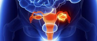
Tumors of the ovary and oviduct In old doves, there are tumors of the ovary and oviduct, merging with each other. Ovariocarcinomas were found in pigeons both as independent lesions and with carcinomas of other organs. Differential diagnostic studies
Ovary
, ovarium
. In it, female reproductive cells (eggs) develop and mature, and female sex hormones entering the blood and lymph are formed.
General structure of the uterus
This internal muscular organ of the reproductive system has a pear-shaped shape that is flattened in front and behind.
In the upper part of the uterus on the sides there are branches - fallopian tubes, which pass into the ovaries. The rectum is located behind, and the bladder is located in front. The anatomy of the uterus is as follows. The muscular organ consists of several parts:
- The fundus is the upper part, which has a convex shape and is located above the line of origin of the fallopian tubes.
- A body into which the bottom smoothly passes. It has a cone-shaped appearance. It narrows downwards and forms an isthmus. This is the cavity leading to the cervix.
- Cervix - consists of the isthmus, cervical canal and vaginal part.
The size and weight of the uterus vary from person to person. The average weight for girls and nulliparous women reaches 40–50 g.
The anatomy of the cervix, which is a barrier between the internal cavity and the external environment, is designed so that it protrudes into the anterior part of the vaginal vault. At the same time, its posterior arch remains deep, and the anterior arch, on the contrary.
Features of the development of cervical diseases, symptoms
The ovaries are located on both sides of the uterus and are defined respectively as right and left. The size of the gland is 3-4 cm in length, 1-1.5 cm in width, but these parameters may vary depending on the woman’s age, condition, for example, pregnancy, menopause. The ovaries are connected to the uterus using a special ligament; their location is asymmetrical; often the gland located on the right is larger than the left.
The maintenance of the gonads in the pelvic area is ensured by the ligamentous apparatus, which relates directly to the ovaries. It allows them to move slightly relative to other organs, which is necessary during pregnancy, when the uterus significantly increases in size.
If we consider the anatomical structure of a woman’s ovaries, the gland consists of various functional layers. Initially there is a junction of the germinal epithelium, followed by a dense protein shell, consisting, among other things, of connective tissue and elastic fibers. It is this layer that forms a kind of stroma or base that helps maintain the shape of the organ.
- Internal or cerebral. It consists mainly of special loose tissue and many vessels.
- Outer layer or cortex. It is more dense in structure, contains connective fibers and maturing follicles.
Mature follicles surround the ovary, their approximate size is 1-2 cm in diameter. An interesting feature of the development of follicles is the dominance of one of them. If it has reached a significant size, larger than the others, then all resources are directed to the maturation of the egg, and the growth of other follicles is suspended.
Pathological changes often occur on the vaginal surface of the cervix, in the “transition zone” of one epithelium to another. When the localization of this zone changes, background diseases arise: ectopia (pseudo-erosion), ectropion, leukoplakia, erythroplakia, polyps and, as a consequence, malignant neoplasms of the cervix.
Ectopic cervix
Ectopia (pseudo-erosion) is a disease of the cervix, characterized by the emergence of columnar epithelium on the vaginal surface of the cervix. Women with irregular menstrual cycles are predisposed to developing this disease.
The clinical picture is somewhat blurred. Such women may complain of the release of blood clots after intercourse and the pain of the process of intercourse itself. The disease can be recognized during a medical examination of the cervix in the mirror: the surface of the organ will not be pale pink, but a bright red hue.
Ectropion is an eversion of the mucous membrane of the cervical canal into the vagina, with the formation of scars and ectopia on the cervix. This disease occurs as a result of unsuccessful diagnostic or surgical intervention on the uterus, as well as after labor or abortion.
Leukoplakia is a disease associated with the appearance of keratinized areas on non-keratinized epithelium. Leukoplakia can be simple or proliferative. The proliferative form is a precancerous condition and when biological material is taken from this area (biopsy), atypical cells are found.
With erythroplakia, the cervix is affected locally, closer to the external os. The pathogenesis of the disease is the thinning of the stratified squamous epithelium and, as a result, tissue atrophy. The etiology of erythroplakia has not yet been studied. The photo clearly shows red spots in the ectocervix area.
Cervical polyps
Polyps are growths of various sizes on the mucous membrane of the cervical canal. According to their structure, polyps are epidermal and glandular. When examined in a speculum, the polyps are oblong and leaf-shaped. It is possible to diagnose and differentiate such polyps only with an ultrasound examination of the pelvic area.
Cervical endometriosis is a disease in which tissue similar in structure to the endometrium is located on the vaginal surface of the cervix. Normally, such tissue should be located in the uterine cavity.
The main reasons for the development of this disease:
- cervical injuries due to violence, childbirth or abortion, as well as medical interventions;
- genetic predisposition;
- hormonal disbalance.
Patients with cervical endometriosis usually do not present characteristic complaints. There is discomfort during sexual intercourse, bleeding after intercourse, as well as before and after menstruation.
Cervical cancer
Cervical cancer (CC) is a malignant neoplasm of the cervix. The location and form of cancer is varied. The main component of the disease is atypical cells that can divide uncontrollably and replace healthy cells. In 90% of all cases, the cause of cervical cancer is the human papillomavirus. The main route of transmission of the pathogen is sexual intercourse.
Factors that increase the risk of developing the disease:
- early onset of sexual activity (before 17 years of age);
- failure to comply with hand and body hygiene rules;
- the presence of precancerous conditions (proliferative leukoplakia, polyps);
- frequent change of sexual partners;
- bad habits;
- infection with the papilloma virus in another way (during medical interventions, transmission from mother to fetus).
With progression, when the malignant disease moves to the next stage and grows, bleeding and nagging pain may appear in the lower abdomen. CC can be suspected only by performing a biopsy of the affected tissue for the presence of atypical cells.
Clubfoot
The foot is shortened and in a supinated position due to subluxation of the ankle joint.
Etiology of clubfoot
Clubfoot can be congenital (among congenital malformations it ranks second - approximately 1-2%) and acquired. More often observed in males. Unilateral and bilateral clubfoot occur with equal frequency. Congenital clubfoot is considered a sex-linked developmental anomaly. Acquired clubfoot can be the result of paralysis and damage to the soft tissues or bones of the foot.
Clubfoot Clinic
The clinical picture has the following 4 types of deformities: - supination position - plantar flexion - metatarsal adduction - pronounced longitudinal arch. The position of supination is of greatest importance; other changes can be expressed to varying degrees. The outer side of the foot is subjected to the maximum load, and in severe cases of deformity, patients stand, even leaning on the back of the foot. The foot cannot be turned inward and the toe cannot be raised. Metatarsal adduction causes patients to walk with their feet turned outward to prevent the toe from sagging. Painful calluses form at the sites of abnormal load. In acquired clubfoot, the listed deformities rarely occur in combination.
Treatment of clubfoot
Treatment for congenital clubfoot should begin immediately after the birth of the child. It is necessary to gradually retrain the leg manually, and then apply a plaster cast. It is especially important to eliminate subluxation in the ankle joint. Redressing plaster casts are initially changed every 3 days, and then the interval increases. After sufficient correction of the shape or position of the foot, the plaster casts are removed and special night splints are used to maintain the achieved effect. Treatment of clubfoot is considered complete if pronation becomes possible and if the foot has a normal shape. If this cannot be achieved by the time the child gets to his feet on his own and tries to walk, then a variety of shoe inserts are needed. Starting from the 3-4th year of life, therapeutic exercises for the foot can be prescribed. If these measures are ineffective, surgical interventions on soft tissues are indicated before the end of growth and formation of the skeleton. In severe cases, interventions on the bones of the foot are necessary, but they should be performed only after the formation and complete development of the skeleton. Treatment for acquired clubfoot
should be carried out in accordance with its cause. If it cannot be eliminated, then operations (arthrodesis of the foot joints) or the provision of orthopedic aids (and orthopedic shoes) are indicated.
Where is the uterus?
The organ is located in the pelvis between the rectum and bladder. The uterus is a very mobile organ, which also has individual characteristics and shape pathologies. Its location is significantly influenced by the condition and size of neighboring organs. The normal anatomy of the uterus in terms of the place it occupies in the small pelvis is such that its longitudinal axis should be oriented along the axis of the pelvis. Its bottom is tilted forward. When the bladder is full, it moves back a little, and when emptying, it returns to its original position.
The peritoneum covers most of the uterus except the lower part of the cervix, forming a deep pouch. It extends from the bottom, goes to the front and reaches the neck. The posterior part reaches the wall of the vagina and then passes to the anterior wall of the rectum. This place is called the pouch of Douglas (recess).
Functions
The functions performed by the ovaries are very important for a woman. The following can be distinguished from them:
- Ovum formation or generative function.
- Maintaining hormonal levels. The gonads are involved in the synthesis of steroid hormones, including estrogens, which are called female, so they are responsible for secondary sexual characteristics and processes characteristic specifically of the fairer sex.
A feature of a woman’s reproductive system is the presence of a certain number of germ cells already in the womb, that is, a newborn girl already has them. Unlike sperm, eggs cannot be formed again, as men do when they reach puberty.
Sprain of the lumbosacral ligaments
Pathology of the sacroiliac region occurs during physical activity of varying intensity. Both athletes and ordinary people face this problem. To do this, it is enough to lift a weight, take an unsuccessful position, bend over or bend your leg excessively at the hip joint.
Main symptoms
The leading sign of damage is intense pain, which intensifies with various movements in the pelvic joint, and decreases with rest. Irradiation of pain to the lower abdomen, inner thigh and buttock is typical. A sign of severe spasm of tendon fibers is a limping gait and curvature of the spine. These symptoms disappear with timely and proper treatment.
Pain in the sacrum (sacrodynia) occurs not only with sprains. Sacrodynia is a leading symptom in pathologies:
- congenital defects in the development of vertebrae;
- osteochondrosis;
- displacement of the sacral vertebrae;
- injuries in the sacroiliac region;
- thrombosis of the blood vessels of the pelvis and sacrum;
- tuberculosis, staphylococcal joint damage.
In women, similar problems arise with gynecological diseases. These can be inflammatory, infectious and oncological diseases:
- parametritis;
- endometritis;
- painful menstruation;
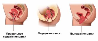
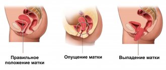
- uterine prolapse;
- thrombophlebitis of the pelvic veins;
- uterine cancer;
Magnetic resonance imaging will help to exclude pathology of the ligaments of the sacroiliac joint from the inflammatory process of the uterus and parametritis. Pain in these diseases has one peculiarity: it does not disappear with rest and has the same intensity.
In men, pain in the sacrum is caused by the following diseases:
- prostatitis, both acute and chronic;
- prostate cancer with metastases to the sacrum;
- for diseases of the rectum and sigmoid colon.
Cancerous lesions of the pelvic bones and spine, as well as arthrosis of the sacroiliac joint, can be diagnosed using computed tomography.
Uterine ligaments: anatomy, purpose
The cervix is the lower segment of the organ and is a continuation of the body of the uterus. The isthmus is the boundary between the body and the neck.
Walls
The wall of the cervix differs from the wall of the uterine body in the presence of more collagenous connective tissue fibers and, to a lesser extent, elastic smooth muscle fibers. The mucous membrane of the cervix is located directly under the connective tissue membrane, represented by columnar epithelium.
Columnar epithelium consists of tall cells capable of secreting mucus. Nabothian glands are derivatives of the mucous membrane of the uterine cavity. At the point where the cervix enters the vagina (the vaginal surface of the cervix), there is a zone where the columnar epithelium is replaced by stratified non-keratinizing epithelium.
The cervix (photo presented later in the article) anatomically consists of: the isthmus, supravaginal and vaginal parts.
- Vaginal surface of the cervix.
- Supravaginal segment of the cervix.
- External os of the uterus.
- Internal os of the uterus.
- Cervical canal.
The vaginal part is the lower part of the uterus. It is in this area that there is a transition zone of the epithelium, which is diagnostically significant for the doctor at the time of the examination. Normally, the entire surface of the vaginal part of the cervix is covered with stratified non-keratinizing epithelium, like the surface of the vagina.
Schematic photo of the cervix
When the transition zone shifts, part of the vaginal surface is occupied by columnar epithelium, which is not normal. In this case, we should talk about pathological changes - the formation of pseudo-erosions or erosions. On the vaginal surface there is an external os, which is limited by the anterior and posterior lips of the cervix.
Between the posterior lip and the vaginal wall there is a “pocket” - the posterior vaginal fornix, and between the anterior lip and the anterior wall - the anterior fornix. These anatomical structures are the site of punctures performed by a doctor for diagnostic purposes. The supravaginal part of the cervix is its upper segment, located between the vaginal section and the isthmus.
The cervical canal is the connection between the vagina and the uterine cavity. The anatomical unit begins with the internal os in the cavity of the body of the uterus, passes into the cervical canal proper of the cervix and exits through the external os into the vagina. Glands that produce a significant amount of mucus form a mucus plug in the canal, preventing foreign infection from entering.
Ligaments and muscles
The ligamentous apparatus is represented by a complex of muscles, ligaments and fascia that perform supporting, fixing and suspending functions.
| Function | Ligaments that perform the function |
| Suspension (attachment of the uterus to the wall of the pelvic ring) |
|
| Fixing (providing the necessary mobility to the pregnant uterus) |
|
| Supportive (separation and support of the pelvic organs in their anatomical position) |
|
The ligamentous apparatus of the cervix includes all 3 functions, but not all ligaments are involved in its attachment.
Normal functioning of the cervix is ensured by:
- wide paired and circular ligaments, which perform a suspensory function;
- the main uterine ligament that performs fixation;
- muscle fibers with a fascial membrane as a supporting apparatus.
internal and external oblique, circular. The latter is also called the average circular. It received this name in connection with the structure. The most obvious is that it is the middle layer of the myometrium. The term “circular” is justified by a rich system of lymphatic and blood vessels, the number of which increases significantly as it approaches the cervix.
Bypassing the submucosa, the uterine wall after the myometrium passes into the endometrium - the mucous membrane. This is the inner layer, reaching a thickness of 3 mm. It has a longitudinal fold in the anterior and posterior region of the cervical canal, from which small palm-shaped branches extend at an acute angle to the right and left.
The rest of the endometrium is smooth. The presence of folds protects the uterine cavity from the penetration of vaginal contents that are unfavorable for the internal organ. The endometrium of the uterus is prismatic; on its surface there are uterine tubular glands with glassy mucus. The alkaline reaction they provide preserves the viability of sperm. During ovulation, secretion increases and substances enter the cervical canal.
In the normal state of the female body, the uterus, ovaries and other adjacent organs are supported by the ligamentous apparatus, which is formed by smooth muscle structures.
The functioning of the internal reproductive organs largely depends on the condition of the muscles and fascia of the pelvic floor. The ligamentous apparatus consists of suspension, fixation and support. Composition of the ligamentous apparatus of the internal reproductive organs
| Apparatus | Functions performed | Ligaments forming the apparatus |
| Suspensory | Connects the uterus to the walls of the pelvis | Paired wide uterine |
| Supporting ligaments of the ovary | ||
| Own ligaments of the ovary | ||
| Round ligaments of the uterus | ||
| Fixing | Fixes the position of the organ and stretches during pregnancy, providing the necessary mobility | Main ligament of the uterus |
| Vesicouterine ligaments | ||
| Sacrouterine ligaments | ||
| Supportive | Forms the pelvic floor, which is a support for the internal organs of the genitourinary system | Muscles and fascia of the perineum (outer, middle, inner layer) |
The anatomy of the uterus and appendages, as well as other organs of the female reproductive system, consists of developed muscle tissue and fascia, which play a significant role in the normal functioning of the entire reproductive system.
The anatomy of the uterus should have suggested its natural purpose - bearing and giving birth to offspring. This process is inevitably accompanied by active contraction, growth and movement of the reproductive organ. In this connection, it is necessary not only to fix the correct position of the uterus in the abdominal cavity, but also to provide it with the necessary mobility. Fixing structures arose precisely for such purposes.
The main ligament of the uterus consists of plexuses of smooth muscle fibers and connective tissue, radially located to each other. The plexus surrounds the cervix in the area of the internal os. The ligament gradually passes into the pelvic fascia, thereby fixing the organ to the position of the pelvic floor. The vesicouterine and pubic ligamentous structures originate at the lower anterior part of the uterus and attach to the bladder and pubis, respectively.
The uterosacral ligament is formed by fibrous fibers and smooth muscles. It extends from the back of the cervix, envelops the rectum on the sides and connects to the fascia of the pelvis on the sacrum. In a standing position, they have a vertical direction and support the cervix.
Prevention
In order to reduce the likelihood of developing endometriosis of the uterosacral ligaments, you must adhere to the following recommendations:
- Justified prescription of any invasive therapeutic or diagnostic manipulations on the pelvic organs;
- Protection from injury to the birth canal during childbirth, proper suturing in case of ruptures of the walls of the vagina and perineum;
- Timely treatment of inflammatory diseases of the genital area;
- Reasonable prescription of COCs.
Women's health depends directly on a woman's attitude to the state of her sexual sphere.
Respect for yourself and your health is the key to a healthy life. Share:
Possible pathologies
The gonads are susceptible to various diseases, leading not only to reproductive dysfunction, but also generally negatively affecting health. The most common ones include:
- Oophoritis. Inflammation of the ovaries of various etiologies, can be one or two-sided. Oophoritis can spread to other tissues, including the fallopian tubes. Complications of the disease affect not only the ability to become pregnant and bear a child, but also the functions of the pelvic organs and the entire body.
- Cystic formations. They are common in women of childbearing age; in advanced stages, they can lead to removal of the ovary. Cysts occur due to various reasons, there are some types that do not require treatment, such as a functional cyst. It spontaneously reduces after several menstrual cycles.
- Polycystic. Dysfunction of the gonads caused by the formation of a number of small cysts, which are immature follicles. The pathology occurs against the background of hormonal imbalance, and in advanced stages it is dangerous for a woman’s health.
- Apoplexy. It occurs due to increased mechanical impact and a widespread acute inflammatory process. It is characterized by rupture of the membrane, which leads to hemorrhage into the abdominal cavity. In such situations, timely surgical care is important.
The structure of a woman’s ovaries implies a direct relationship between the uterus and the pelvic organs, therefore, if unpleasant pain is detected, the entire area must be examined at once. This can be done using ultrasound for both diagnostic and preventive purposes.
Horse foot
The foot forms an obtuse angle with the axis of the lower leg and cannot move in the dorsal direction.
Etiology of equine foot
In most cases, equine foot develops as a result of flaccid paralysis of the triceps surae muscle. In spastic paralysis, it occurs as a result of the functional predominance of the strength of the toe flexors. It may be a consequence of improper immobilization of the leg or, for example, pressure of a blanket on the toe of the foot during prolonged bed rest. In this case, the triceps surae muscle and finger flexors are stretched.
Equine Foot Clinic
The foot cannot be actively brought into a position that makes a right angle with the axis of the lower leg. Depending on the cause of this pathology, as well as on the duration of suffering, this sometimes cannot be done passively (overextension and contracture of antagonist muscles). When moving, the patient stumbles, clinging to the floor with his sagging toe.
Treatment of equine foot
For fresh paralysis, along with conventional treatment, it is necessary to use orthopedic devices that fix the position of the foot at a right angle to the axis of the leg (night splints for heel feet). If it is impossible to carry out the correction passively, you should try to gradually eliminate the pathological position of the foot using redressing plaster casts, and then use night splints. Heel traction, heel splints, orthopedic shoes or braces are used for walking. By surgically lengthening the calcaneal tendon, restoration of muscle balance can be achieved. It is also possible to perform arthrodesis of the ankle joint to give the foot the most functionally favorable position.
Characteristics of the hanging apparatus
The suspensory apparatus consists of paired ligaments of the uterus, thanks to which it is “attached” at a certain distance to the walls of the pelvis. The broad uterine ligament is a transverse fold of the peritoneum. It covers the body of the uterus and the fallopian tubes on both sides. For the latter, the structure of the ligament is an integral part of the serous covering and mesentery.
The own ligaments of each of the ovaries originate from the uterine fundus on the posterior side below the branch of the fallopian tubes and reach the ovaries. The uterine arteries and veins pass inside them, so the structures are quite dense and durable.
One of the longest suspensory elements is the round ligament of the uterus.
Its anatomy is as follows: the ligament looks like a cord up to 12 cm long. It originates in one of the corners of the uterus and passes under the anterior sheet of the broad ligament to the internal opening of the groin. After which the ligaments branch into numerous structures in the tissue of the pubis and labia majora, forming a spindle. It is thanks to the round ligaments of the uterus that it has a physiological inclination anteriorly. Anatomy of the female uterus - structure of the pelvic floor
| Layer | Muscles | Characteristic |
| Outer | Ischiocavernosus | Steam room, located from the ischial tuberosities to the clitoris |
| Bulbous-spongy | The steam room wraps around the entrance to the vagina, thereby allowing it to contract | |
| Outdoor | Compresses the anus with a “ring”, surrounds the entire lower rectum | |
| Superficial transverse | Poorly developed paired muscle. Originates from the ischial tuberosity on the inner surface and attaches to the perineal tendon, connecting with the muscle of the same name coming from the reverse side | |
| Middle (genitourinary diaphragm) | m. sphincter urethrae externum | Compresses the urethra |
| Deep transverse | The steam room is located between the symphysis, pubis and ischium. | |
| Internal (pelvic diaphragm) | Pubococcygeal | Paired branches m. levator ani, which raises the anus. Well developed. |
| Iliococcygeal | ||
| Ischiococcygeal |
The normal anatomy of the uterus and appendages is ensured precisely by the pelvic floor, which is the main support of the internal organs of the genitourinary system. The correct location of organs is the key to their healthy functioning. Damage and significant weakening of the pelvic floor muscles threatens prolapse and even loss of organs.
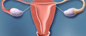
The anatomy of the uterus and ovaries are reproductive organs connected to each other through the fallopian tubes. The ovaries are the sex glands located on either side of the uterus. Inside them, during the menstrual cycle, eggs mature, which then enter the uterine cavity through the fallopian tubes.
The ovaries are supported by the suspensory ligament and mesentery. Unlike the uterus, they are not covered with peritoneum. The structure of the ovary is based on the medulla and cortex. The latter contains mature follicles. On the inside, a granular layer is adjacent to the wall, in which the egg lies. It is surrounded by a radiant crown and a transparent zone.
During ovulation, the follicle approaches the outer layer of the ovary and bursts. This releases the egg and enters the uterine cavity through the fallopian tube. The burst follicle replaces the corpus luteum, which gradually disappears in the absence of pregnancy. If fertilization occurs, the corpus luteum continues to exist throughout the entire period to perform intrasecretory functions.
The surface of the ovaries is covered with a white membrane formed by connective tissue. Each ovary is surrounded by appendages, which have a convoluted shape and consist of longitudinal and transverse tributaries. They are considered to be vestigial formations.
What function do the ovaries perform in women?
The main function of the ovaries in a woman’s body is to produce oocytes (eggs) for fertilization and secrete sex hormones, estrogen and progesterone.
There are several similarities between the functions of the ovaries and testes. Testes and ovaries during ontogenesis develop from one germ layer. Leydig cells (sperm producing cells), seminiferous tubules and testicular interstitium in the ovaries are represented by granulosa cells, primary follicles and stroma, respectively. The functions of the ovaries and testes are equally controlled by gonadotropins produced by the pituitary gland.
Hormonal function
As already mentioned, the ovaries are a component of the endocrine system and are an endocrine gland. They are involved in the production of steroid hormones. Depending on the phases of the menstrual cycle, hormone levels vary.
Let us consider in detail the groups of steroid hormones.
Estrogens (these include hormones such as estriol and estradiol). This group of hormones is the main one, produced by the dominant ovaries in huge quantities before the onset of ovulation. It is these hormones that affect the condition of the vagina, uterus, and also cause the mammary glands to grow. In addition, thanks to them, the exchange of minerals and carbohydrates occurs in the body.
A group of gestagens (this includes 17-hydroxyprogesterone and progesterone). They are produced mainly in the corpus luteum, after which the placenta begins to produce them. These hormones are also very important for the female body. The possibility of conception depends on progesterone, since it supports the life of the embryo as it moves through the fallopian tube from the ovary to the uterus. Gestagens are necessary for the embryo in the first three months of pregnancy.
The next hormonal group is androgens. This includes testosterone. So far, the effect of these hormones on the female body has not been fully studied. They do not affect it as strongly as the previous two groups of hormones, but their lack leads to dysfunction of the menstrual cycle, which subsequently leads to infertility.
As can be seen from all of the above, the ovaries are complex in structure and are of invaluable importance for a woman’s body, despite their miniature size. With ovarian dysfunction, problems occur in the menstrual cycle, lack of pregnancy, even infertility, early menopause and aging women. Therefore, it is necessary to make it a rule and regularly visit a gynecologist to monitor your health and timely treatment.
The generative function of the ovaries in women is that they produce an egg ready for fertilization in the middle of each menstrual cycle. Typically, only one oocyte is released from one ovary during one menstrual cycle, and the ovaries normally work alternately.
A girl baby at birth already carries in her ovaries all the eggs she will ever have, unlike a boy baby who will only begin to produce sex cells after puberty.
The number of eggs initially provided by nature is estimated to be about two million, but by the time a girl reaches puberty, this number is reduced to 400,000 cells stored in her ovaries. From puberty to menopause, only about 400-500 oocytes reach maturity, are released from the ovary (in a process called ovulation), and can be fertilized in the fallopian tubes/fallopian tubes of the female reproductive tract.
The process of gamete formation in the ovary is called oogenesis and involves the formation of an egg from primordial germ cells through a series of morphological, genetic and physiological changes. These changes consist of oocyte maturation, cytoplasmic maturation, and meiotic division.
Folliculogenesis is a process involving the maturation of the ovarian follicle. It describes the progression of a number of small primary follicles into large preovulatory follicles. In the ovary, all oocytes are initially enclosed in a single layer of cells known as a follicle. Over time, the eggs begin to mature and one is released from the ovary each menstrual cycle. As the oocyte matures, the cells in the follicle rapidly divide and the follicle becomes larger. Many follicles lose their ability to function during this process, but one dominates each menstrual cycle and the contained oocyte is released at ovulation.
The hormonal function of the ovaries is that as the follicles grow, they produce the hormone estrogen. Estrogen is necessary for the development of secondary sexual characteristics such as breasts, the growth and development of reproductive organs, and female-pattern hair growth. After the egg ruptures and leaves the follicle at ovulation, it becomes the corpus luteum. The corpus luteum, in turn, produces the hormone progesterone. Progesterone is needed to prepare for the potential implantation of a fertilized egg and pregnancy. If the egg was not fertilized in this cycle, the corpus luteum regresses, and the secretion of estrogen and progesterone stops. Since these hormones no longer exert their stimulating effect on the endometrium of the uterus, it begins to slough off and is removed from the body in the form of menstruation. After menstruation, another cycle begins.
When analyzing the endocrine function of the ovaries, it is worth considering the ovarian appendages, the function of which is to move the egg to the uterine cavity, and fertilization also takes place in them. After ovulation, the oocyte, surrounded by several granulosa cells, enters the abdominal cavity. The nearby fallopian tube, left or right, captures the oocyte. Unlike sperm, oocytes do not have flagella and therefore cannot move independently. High concentrations of estrogen during ovulation cause contractions of the smooth muscles of the fallopian tube, as well as coordinated movement of the fimbriae of the ampulla of the fallopian tube. As a result of these mechanisms, the oocyte enters the fallopian tube and slowly moves towards the uterus.
If the oocyte is successfully fertilized, the resulting zygote will begin to divide into two cells, then four, and so on as it moves down the fallopian tube into the uterus. There it implants into the wall of the uterus and continues to grow under the supportive influence of progesterone.
If the egg is not fertilized, it simply regresses.
Another function of the female ovaries is the production of testosterone and its secretion directly into the bloodstream. In women, testosterone is necessary for the growth of bones and muscles, maintaining energy and activity, and sufficient libido.
Since the number of ovarian follicles decreases with age, the reproductive and endocrine functions of the ovaries in a woman’s body fade away and menopause occurs at the age of about 50 years. Menopause is a state when there are no more follicles and eggs, and the ovary no longer secretes the hormones estrogen, progesterone and testosterone that regulate the menstrual cycle. As a result, menstruation stops.
Considering the endocrine functions of the ovaries in the female body, it is obvious that sex hormones are not vital for the functioning of the person as a whole, but are absolutely necessary for the continuation of the human race.
10709 0
In the lower sections, the parietal peritoneum does not reach the inguinal ligaments at a distance of 1 cm, forming a cellular space containing the external iliac vessels. Here the peritoneum forms several folds: the middle one (pl. umblicalis mediana) contains an overgrown urinary duct (urachus), running from the top of the bladder to the umbilical ring. More lateral are the folds (pl. umblicalis medialis), containing obliterated umbilical arteries extending from a. iliaca interna.
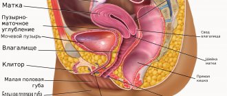
Peritoneal cover of the pelvic organs:
1 - median umbilical fold;
2 - supravesical fossa; 3 - middle umbilical fold; 4 - middle inguinal fossa; 5 - lateral umbilical fold; 6 - lateral inguinal fossa; 7 - bladder; 8 - uterus; 9 - rectum; 10 - ovary; 11 - fallopian tube; 12 - infundibulopelvic ligament; 13 - sacrouterine ligament; 14 - uterorectal fold; 15 - rectal-uterine cavity; 16 - uterovesical recess; 17 — terminal line; 18 - round ligament Even more lateral are the pl.
umblicales laterales, in which the lower epigastric vessels pass. The folds of the peritoneum form the inguinal fossae. The lateral is located outward from pl. umblicales lateralis above the internal opening of the inguinal canal, where the round ligament of the uterus enters. The medial inguinal fossa is a blind recess, as is the supravesical fossa, located between the median and medial umbilical folds. The parietal peritoneum, passing from the anterior abdominal wall to the pelvic organs, covers the bladder on top. Next, the peritoneum passes to the anterior surface of the isthmus and body of the uterus, where it forms a small vesicouterine cavity. The bottom of the notch is a horizontally located vesicouterine fold. Covering the anterior wall and fundus of the uterus, the peritoneum passes to the posterior surface of the body of the uterus, the supravaginal part of the cervix and covers part of the posterior vaginal vault (for 1.5-2 cm), after which the peritoneum rises up and covers the anterior and lateral walls of the upper third of the rectum intestines.
In the space between the posterior wall of the uterus and the rectum, the peritoneum, covering the rectal-uterine and sacro-uterine ligaments, forms two longitudinal rectal-uterine folds. These folds limit the sides of the deepest peritoneal recess - the rectouterine (Douglas) space.
On the sides of the uterus, the peritoneum forms peritoneal duplications - wide uterine ligaments. Along the anterior border, the peritoneum creates arched folds corresponding to the passage of large uterine ligaments.
The highest part of the broad ligaments is the duplication of the peritoneum; the fallopian tubes are located in it. The posterior leaf of the broad ligament forms a duplication of the peritoneum - the mesentery of the ovary (mesovarium), on which the ovary is suspended. The space between the base of the ovarian mesentery and the fallopian tube is called the mesosalpinx. The free lateral edge of the broad ligament, the infundibulopelvic ligament, contains the ovarian vessels.
The ovaries are usually located on the side wall of the pelvis, in the recess of the parietal peritoneum - the ovarian fossa. The edges of the recess are limited by retroperitoneal anatomical structures. The upper and posterior walls are formed by the bifurcation of the common iliac artery, dividing into the external and internal iliac arteries, and the ureter and ovarian vessels passing through the division of the common iliac artery. In front, the ovarian fossa is limited by the posterior layer of the broad ligament. Its lower border is formed by the umbilical and uterine arteries, and the medial edge is formed by the ureter.
The female reproductive organ is a paired formation and performs generative and secretory functions. The ovaries in women have a suspended position; they are located in the middle of the pelvic organs and are held in place by three ligaments. They have an almond shape, a whitish blue-pink color and a lumpy surface.
The fallopian tubes
A paired organ through which an egg from the abdominal cavity enters the uterus. The fallopian tubes are oval-shaped ducts that run at the top of the broad ligament of the uterus. Their length can be up to 13 centimeters, and their diameter is 3 mm. Transportation of the egg is carried out using the uterine and abdominal openings, the name of which corresponds to the cavities into which it exits.
The fallopian tubes consist of:
- uterine part - located in the thickness of the uterus;
- isthmus - the narrowest part with thick walls;
- ampoules;
- funnels - through their lumen the egg enters the fallopian tube;
- fimbria - they direct the egg to the funnel.
The inside of the tube is lined with a mucous membrane with ciliated epithelium and longitudinal folds, the number of which increases as it approaches the abdominal opening. On the outside, the fallopian tubes are covered with a serous membrane.
Main types
There are several types of ligaments, they are:
- wide;
- round;
- sacrouterine;
- cardinal.
- The broad ligament of the uterus is considered double, it is frontal and is located in the pelvic area. Located in front and behind. The fibers intertwine with the uterine artery and are attached on both sides, helping to maintain the organ’s mobility.
- The round ligament of the uterus is 15 cm long, double and ends in the labia minora. They extend from the lateral surface of the reproductive organ, which makes it possible to fix its position. The presence of such fibers in the device allows the uterus not to fall back. The organ has a curved position, for this reason a significant amount of fibers is required to fix it.
- Cardinal. At their core, they are part of the broad ligaments. Located in the lower part of the small pelvis. They support the cervical canal, its walls and the vagina. These fibers are intertwined with the vessels of the organ and the ureters, they allow the reproductive organ to move back and forth. This is a feature of the ligamentous apparatus as a whole.
- Sacrouterine. More significant for the ovaries, they support them, consist of 2 types of fibers, smooth muscle and connective fibers. They are considered special because they do not affect the organ itself, do not support it and do not affect the attachment.
- The rib of the uterus is the lateral surface of the reproductive organ. Under certain circumstances, the ovary may be pressed against the rib. This is not considered a pathology, since it does not affect the functioning of the organ. An ovary pressed against the rib of the uterus indicates a weakening of the uterine ligaments - they weaken, as a result of which the ovary is pressed against the rib.
It is worth noting that over time, the fibers weaken, the muscles lose their elasticity and firmness, which affects the state of the reproductive system. Over time, the ligaments weaken and become rougher; this also occurs due to a decrease in the level of hormones in the blood.
Structure of the circulatory system
The blood supply to the reproductive organ occurs thanks to the uterine artery, which is a branch of the internal iliac artery. The anatomy of the uterus and fallopian tubes involves the outflow of blood from two sides, so the artery has two branches. Each of them is located along the broad ligament, then dividing into smaller vessels going to the anterior and posterior surfaces of the organ. Near the uterine fundus, the vessel branches again to provide blood flow to the fallopian tubes and ovaries.
The uterine veins are formed from the venous plexus, into which venous blood drains. From here the veins originate, which then flow into the internal iliac, ovarian veins and the plexuses of the rectum. Venous drainage after the uterine and ovarian veins passes to the iliac and inferior vena cava.
Blood supply to the uterus
It is carried out by two uterine arteries (from a. iliaca interna) and ovarian arteries (aa. ovaricae from the abdominal aorta).
The beginning of the uterine artery is covered from above by the ureter. 4-5 cm downward from the point of origin, the uterine artery passes at the base of the broad ligament, and, not reaching 1.5-2 cm to the cervix, crosses the ureter from above. At the lateral edge of the uterus, the artery gives off the vaginal branch (ramus vaginalis), rises up to the lateral edge of the uterus and widely anastomases in the broad ligament with the ovarian artery. The veins of the uterus form the uterine venous plexus, plexus venosus uterinus, located in the area of the lateral walls of the cervix and periuterine tissue. The outflow of venous blood goes through the uterine veins into the hypogastric and through the ovarian veins into the inferior vena cava. Lymphatic drainage from the body of the uterus occurs in the lymph nodes located in the vicinity of the arteries and the inferior vena cava. Lymph partially flows from the fundus of the uterus along the round ligament to the inguinal lymph nodes. The uterus is innervated by the utero-vaginal nerve plexus, located along the uterine artery (from the plexus hypogastricus superior et inferior).
V. D. Ivanova, A. V. Kolsanov, S.S. Chaplygin, P.P. Yunusov, A.A. Dubinin, I.A. Bardovsky, S. N. Larionova
Outflow of lymph from the internal genital organs
The lymph nodes to which lymph is directed from the body and cervix are iliac, sacral and inguinal. They are located along the passage of the iliac arteries and on the anterior part of the sacrum along the round ligament. Lymphatic vessels located at the bottom of the uterus reach the lymph nodes of the lower back and groin area. The common plexus of lymphatic vessels from the internal genital organs and rectum is located in the pouch of Douglas.
The internal genital organs are innervated by the sympathetic and parasympathetic autonomic nervous system. The nerves going to the uterus are usually sympathetic. On their way, spinal fibers and structures of the sacral nerve plexus are attached. Contractions of the uterine body are regulated by the nerves of the superior hypogastric plexus.