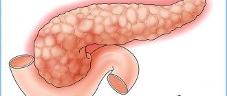
The formation of stones (concretions) in the gallbladder is a common phenomenon that occurs as a result of the progression of concomitant diseases of the organ. The diagnosis is made when dense structures of various shapes and sizes are detected in the cavity of the bladder. Depending on the stage of the disease and the individual characteristics of the patient, the doctor selects a treatment regimen. You should find out how dangerous gallstones are, what types they are and how to treat the pathology.
Causes
Why do stones form in the gallbladder? The most common causes of stones are:
- inflammatory processes;
- cholestasis caused by impaired contractility of the organ;
- a significant amount of calcium, cholesterol, and insoluble bilirubin in bile;
- obesity;
- course intake of estrogen-containing drugs;
- predisposition at the genetic level;
- taking medications such as Cyclosporine, Octreotide, Clofibrate;
- non-compliance with diet;
- diabetes;
- hemolytic form of anemia;
- cirrhosis of the liver;
- surgery on the intestines;
- alcohol abuse.
How to treat cholelithiasis without surgery
Conservative (without surgery) treatment of cholelithiasis is effective if the stones are cholesterol in nature. If you notice symptoms of this disease, you should consult a gastroenterologist who can prescribe different treatment methods:
- Contact dissolution of stones involves the injection of methyl tert-butyl ether (MTBE) into the gallbladder. The operation is complex and dangerous.
- Shock wave therapy (lithotripsy) - stones are crushed by sound waves, this procedure is carried out in the presence of stones no more than 2 cm in diameter.
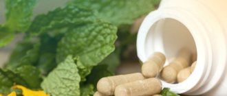
- Folk remedies in the form of herbal preparations, decoctions and juices.
- Teas based on herbal infusions of immortelle flowers, peppermint leaves, coriander fruits, chamomile, lemon balm pour boiling water, wormwood, horsetail, sandy immortelle, buckthorn, chicory knotweed, St. John's wort. Decoctions from marshmallow root, rose hips, lingonberry and strawberry leaves, beets and potatoes. The most effective is a decoction of the birch chaga mushroom.
- Alternative ways:
- With the help of herbal medicines (Rovachol, course of treatment – 6 months).
- Acupuncture - acupuncture (relieves pain, eliminates bile stagnation, stabilizes the functioning of the liver and gallbladder).
- Massage (relaxes and makes the organs work actively).
Medicines
Several types of medications are used to treat the disease:
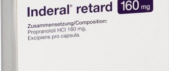
- Choleretic drugs, which accelerate the excretion of bile, eliminate the inflammatory process in the gallbladder and ducts, and relieve intoxication with bile acids. These include “Allohol”, “Urolesan”, “Holosas”, “Flamin”, “Holagol”. The medications should be taken after every meal. Each drug has contraindications.
- Antispasmodics are used to relieve pain - relieve spasm of smooth muscles. These are “No-Shpa”, “Bespa”, “Drotaverin”, “Spazoverin”, “Pakovin”, “Spazmalgol”, “Duspatalin”.
- Anti-inflammatory and painkillers are prescribed for relapses. These are the well-known “Paracetamol”, “Ibuprofen”, “Nurofen”, “Analgin”, “Diclofenac”, “Indomethacin”. Taken after meals.
- Ursodeoxycholic acid is a natural component of bile; it thins bile, breaks down stones, and protects the liver. These are “Holacid”, “Destolit”, “Urdoxan”, “Solutrat”, “Ursahol”, “Urososan”, “Urzofalk”.
- Antibiotics (only as directed by a doctor).
Mechanism of development of cholelithiasis
What causes stones to appear in the gallbladder cavity? Doctors consider two mechanisms as leading ones. These are vesical-inflammatory and hepatic-metabolic. The cause of the pathology developing according to the first scenario is inflammation, leading to acidification of bile. Against this background, the protective function of protein fractions decreases. The result is the formation of bilirubin crystals, followed by the layering of foreign components on the surface of the primary center: epithelial cells, mucous particles, bile.
This is what extracted gallstones look like
The development of the liver-metabolic mechanism is promoted by:
- content in the diet of a significant amount of coarse fats - lamb, pork and beef;
- abnormalities in the functioning of the endocrine system, in particular, hypofunction of the thyroid gland;
- lesions of the liver parenchyma of infectious-toxic origin;
- sedentary lifestyle;
- changes due to a person's age.
Important! It takes quite a long time for stones to form, sometimes several years. On average, over 1 year the diameter can increase by three to five millimeters.
Diet and nutrition for cholelithiasis
An important place in the treatment of gallstone disease is given to nutrition. The doctor prescribes treatment table No. 5, which improves the functioning of the liver and biliary tract, stimulates bile secretion, and improves the functioning of the digestive system.
Dietary recommendations
Diet therapy consists of eating the following foods:
- vegetable soups;
- porridge with water;
- vegetables and fruits with non-acidic pulp;
- lean meats and fish;
- crackers, day-old bread;
- low-fat fermented milk products.
Vegetable and animal fats, oxalic acid, salt and coarse fiber should be excluded from the diet. It is recommended to consume sufficient amounts of proteins and carbohydrates. You need to adhere to a strict diet until complete recovery. Patients should be fed in small portions from 5 to 6 times a day. Food should be steamed or served boiled.
What is prohibited
It is strictly forbidden to use:
- fatty meats;
- seasonings;
- smoked meats;
- dairy products;
- sweets;
- pickled products;
- pickles;
- sausages;
- baked goods;
- alcoholic drinks.
Any errors in nutrition or consumption of prohibited foods can cause an attack of biliary colic. If a person takes medications, but at the same time consumes foods that are prohibited, the treatment will have no effect. Only an integrated approach will help reduce the manifestations of the disease and reduce the risk of possible complications.
Types of gallstones
What types of gallstones exist? Depending on the composition, it is customary to distinguish:
Symptoms of calculous cholecystitis
- Cholesterol. On an x-ray, they appear as round formations with a diameter of 15–18 mm. They are formed as a result of metabolic disorders. Cholesterol stones are diagnosed in overweight people. Located directly in the gall cavity. There are no signs of inflammation.
- Bilirubin (pigment). Where do these types of stones come from? The process of stone formation is based on changes in the protein composition of the blood and diseases in which active breakdown of red blood cells occurs. They have specific biliary colors - red, brown, mixed. Numerous, but small in size. They are determined both in the lumen of the biliary tract and in the cavity of the organ, in particular the cervix.
- Calcareous. The reason for the formation of stones is the increased calcium content in the bile. They are rarely diagnosed and develop as a result of an inflammatory process.
- Mixed. Initially, pigment or cholesterol stones form in the gall cavity. Later, calcifications are layered on their surface. A layered structure is typical for this type of stone. You can only get rid of them surgically.
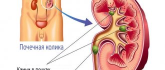
How to relieve an attack of biliary colic at home
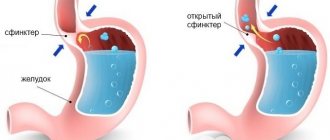
An attack of biliary colic means the appearance of sudden acute pain that intensifies with any movement, coughing or breathing. Appears as a result of spasm of smooth muscles in the walls of the gallbladder against the background of displacement of stones. Stones often get stuck in the neck of the bladder, blocking the outflow of bile, which begins to accumulate and flows directly into the intestines.
In case of severe and acute pain, vomiting of bile, high body temperature, it is recommended to call an ambulance as soon as possible.
Before the ambulance arrives
Before the arrival of specialists, the patient can be given first aid, which will help relieve or reduce the attack:
- take a horizontal position, lie on your back;
- maintain bed rest;
- put an ice pack on the abdominal area, never a heating pad;
- drink plenty of warm water in small portions;
- no food can be taken, complete fasting;
- if the temperature rises, cover the patient with a blanket;
- It is not advisable to take antispasmodics.
After completing all the steps, the patient must be taken to the hospital, even in cases where relief has occurred. Taking antispasmodics can only blur the clinical picture and temporarily improve your well-being. The danger of biliary colic is that there is a risk of rupture of the bile duct, which can lead to the death of the patient.
Help in hospital
Relieve pain with injections of Papaverine or Dibazol. No-Shpu or Eufillin is administered intramuscularly. Analgesics are used as auxiliary painkillers.
If these medications do not help, a potent drug is administered, for example, Tramal, Atropine, etc. If vomiting does not stop, use Cerucal. To replenish fluid losses, a drink based on a solution of Regidron or Citroglucosolan is prescribed.
Symptoms
Small stones are not accompanied by any pathological changes. And only after an increase in their total number and size can characteristic symptoms of the disease appear. Large stones put pressure on the walls, which is accompanied by the development of pain. Developed inflammation can cause colic and require medical help. When the stone passes through the bile duct, the patient’s condition improves, and the formation itself leaves the intestine along with the feces.
The photo shows a gallbladder that is clogged with stones
Quite often, stones get stuck, completely blocking the lumen of the duct. In this case, the patient develops jaundice and cholecystitis. Potential symptoms of gallstones: heaviness in the right hypochondrium, rotten belching, attacks of nausea, pain that occurs due to overeating and physical activity, bitter taste in the mouth.
Important! Sometimes the only sign of gallstones is yellowing of the sclera of the eyes.
With calculous cholecystitis, a person develops the following symptoms: increased body temperature to high levels, unpleasant taste, anorexia (refusal to eat), pain in the right hypochondrium, general weakness. The movement of stones along the bile ducts is accompanied by the development of colic.
This condition is accompanied by acute pain of a cutting nature, radiating to the lower back, shoulder blade and area of the right forearm (sometimes the pain localization area is behind the sternum), nausea accompanied by vomiting (it does not bring relief to the person), belching, increased gas formation. The cause of colic in most cases is the presence of a provoking factor.
Clinical picture of cholelithiasis
In most cases (60–80%), cholelithiasis is asymptomatic and is detected by ultrasound or x-ray. Stone-carriers may not know about their disease for years and note only periodic heaviness/a feeling of fullness in the right hypochondrium and temporary loss of appetite, until the first attack of biliary colic occurs - stones enter the duct and clog it. It can be triggered by a festive feast or driving with shaking (by car on a dirt road, by bicycle).
Symptoms of cholelithiasis in women often occur after wearing shapewear, which disrupts blood flow and prevents the flow of bile. An attack of colic usually occurs suddenly, often in the evening or at night, and lasts from 20–30 minutes to 3–8 hours. Symptoms of biliary colic:
- sharp pain in the right hypochondrium with possible irradiation to the right arm and shoulder blade;
- increased sweating;
- belching;
- bloating;
- nausea, vomiting without relief;
- unstable stool (dyspeptic form);
- bitterness in the mouth.
Small stones usually pass independently through the ducts and, once in the duodenum, are subsequently excreted in the feces. Painful symptoms disappear on their own, but this does not mean that the process of stone formation has stopped. But in only 50% of patients, exacerbation of cholelithiasis recurs in the next year.
An attack that lasts 12 hours or more indicates persistent blockage of the ducts and the development of acute cholecystitis. Often the temperature rises, mechanical jaundice is possible (yellowish spots on the palms and yellowness of the sclera), discoloration of stool (stool looks like white clay) and some darkening of the urine (excretion of bilirubin through the kidneys).
Treatment
Next, the main methods of treating gallstone disease are discussed.
Non-surgical treatment
In many cases, surgery can be done without surgery. Conservative treatment involves taking medications. Prescribed medications that normalize the composition of bile, enzyme agents that improve the course of digestive processes, in particular, for better digestion of fats, painkillers from the group of muscle relaxants, stimulants for the production of bile acids.
Conservative organ-preserving treatment is carried out in three directions:
- dissolution of identified stones;
- crushing formations using ultrasound/laser;
- percutaneous cholelitholysis.
Most often, patients are prescribed non-surgical treatment
Dissolution of stones (litholytic therapy)
You can fight gallstones with the help of medications. To dissolve the formation, drugs containing ursodeoxycholic or chenodeoxycholic acid are used. The indications for this form of treatment of the disease are small-sized stones that occupy no more than half of the organ cavity, the absence of disturbances in the contractility of the organ with good patency of the bile ducts, and the diagnosis of cholesterol stones.
The litholytic agents used solve the following problems: reduce cholesterol levels, increase the volume of bile acids. Litholytic therapy will be effective only at the very beginning of the development of the disease and cholesterol stones that are small in size. The dosage and duration of taking the drugs is determined by the attending physician individually, taking into account the ultrasound data.
Splitting up
You can remove gallstones using the wave technique. Shock wave lithotripsy helps break down stones into grains of sand, which are removed from the organ naturally. Contraindications include problems with blood clotting and chronic gastrointestinal pathologies. Side effects of the technique are the likelihood of blockage of the bile ducts, traumatization of the walls of the organ by fragments formed during crushing.
Lithotripsy can be done in the following cases:
- when identifying cholesterol stones no larger than 3 cm in size;
- in the absence of manifestations of bile duct obstruction.
Percutaneous cholelitholysis
The technique is used in rare cases. A catheter is inserted into the human gallbladder, through which special mixtures of medications enter the organ cavity. The procedure must be repeated: within a month, approximately 90% of all formations can be dissolved.
Cholelitholysis is used to eliminate all types of gallstones, regardless of the total number and size
Important! The technique is used not only for the asymptomatic form of the disease, but also in the presence of characteristic symptoms of cholelithiasis.
Diet
There is no point in treating a disease without revising nutritional principles. The main principle is to limit foods containing a high percentage of cholesterol. The following are prohibited:
- cheese;
- brain;
- egg yolk;
- sausages;
- liver;
- high fat dairy products;
- pork;
- duck;
- goose.
It is necessary to completely exclude carbonated drinks, chocolate, ice cream, as well as fatty foods and foods from the menu. If you are overweight, you should completely avoid easily digestible carbohydrates. Authorized products:
- Protein products. The menu should include boiled meat, fish, poultry, and rabbit. When roasting poultry, be sure to remove the skin.
- Cottage cheese with low fat content.
- Vegetable salads. It is recommended to use vegetable oils for dressing.
- Homemade crackers or dried bread.
- Vegetables.
During the day you need to drink as much fluid as possible to speed up the elimination of toxins and maintain the normal course of metabolic processes. Meals should be fractional up to 7 times a day, and the portions should be small.
Surgical treatment of cholelithiasis
But conservative methods do not always bring the desired result. Sometimes surgery is the only possible way to treat the pathology. Surgical removal of stones is indicated in the following cases: frequently recurring colic, non-functioning gallbladder, large stones, regular exacerbations of cholecystitis, and associated complications. There are two types of operations: laparoscopy, intracavitary - cholecystolithotomy, cholecystectomy, papillosphincterotomy, cholecystostomy.
The choice of intervention method is individual in each specific case.
Postcholecystectomy syndrome
In most cases, surgery helps eliminate the disease. But in rare cases - 2 out of 10 - the patient develops residual effects. These include:
- disorders of the bile ducts not associated with organ damage, in particular, damage to the sphincter of Oddi;
- postoperative complications - adhesive disease, hernias, injuries of the biliary tract, the formation of stones in the stump of the gallbladder and others);
- development of hepatitis, biliary pancreatitis.
Traditional methods of treating cholelithiasis
As an auxiliary therapy, many use traditional methods of treatment that will reduce the symptoms of the disease and improve the functioning of the digestive system. Herbs are often used as medicinal raw materials, from which decoctions and infusions are prepared.
Recipe No. 1 . To prepare, you need to take equal parts of immortelle flowers, peppermint leaves and coriander fruits. Mix everything, pour boiling water, leave for 2 hours, strain, take 50 ml 2 times a day.
Recipe No. 2 . To prepare the following recipe, you will need wormwood herb, immortelle flowers, dandelion root, madder root. Herbs are taken in equal parts, 10 g is poured with boiling water, left for 1 hour, filtered and taken 50 ml twice a day.
Recipe No. 3 . For preparation, take chamomile flowers, immortelle flowers, peppermint leaves, dandelion root and buckthorn bark. All herbs are poured into 0.5 l. boiling water, put on low heat for 10 minutes. Then infuse, filter and take one glass every day in the morning and before bed.
Various pharmaceutical preparations will be beneficial for cholelithiasis, but they should be taken, like any traditional medicine recipe, after prior consultation with a doctor.
Prevention of recurrent stone formation
In order to prevent the re-development of the pathology, it is necessary to immediately eliminate the factors leading to stagnation of bile and disruption of metabolic processes. People who are at risk for the formation of gallstones, as well as those who have problems with the digestive system, need to strictly monitor their diet. From the menu you need to exclude as completely as possible foods and dishes that are not recommended by the therapeutic diet.
Important! It is necessary to limit the consumption of “fast” carbohydrates, since cholesterol is produced precisely from glucose.
It is necessary to normalize the stool, eliminating constipation, and stop wearing tight belts. It is important to lead an active lifestyle. Since the increase in the diameter of stones occurs at night, before going to bed it is necessary to empty the gallbladder sac as completely as possible. Two hours before bedtime, you need to drink mineral water, tea or kefir, adding a little honey. If a patient is diagnosed with a latent course of cholelithiasis, then he needs to be examined by a gastroenterologist at least once a year.
Causes of stone formation
Why do gallstones form, where do they come from, and what triggers the development of cholelithiasis? Today there is no exact answer to this question. Experts can only name possible causes for the formation of gallstones. The main one is stagnation of bile as a result of impaired motility in combination with errors in nutrition and other predisposing factors.
The nature of nutrition plays a major role in the condition of the biliary tract. Rare snacks and violations of the principles of a healthy diet lead to thickening of bile, an increase in the amount of cholesterol in it and a violation of the ratio between its main components. This leads to the formation of sediment, the formation of flakes, when they merge, stones of different shapes and sizes appear.
The main causes of gallstones are:
- high cholesterol content, as a result of which the bile becomes lithogenic, causing the formation of cholesterol stones;
- metabolic disorders, diseases associated with increased cholesterol levels in the blood (obesity, atherosclerosis);
- reduction in the level of phospholipids, which prevents the hardening of cholesterol and bilirubin with the formation of sediment;
- thickening of bile due to large gaps between meals, poor nutrition;
- bending of the gallbladder and other developmental anomalies;
- decreased liver functionality, infectious and inflammatory processes accompanied by the death of liver cells (hepatocytes);
- low level of physical activity.
In addition to the main reasons for the formation of gallstones, there are a number of predisposing factors. Stones do not always appear only due to poor diet and diseases of the biliary organs.
Predisposing factors
The following unfavorable factors contribute to the formation of gallstones:
- long-term use of medications that can disrupt the metabolism of cholesterol and bilirubin (estrogen-containing drugs, ceftriaxone, fibrates);
- prolonged fasting due to serious diseases of the digestive tract;
- multiple pregnancies;
- development of diabetes mellitus;
- the presence of adhesions, an increase in the size of the liver and other nearby organs;
- development of allergies and autoimmune diseases.
No person is immune from the appearance of stones. The absence of symptoms of gallstones does not mean that you do not have cholelithiasis. The sooner the disease is diagnosed, the sooner everything necessary can be done to prevent complications. Regular diagnostics are especially necessary for those who have a family history. If among your close relatives there are people with cholelithiasis and cholecystitis, there is a high probability that stones may form in you.
At risk are people with obesity, diabetes, high blood cholesterol, and atherosclerosis. These diseases are incredibly common among modern people. And every year the number of such pathologies is growing, as is the number of patients with stone-carrying stones and cholecystitis.
Possible complications
In the absence of adequate therapy, the development of serious complications cannot be ruled out. This:
- inflammation of the gallbladder (cholecystitis);
- attack of colic;
- bile duct dyskinesia;
- biliary peritonitis - a violation of the integrity of the organ wall caused by its thinning with further spillage of contents into the abdominal cavity;
- biliary pancreatitis – caused by an increase in pressure in the lumen of the biliary tract and the reflux of bile into the pancreatic ducts, which is accompanied by their damage;
- development of sepsis caused by secondary bacterial infection;
- oncopathology that developed as a result of regular injury to the walls of the bladder.
Gallstones are a disease that requires adequate treatment for the condition. That is why, in the event of the development of characteristic symptoms and simply suspicions, a person needs to consult a qualified specialist and undergo a comprehensive examination.
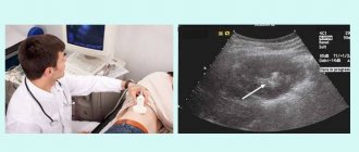
Diagnosis of cholelithiasis
If you suspect stones in the bile ducts and bladder, you should contact a gastroenterologist. The diagnostic complex includes:
- general blood test (signs of inflammation - leukocytosis, elevated ESR);
- biochemistry (high liver parameters);
- duodenal intubation;
- cholecystocholangiography - x-ray with a contrast agent administered orally or intravenously;
- retrograde cholangiopancreatography (endoscopic examination with the possibility of removing small stones from the ducts);
- computed tomography with contrast injection.
Diagnostics
Timely diagnosis of gallstone disease will allow you to avoid surgical intervention in the body and limit yourself to conservative treatment methods.
| Diagnostic method | Peculiarities |
| Ultrasound examination | Determines the presence of stones, the area of damage to the gallbladder and ducts. It is carried out after breakfast - time in the gastrointestinal tract, which allows you to correctly assess the functioning of the gallbladder and the degree of bile outflow |
| Preoral cholecystography | The patient must take medications containing iodine. At the same time, an x-ray is taken to show calcification of the stones. |
| Intravenous cholegraphy A gentle diagnostic method. | A contrast solution is injected intravenously to clarify the scope of functioning of the bile ducts and the presence of stones, the degree of narrowing and expansion of the ducts. |
| Hepatobilioscintigraphy | A radioisotope method for introducing a radiopharmaceutical and recording its movement using a gamma camera in order to clarify the indicators of the movement of bile through the ducts into the intestines. |
| CT scan | The patient undergoes a detailed examination using X-rays. They make it possible to determine the density of stones and their nature of origin in order to select a treatment method (cholesterol stones are dissolved with the help of medications, pigment stones are crushed by a shock wave). |
| Magnetic resonance cholangiography | An examination using an electromagnetic field that allows you to obtain a complete image of the organ. Allows you to accurately identify a duct clogged with stones. |
A little history
Biliary colic, provoked by the movement of stones in the bladder, was mentioned in the treatises of medieval doctors. It was believed that the stones formed in the liver and from there migrated to the gallbladder. Until the end of the 19th century, this very inconvenient disease in all respects was treated in one way - Carlsbad waters and herbal decoctions, consumed internally and externally.
The effectiveness of these methods from the standpoint of modern medicine was zero, and in the case of spasm of the bile ducts and the development of jaundice or acute inflammation, patients often died.
Only in the 80s of the 19th century, the German surgeon Langenbuch invented a very effective method for eliminating problems with a gallbladder filled with stones - its complete removal through a 30-centimeter incision in the anterior abdominal wall. It was then that the era of successful treatment of cholelithiasis began, which, by the way, did not lead to total relief from it, since no one suspected the need to follow a special diet after cholecystectomy.
Prevalence of the disease and its significance
Currently, at least 15% of the entire world population suffers from cholelithiasis.
Most of them are adults, especially women. Cholelithiasis is also diagnosed in children, but these cases are very rare and are associated with more complex problems, such as significant disruptions in metabolic processes.
Gallstone disease has an uneven distribution, since its development requires certain conditions of human existence. The disease is most often diagnosed in residents of Europe and North America. Aborigines of the African continent and Southeast Asia suffer least often from calculous cholecystitis.
Gallstone disease often provokes the occurrence of other pathologies, but in itself it is a consequence of changes that are difficult to treat. Timely treatment, and even better, prevention of the disease, allows you to avoid significant and irreversible changes in physiological processes.
Risk factors
Literally every person has a risk of developing calculous cholecystitis, since the list of phenomena that can trigger the onset of the formation of stones from the bile fraction consists of a dozen points:
disruptions in the functioning of the endocrine system;
- metabolic changes;
- hormonal changes;
- improper (unbalanced) nutrition;
- pathology of the pancreas;
- chronic intestinal diseases;
- decreased contractile functions of the bile ducts;
- excess body weight;
- hormone replacement therapy;
- physical inactivity.
Also, the occurrence of gallstone disease can be affected by pregnancy. During the growth of the gradual enlargement of the uterus, the gallbladder is displaced, and the hormonal background changes.
In childhood, stones in the gallbladder are formed mainly due to genetic defects and congenital anomalies in the structure of the gallbladder and gastrointestinal tract.
Treatment with unconventional methods
Folk remedies are ineffective, however, they play a certain role in therapy.
Positive effects include relaxation of smooth muscles, a decrease in bilirubin concentration, and normalization of the process of bile discharge.
Before using any folk remedy, it is necessary to check the body’s reaction - start with a tiny dosage. If your health has not worsened and there are no harmful symptoms, treatment is permitted.
Folk remedies:
| Alternative Treatment | Recipe and features of reception |
| Radish juice + honey | Mix in equal quantities and consume a tablespoon several times a day. The course lasts a month. |
| Rowan berries | Pour 30 g of berries into 500 ml of boiling water and leave for several hours. Drink 120 ml 3 times a day, treatment for 3 weeks. |
| Mint + celandine | Mix in equal proportions. Next 2 tbsp. pour 1000 ml of boiling water and leave for 10 hours. Drink 1 glass per day. The shelf life of the finished “medicine” is no more than 3-4 days in a cold place. |
Symptoms of possible complications with choledocholithiasis
The course of gallstone disease can be complicated by the following conditions:
- phlegmon of the gallbladder wall;
- biliary fistulas;
- Mirizzi syndrome (compression of the common bile duct);
- gallbladder perforation;
- biliary pancreatitis;
- acute and chronic cholecystitis;
- hydrocele of the gallbladder;
- intestinal obstruction;
- gallbladder cancer;
- acute purulent inflammation (empyema) and gangrene of the gallbladder.
Gallstone disease treatment without surgery (conservative treatment of cholelithiasis without surgery)
Relatively recently, stones began to be crushed without violating the integrity of the skin and internal organs. This method is called shock wave lithotripsy. Drops crushed into many fragments or even turned into sand can enter the intestines with a current of bile and leave the body.
This method can be either self-sufficient or serve as the first stage of the treatment process. In this case, the second stage is drug treatment.
Drug treatment also follows surgical intervention, but can act as independent therapy (this is possible in the case of normal patency of the ducts, the absence of complications of cholelithiasis, stones of a certain composition that are not very large in size, etc.).
Principles of drug treatment:
- normalization of bile movement;
- relief and prevention of inflammation;
- anesthesia;
- harmonization of metabolism.
An illustration of the latter principle within the framework of drug therapy is treatment with ursodeoxycholic acid (a natural component of our bile). How do they work? They dissolve existing stones (we repeat - not all types of stones), prevent their re-formation, and have a choleretic effect. They also protect the liver and affect the functioning of the immune system.
There are also universal treatment measures. First of all, it's nutrition. In the early stages of the disease, nutritional adjustments can be a very effective and self-sufficient measure. Moreover, both the correct choice of products and the mode of their consumption are important. There are universal prohibitions regarding, for example, fatty, fried, spicy, alcohol, etc. But it is important to remember that there are many options for metabolic disorders that lead to thickening of bile. And the more individually (taking into account specific reasons) the diet is formed, the better the therapeutic effect.
But the therapeutic (and preventive) lifestyle is not limited to nutrition. Motor activity and psycho-emotional stability play an equally important role.
In psychosomatics, gallstones are called “frozen aggression.” Bile must flow and energy must move. Petrified aggression, which has not reached consciousness and has no way out, destroys from the inside. Does this mean that we should not hold back, but give free rein to our feelings, even if they are aggressive? It all depends on the form of their expression. The main thing is to be aware of the internal forms of conflict and discontent and look for “ecological” types of resolution.
The methods of choice in the treatment of gallstone disease, a reflection of systemic disharmony, can be holistic methods of traditional medicine - acupuncture, qigong therapy, osteopathy, herbal medicine, homeopathy. Depending on the individual medical history and mood of the person, they can be an alternative to generally accepted therapeutic effects, or may accompany them.
Preventive measures
Preventive measures mainly come down to eliminating all sorts of factors and causes that influence the occurrence of gallstones. Pay special attention to a healthy lifestyle and a balanced diet.
In addition, as preventive measures it is necessary:
To refuse from bad habits
- Monitor your weight, avoid obesity and sudden weight loss.
- If you are overweight, choose a set of sports training and a diet that does not harm the body.
- Women are advised to reduce or eliminate the use of hormonal drugs.
- To refuse from bad habits.
- Monitor the condition of the stool and urine; if the bile ducts are blocked, the stool will be light and the urine will be dark.
It is recommended to have regular laboratory blood tests to monitor cholesterol, bilirubin and enzyme levels. It is important to have timely treatment and examination of the gallbladder when symptoms of the disease appear.
Main causes and risk factors
In recent years, the problem of cholelithiasis has been especially relevant. Gallstones are found in 40% of the adult population. There are many reasons for the development of gallstone disease; the process of stone formation is not fully understood. Based on long-term observations in medicine, factors that precede the development of gallstone disease have been identified:
- Gender – among patients under 50 years of age, women are found 2 times more often than men. This is due to incomplete emptying of the gallbladder in late pregnancy.
- Excess weight – obesity is accompanied by increased cholesterol formation. The exception is vegetarians, in whom stones are rarely diagnosed, even with increased body weight.
- Hereditary factor - in relatives of patients with cholelithiasis, stone formation does not depend on diet, lifestyle or physique.
The root cause of cholelithiasis is a violation of the correct ratio of bile components. Changes in the chemical composition are affected by the use of medications (oral contraceptives for women), infectious pathogens that enter the gallbladder through the bloodstream, helminths, and single-celled parasites.
The catalyst for the development of cholelithiasis is bile stagnation. Obstacles to the outflow of secretions can be:
- tumor formations;
- formation of scars after operations;
- bending of the gallbladder (congenital or acquired);
- strictures (narrowing) of the bile ducts;
- enlarged lymph nodes located near the bladder and bile ducts;
- swelling caused by inflammation;
- dysfunction of the sphincter of Oddi and biliary tract with dyskinesia.
The reason for the increase in cholesterol concentration is the consumption of food oversaturated with animal fats. Over time, it is absorbed by the mucous membrane of the gallbladder. Cholesterol deposits in the walls reduce the contractility of smooth muscles. As a result, atony of the gallbladder and stagnation of bile appears.
Medical reasons are:
- diabetes;
- hormonal imbalances in women during menopause;
- autoimmune disorders;
- Crohn's disease;
- metabolic disorders;
- allergic reactions;
- biliary tract infections;
- diverticula of the gallbladder and ducts.
The work of the biliary chain is under hormonal and reflex control. Cholecystokinin is formed in the small intestine, which is responsible for activating the gallbladder during food digestion. Diseases affecting the small intestine lead to disruption of cholecystokinin synthesis and regulation of the process of bile excretion.
Cholelithiasis is often suffered by people who lead a sedentary lifestyle due to their own laziness or due to sedentary work. Those at risk include those who have lost weight dramatically or are rapidly gaining weight, as well as patients who were forced to switch to parenteral nutrition.
How do you know if you have gallstones?
In order to diagnose a patient with cholelithiasis, the doctor carefully listens to the patient’s complaints and takes into account all the symptoms and sensations. But since no preliminary conclusions can be drawn without diagnostics, the following procedures are carried out:
- The patient's blood is taken for a general analysis, which will reveal the stage of the disease and existing inflammatory processes;
- the blood is also sent for biochemical analysis, which will reveal the activity of substances involved in metabolic processes;
- cholecystography is performed, showing possible enlargement of the organ;
- The patient is necessarily sent for an ultrasound of the abdominal cavity, where the presence of stones, their size, their movement along the ducts, as well as the presence of pathologies is determined.
After all the tests have been completed, the doctor proceeds to prescribe treatment.
How do stones appear?
Sedimentation and crystallization of the leading components of bile is how stones appear in the bile-depositing organ. This course of cholelithiasis is accompanied by:
- restructuring of bile composition;
- presence of cholecystitis;
- bile stasis.
Mostly stones originate in cholecystis, rarely in the bile, hepatic ducts and intrahepatic bile ducts.
The mechanism of formation of stones in cholecystis is as follows:
- with hypercholesterolemia, the cholesterol content in bile also increases;
- the balance of factors that prevent cholesterol from settling into grains of sand is disorganized;
- further exposure to provoking factors on the wall of the bladder provokes its inflammation and mucoids begin to be intensely released;
- excess cholesterol is deposited in their clots;
- the combination of such clots leads to the formation of cholesterol stones; subsequently, crevices appear in these stones, into which pigments penetrate, and thus its core is formed.
The infection initiates the formation of a stone in the organ that stores bile. The transformation of conjugated bilirubin into unconjugated bilirubin is carried out under the influence of certain enzymes that are secreted by microorganisms. Such bilirubin, synthesized in excess, binds to calcium ions, forming calcium bilirubinate - an element of pigment stones.
Stones in the liver without the organ itself also deserve attention. This phenomenon occurs when congestion forms in the hepatic ducts after cholecystectomy.
Cholelithiasis
What are gallstones?
Cholecystis is an organ that stores bile, which is involved in digestion and is produced continuously throughout life. Let's look at what it is - gall bladder stones and their pathogenesis - in detail.
During meals, bile moves into the duodenum (duodenum) and helps assimilate nutrients. 90% of bile is water, another 10% is represented by:
- pigments;
- bile acids;
- cholesterol and other substances.
Disorganization of the biochemical composition of bile is the main cause of cholecystis stones.
However, for the development of a stone in the gall bladder there must be some prerequisites, among them are:
- pathology of metabolism, mostly cholesterol;
- weakness of the wall of the bladder and ducts;
- deviation from the normal anatomy of the biliary tract in the form of kinks, adhesions, cicatricial deformities;
- the presence of an obstacle to the contents exiting the bubble;
- diathesis.
Accumulated sand in the gallbladder, forming conglomerates of different sizes, is the stones of the bile-storing organ.
What types of stones are there?
Let's look at some variants of gallstones and their symptoms to represent:
- why therapy is not always effective and sometimes dangerous without prior consultation with a doctor;
- ways to excrete gallstones from the body.
The following types of stones in cholecystis are distinguished.
- Monocomponent
Cholesterol stones:
- metabolic pathology is the main factor in their occurrence;
- in overweight patients;
- there are no signs of cholecystitis;
- size from 3–5 to 15 mm;
- X-ray negative;
- pale yellow;
- localized in cholecystis;
- single;
- round or oval configuration, as well as flat stones;
- so light in weight that they do not submerge in liquid;
- When burning they glow well.
Bilirubin (colored) stones:
- increased hemolysis is the main cause of such stones;
- localized in the bladder and ducts;
- composed of bilirubin and lime;
- have a variety of shapes;
- often small stones, multiple;
- black with a green tint;
- consistency is strong, but fragile.
Calcifications are an uncommon phenomenon:
- consist of calcium carbonate.
- What other types of gallstones are there? 80% of all stones are combined in composition. Their center is composed of an organic substrate, around it there are layers of:
- cholesterol;
- bilirubin;
- calcium salts.
Such concretions:
- difficult to burn;
- immersed in liquid;
- mostly small and numerous.
- About 10% of stone-carrying cases are complex stones; they are represented by a combination of two forms:
- the base is represented by cholesterol, and the shell is of combined genesis;
- are formed with concomitant cholecystitis and cholangitis.
Types of gallstones
Are sand and stones the same thing?
When, under the influence of provoking factors, the structure of bile changes, it settles, which is how sand is formed in the cholecystis and ducts, and then stones. It becomes clear what stones in the bile-accumulating organ are and where they originate.
Sand in the gallbladder and ducts, with its mild symptoms, makes it difficult to diagnose.
Sometimes the following symptoms appear:
- asthenoneurotic condition;
- persistent dermatitis;
- aversion to fried, fatty foods and alcohol.
Examination using ultrasound sensors is a diagnostic method that helps detect sand in the bile ducts.
Asymptomatic carriage of sand in cholecystis is always dangerous. If it is detected, therapy should be started as early as possible to avoid the subsequent formation of stones in the bile-storing organ.
Treatment of gallbladder with stones
Splitting stones according to the use of drugs does not always give the expected effect, since it requires indefinite therapy and does not often give positive dynamics. It is necessary to eliminate the very reason for the formation of stones.
The areas of treatment for cholelithiasis are listed below:
- Lysis of stones using special medications; this method is considered in the following cases:
- the presence of X-ray negative stones;
- when the stone size is less than 20 mm;
- in the absence of signs of acute cholecystitis.
- Crushing large stones (more than 30 mm) with a special ultrasonic beam for the purpose of their further removal.
- Operative - cholecystectomy by laparotomy or laparoscopic approach.
Removing the organ itself does not lead to a significant increase in the life expectancy of patients.
Stones in a child
The appearance of biliary tract stones in childhood is a rare disease and occurs in less than 1% of cases of biliary tract pathology.
Until the age of 7, boys get sick 2 times more often than girls; at the age of 10–12 years, the situation changes and girls get sick 2 times more often than boys; in adolescence, girls get sick three times more often.
The main reasons for the appearance of stones in cholecystis in children:
- hereditary predisposition to metabolic diseases. Because of this, bile stasis occurs, inflammation occurs in the biliary tract and, as a result, destabilization of the biochemical composition of bile;
- abnormalities of the biliary tract;
- history of viral hepatitis;
- diseases with increased hemolysis (hemolytic anemia).
It should be noted conditions that increase the risk of stones in children:
- family history of biliary tract diseases;
- anomalies in the development of the gallbladder;
- early transfer to artificial feeding;
- physical inactivity;
- nutritional features.
What types of stones do children have?
- in young children, stones are predominantly pigmented;
- Stones in older children are often cholesterol.
In the clinical picture of cholelithiasis in children, there are two variants of the course:
- Hidden when children note the following symptoms of gallstones:
- pain with stones - peri-umbilical or without clear specification, aching in nature. After eating, it takes from 5-10 minutes to half an hour before symptoms appear;
- heaviness in the upper abdomen;
- dyspeptic disorders.
There is no association between consumption of fatty and fried foods and symptoms in children.
- The obvious, or typical, course is characterized by the manifestation of symptoms of hepatic colic. The so-called “vagus phenomenon” appears when, with tachypnea, the pulse becomes slower.
When diagnosing stones in the bladder, in addition to a detailed collection of anamnesis and symptoms of cholelithiasis, laboratory methods are used:
- general blood analysis;
- general urine analysis;
- biochemical blood test.
Instrumental methods that are most preferable in pediatric practice:
- Ultrasound diagnostics.
- Cholecystography.
X-rays are suitable for detecting mixed stones because they contain lime. As a method for identifying cholesterol and pigment stones, x-rays are not informative.
The basic principles of treatment for cholelithiasis are similar to those for adults. Conservative treatment includes:
- diet No. 5;
- taking medications that stop the inflammatory process;
- prescription of antispasmodics;
- painkillers;
- preparations of cheno- and ursodeoxycholic acids.
Recurrent course with continuous colic - evidence for surgical treatment.
Children with cholelithiasis should undergo an annual medical examination in order to:
- preventing recurrence of exacerbation of the disease;
- prevention of complications.