Having discovered a lump in the breast, women tend to panic. The first thing that comes to their mind is the thought of oncology. In fact, cancer is not the only cause of such tumors. True, most of them are also pathological conditions.
Still, sometimes lumps palpable in the mammary glands are just a sign of hormonal imbalance. It can be caused, for example, by taking oral contraceptives. Some girls discover lumps during menstruation. With the end of menstruation everything goes away. Hormone surges during pregnancy also provoke the appearance of tumors in the breast. A similar phenomenon occurs in almost every woman during menopause due to a lack of estrogen.
Even if you are sure that the problem is hormones, you should still consult a doctor. It is impossible to find out the cause on your own, and there is a risk of missing a dangerous disease.
Mastopathy
Pathological proliferation of breast tissue. It is considered a precancerous condition. It occurs due to an increase in estrogen levels and a lack of progesterone. The content of prolactin, which affects the growth and development of the mammary glands, also increases. The disease manifests itself in the formation of nodules in the connective tissue. Sometimes the growths reach the size of a walnut. There is pain in the chest, radiating to the arm. Local lymph nodes swell. The nipple becomes covered with cracks, serous fluid mixed with blood is released from it.
https://youtu.be/5zxcfRO5h7s
Symptoms of mastopathy
A woman can see the very first signs of this disease during self-examination. You need to urgently visit a doctor to clarify the diagnosis if you find:
- changing the shape and contours of the breast;
- skin wrinkling;
- retraction of the nipple inward;
- enlarged lymph nodes;
- peeling, redness of the skin;
- pain when palpating;
- bleeding when pressed;
- enlarged lymph nodes;
- temperature increase.
If the following signs appear, you should immediately consult a doctor:
- The appearance of a large number of bumps of different sizes.
- External change in one breast.
- When you raise your arms, you can see a depression in the skin.
- Pus or bloody fluid appears from the chest when pressed.
- There is constant pain in the area of the nodules.
- Enlarged axillary lymph nodes.
- If the breast lump is motionless and does not have clear contours.
- The skin around the nipples is rough. The nipple itself is retracted or has changed its shape.
Bursting pain is a symptom of breast cancer.
There is no need to wait for everything to work out, and when, in addition to the above signs, the following are observed:
- frequent anovular cycles;
- chronic diseases of the genitourinary system;
- the second phase of the menstrual cycle is shortened.
If any of these appear, contact a specialist immediately. Only he can make the correct diagnosis.
Mastopathy occurs at different periods of a woman’s life. With timely treatment, it usually does not lead to cancer. There is diffuse mastopathy. With it, the pain is mild and appears a week before the critical days. But if the disease progresses, it intensifies and becomes permanent.
With nodular mastopathy at the initial stage, pain appears before menstruation. She's aching, dull. Sometimes it can be very strong, such that you can’t touch your chest. The discharge is small, but after a while its quantity increases. The cones are dense, their size starts in millimeters and ends in centimeters.
Cystic mastopathy is accompanied by intense pain and purulent discharge. They may be clear or brown. The lymph nodes swell and the breasts become enlarged. The boundaries of the tumor are clear and elastic.
If a woman has a lump in her breast, this may indicate that she has a disease such as mastopathy. The main reason for its occurrence is hormonal disorders. During this disease, connective tissues grow, and some sacs with fluid, called cysts, begin to form. Fibrocystic disease can be of two types:
- Diffuse. This is a large number of nodules that can spread throughout the chest.
- Nodal. A particularly dangerous disease that can develop into cancer. It is important to say that this neoplasm cannot be palpated in the “lying on your back” position.
Fat necrosis
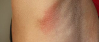
Death of adipose tissue due to poor circulation. Occurs against the background of bruises, operations, and inflammatory processes. It can also be caused by radiation, hormonal disorders, or sudden weight loss. The disease is characterized by the appearance of a hard lump in the chest. The affected area turns red and becomes hot. Patients are concerned about pain and fever. The axillary lymph nodes swell. In advanced cases, ulcers and cracks appear. The tumors are considered benign, but they accelerate the growth of cancer cells.
Diagnostic methods
A woman can suspect the presence of a lump in the mammary gland on her own. Subjective changes may indicate pathology. These include soreness that may radiate to the armpit and arm on the right or left, and a feeling of heaviness in the chest. Also, a woman should periodically conduct a self-examination, including examining the mammary glands in the mirror, as well as palpating the tissue, during which a lump may be felt.
- Mammography is an X-ray technique that makes it possible to detect gross changes in tissues and is used for preventive screening examinations for the early detection of possible malignant neoplasms in women over 40 years of age.
- Ductography is a variant of x-ray examination in which a special contrast compound in the form of a solution is injected into the ducts of the mammary gland.
- Ultrasound examination (ultrasound) is a safe method, since during its conduct the woman’s body does not experience radiation exposure. Ultrasound makes it possible to clearly visualize cavity fluid formations.
- Computed tomography (CT) is an X-ray layer-by-layer scanning of tissues with high resolution and visualization of minimal changes in them.
- Magnetic resonance imaging (MRI) is a layer-by-layer scanning of tissue, in which visualization is carried out due to the physical effect of nuclear resonance in a strong magnetic field. MRI is necessary to study large areas of the body to detect metastases in breast cancer.
- Biopsy is the intravital removal of a small piece of tissue directly from the formation, which is then examined under a microscope to determine the tissue structure. The method is necessary to verify various tumor formations and determine their tissue origin. To avoid pain during the puncture, local anesthesia is performed. You may then experience some pain in the area where the biopsy was taken for a few hours.
If a woman has not seen a mammologist before, you can go to a family doctor or local physician. He will tell you where to go for further examination.
A neoplasm can appear in the mammary gland at any age. With timely consultation with a doctor, correct diagnosis and therapy, the chances of recovery increase, including for cancer.
To make a diagnosis, methods such as:
- X-ray of the breast (at a young age, when glandular tissue predominates in the breast, neoplasms are not clearly visible under X-rays);
- Ultrasound;
- galactography, which is a type of mammography, during the examination, radiopaque agents are injected into the milk ducts, with their help intraductal seals are determined;
- biopsy (puncture) is a method that allows you to obtain breast tissue for further research, with its help it is possible to determine the nature of the neoplasm (cyst, oncology or fibrocystic disease).
Cyst
When the mammary duct enlarges, a cavity is formed in it filled with liquid contents. This is a cyst. The neoplasm is usually small, but can reach 5 cm in diameter. Is round, oval or irregular in shape. Most often, such seals do not cause physical discomfort, but when they become inflamed, unpleasant symptoms appear. The gland becomes deformed and begins to hurt. The skin turns red or takes on a bluish tint. Lymph nodes enlarge. Body temperature rises. The pathology rarely turns into cancer, but the risk still exists.
Classification of breast lumps
Bilateral pain in the chest area that goes away after menstruation indicates mastalgia. The disease can bother a woman for both a short period and the entire reproductive period of life.
Diagram of the female breast.
Women aged 20-30 years old, when feeling their breasts, can detect a round, painless and mobile lump - this is a fibroadenoma. The diffuse form of the disease is characterized by many granular small balls located inside both mammary glands. If the formation is single and has clear outlines, then it is a localized form.
There are also focal (condensed area), as well as nodular (nodes) and cystic (cysts). If, during an independent examination of the upper part of the chest, oval or round shaped lumps, dense in structure, are felt, then this is a phyllodes form of fibroadenomatosis. This type of disease can develop into breast cancer, and therefore requires prompt treatment. A mature woman may develop a ductal cyst - a papilloma located inside the milk duct.
The deposition of calcium salts leads to the formation of calcifications inside the ducts of the mammary glands. They are diagnosed only during mammography and are a precancerous condition.
After trauma to the mammary gland or sudden weight loss, as well as receiving large doses of radiation, dead fat cells disintegrate and can form a cystic capsule - lipogranuloma.
Lactostasis
It occurs due to blockage of the milk duct due to stagnation of milk. Many nursing mothers suffer from the problem. The reason for the deviation is improper attachment of the child, long intervals between feedings, and tight clothing. Congestion leads to the formation of a small lump, and the lump in the chest hurts. The skin over the affected area turns red and its temperature increases. The sensitivity of the gland increases, a small white bubble appears on the nipple. The rate of milk release from the damaged gland is reduced. If measures are not taken, inflammation occurs.
Diagnosis of seals
Usually a woman herself finds a lump in her breast. It is necessary to learn how to competently conduct self-examination; inept actions can cause injury. The procedure consists of several stages:
- Visual inspection. You need to undress to the waist, in front of the mirror and lower your arms. Normally, the mammary glands may differ slightly in size from each other, but if the difference becomes larger, this may be a cause for concern. The mammary glands should be located symmetrically. When standing and placing your hands behind your head, bending your torso, both breasts should move evenly. It is important to pay attention to whether there is any fixation of one breast when raising your arms.
- Condition of the skin. You need to check whether the skin folds well, whether there are redness, diaper rash, rash, or “lemon peel.” Has the pattern of the veins become stronger, is there any wrinkling of the skin, tubercles, retractions, or dimples?
- Palpation of the breast. To examine the right breast, you need to raise your right hand up and behind your head. With your left hand you need to press lightly on your chest, avoiding painful sensations. It is advisable to use a rich cream to prevent injury to the skin. Palpation is carried out only with the pads of the fingers; it is better to make circular movements, centimeter by centimeter examining the entire mammary gland. Start from the nipple area and gradually move to the armpit.
- Nipple examination. You need to pay attention to whether they have changed color, shape, whether there are any retractions or cracks; after gentle pressure, see if there is any discharge from them.
- Examination of the breast in the supine position. To do this, lie on your back and place a small pillow or a towel cushion under your left shoulder blade. Each mammary gland must be felt in 3 positions: the hand lies along the body, the hand is moved to the side, the hand is placed behind the head. Visually divide the mammary gland into 4 sectors and feel them in a spiral.
Self-examination should become a habit and be done regularly, and can be done while taking a shower. Fingers will glide well over the soaped skin, and the examination will be of better quality.
If a lump is detected in the mammary gland, a woman should immediately make an appointment with a mammologist. At the first appointment, the doctor will interview the patient, find out the symptoms that are bothering her, and conduct a visual examination and palpation.
Oncology
All of the above diseases do not pose a threat to a woman’s life, but significantly increase the risk of developing malignant neoplasms. There are several types of breast cancer. Each has specific characteristics. Typically, oncology manifests itself in the form of compactions of various sizes and hardness. The breast increases or decreases, shifts relative to its normal position. The skin changes color and texture. Pathological discharge from the nipple, the appearance of cracks, ulcers and erosive areas are possible.
Do not panic when a lump appears in the mammary gland. But you still need to be wary. Although in most cases neoplasms are relatively harmless, each of them brings them closer to oncology. If you have the slightest concern, you should contact a mammologist.
https://youtu.be/wyEQahWsVjE
What types of seals are there?
For the most part, various lumps in the mammary gland are benign formations that can lead to tumors. Among them are the following types:
- Lipoma. It appears as a benign tumor in adipose tissue. They come in various sizes, do not cause pain, and can be located both singly and multiple times. Sometimes they do not require medical treatment.
- Abscess. Becomes common during breastfeeding. It is a pocket that is filled with pus and is located in the mammary gland. All this is accompanied by a slight lump in the chest, severe pain, redness of the skin and an increase in temperature in the area of the affected chest. Here, only surgical intervention is prescribed, when pus is removed from the formed compaction and a course of antibiotic therapy is administered.
- Thrombosis. It occurs due to blockage of the veins in the chest area. Swelling of the mammary gland, hyperthermia, compaction in the area where the vein drains, pain, and redness of the skin appear. First, conservative treatment is carried out, and then surgical treatment if the previous one does not help.
- Cyst. It often occurs in women aged 35-50 years due to a hormonal imbalance in the body’s level of estrogen, the main female hormone. They are provoked by the parallel development of inflammatory processes in the reproductive system. The woman feels discomfort in her chest, and a moving lump is palpable due to its being filled with secretions. Pain may or may not be present.
- Fat necrosis. This is when healthy cells in the body transform into rounded shapes that are very sensitive and painful. The skin over them is changed and has a bluish tint. They are usually caused by trauma to the chest shortly before they appear. They usually resolve on their own, but there are cases of scarring. They feel like painless lumps to the touch.
Often these lumps are accompanied by symptoms such as chest pain and nipple discharge. If such discharge becomes cloudy, brown or bloody, then you should definitely consult a doctor, as this may indicate the development of cancer.
Depending on whether lumps are felt in one or both breasts, they are unilateral or bilateral. They are also divided into cyclic and acyclic:
- Cyclic compactions are also called physiological and inherent in hormonal levels. In the female body, the hormonal background is constantly in flux. If such lumps are a consequence of the onset of menstruation, then you should not pay attention to them, they will go away on their own.
- Acyclic compactions do not depend on the menstrual cycle, so they occur at any time. It is these seals that require medical examination and treatment. Sometimes this may involve taking hormonal contraceptives, which affects hormonal levels.
go to top
Diagnostic methods
Depending on the nature of the compaction, therapy will vary. Therefore, if a neoplasm is detected, you should not self-medicate, but should immediately contact a specialist.
For lactostasis, the doctor may prescribe various compresses on the chest, for example with camphor oil or balsamic liniment according to Vishnevsky.
For mastitis, antibiotics are indicated; if an abscess develops, surgical treatment is used.
If a woman is diagnosed with mastopathy, she is recommended to visit a doctor every six months to monitor the disease over time.
Young women should have an X-ray and ultrasound of the breast done once every two years, and older women once a year.
Hormonal and non-hormonal drugs are prescribed for treatment. For diffuse mastopathy, therapy is aimed at eliminating the main cause of the pathology, which caused dysfunction of the pituitary gland and ovaries. Treatment often begins with the treatment of diseases of the reproductive organs, normalization of the liver and nervous system.
For nodular mastopathy, specific immunotherapy drugs are prescribed: an allergen vaccine is administered, gradually increasing the dosage to achieve stable remission.
In most cases, surgical treatment is indicated, which consists of excision of nodes. Non-hormonal methods include diet and vitamin therapy, diuretics, non-steroidal anti-inflammatory drugs, drugs that improve blood circulation, Bromcamphor, and Potassium iodide are prescribed. Antioxidants are prescribed, such as B-carotene, phospholipids, zinc, selenium, which have a positive effect on liver function. It is also important to choose the right bra, avoid visiting saunas and solariums, and reduce your time in the open sun.
If small cysts with a diameter of no more than 0.5 mm are detected, conservative treatment is prescribed, which is primarily aimed at normalizing hormonal levels.
To resolve a single-chamber cyst, a puncture is performed. The specialist makes a puncture, pumps out the liquid and injects a solution that destroys the capsule. In cases of an atypical cyst, surgery is performed, and the excised tissue is sent for histological examination.
Aspiration is also used to treat cysts.
A thin cannula is inserted into the cyst cavity with the help of which the fluid is pumped out. If traces of blood are present in the cystic fluid, additional examination and treatment are required.
Drug therapy consists of prescribing hormonal agents and drugs aimed at strengthening the immune system.
Depending on the severity of breast thrombophlebitis, treatment can be conservative or surgical. Conservative therapy requires bed rest, wearing elastic bandages, physiotherapy, taking medicinal herbs, compresses with ointments or alcohol.
Breast lipoma is treated only surgically.
If the lump in the chest is a malignant neoplasm, then radical, auxiliary, and palliative treatment is prescribed. Radical or surgical treatment consists of removing the tumor. Auxiliary or conditionally radical treatment is prescribed before, during and after surgery, and in rare cases it is carried out instead.
This could be radiotherapy, chemotherapy, molecular targeted therapy, immunotherapy. If the disease is diagnosed later, there is no point in performing surgery. In the presence of metastases, palliative treatment is prescribed, which will help alleviate the patient’s well-being and prolong his life.
Treatment of breast lumps can be conservative, surgical or complex. It depends on the cause and type of tumor.
- Surgery is also indicated for rapidly growing benign breast tumors.
- Malignant neoplasms of the mammary gland, in addition to mandatory radical surgery, require additional treatment in the form of a combination of radiation and chemotherapy.
- Treatment of various options and types of benign mastopathy, which is prescribed, carried out and monitored by a mammologist, consists of changing the usual lifestyle, using modern hormonal drugs, multivitamins, immunostimulating, decongestant and anti-inflammatory drugs, as well as analgesics.
Nota bene!
If you are concerned about a lump in the mammary gland, we recommend that you consult a mammologist-oncologist for diagnosis and treatment. Lumps in the breast often threaten a woman’s life and can cause cancer.
If a lump in the chest hurts, grows quickly, and is accompanied by the appearance of other clinical manifestations, diagnostics are required, as well as subsequent treatment if necessary. The direction of therapeutic measures depends on the nature and severity of the diagnosed pathological process that led to the formation of the compaction:
- Conservative treatment – for mastopathy, hormonal drugs are used, including oral contraceptives. In case of infectious origin of the disease, broad-spectrum antibiotics are prescribed. For breast cancer, cytotoxic drugs and radiation therapy are additionally used. After proper therapy, the chest may stop hurting, which indicates a decrease in the severity of the pathological process.
- Surgical intervention - an operation is performed to remove the identified formation, which continues to grow and poses a danger to the woman. If a malignant tumor is diagnosed, partial or complete removal of the mammary gland en bloc with regional lymph nodes can be performed.
The operation is performed according to strict indications, especially in cases where the expectant mother is going to give birth and breastfeed the child. If there are no health-threatening conditions, conservative therapy is carried out. Pregnancy and breastfeeding contribute to the reversal of mastopathy.
Based on the clinical picture, it is impossible to reliably make a conclusion about the nature and severity of the pathological process, therefore additional objective diagnostics are required.
Possible reasons
A painful lump in the mammary gland is not an independent disease. It refers to the manifestations of a large number of pathological processes. The most common ones include:
- Lactostasis is stagnation of milk in individual lobules of the mammary gland, which occurs during lactation associated with breastfeeding.
- The formation of a cyst of various origins, which is a limited cavity filled with fluid.
- Adenoma is a benign tumor that originates from glandular cells. Depending on the histological structure, fibroadenoma and fibrolipoadenoma are distinguished, which also contain connective and adipose tissue.
- Lipoma is a benign neoplasm originating from adipose tissue.
- Intraductal papilloma is a tumor, the manifestation of which is provoked by the activity of certain viruses. It usually has a benign course and can lead to lactostasis during breastfeeding.
- A galactocele is a specific cyst that contains fluid inside that resembles breast milk. It can usually form due to blockage of the excretory duct.
- A leaf-shaped or phylloid tumor is a variant of fibroadenoma, which, unlike other benign tumors, can degenerate into cancer.
- Malignant neoplasm or breast cancer.
- Mastopathy is the appearance of growths of glandular tissue, provoked by changes in a woman’s hormonal levels. In a small number of cases, a girl before puberty, including a child or teenager, may develop mastopathy. The appearance of a knot can provoke a blow to the chest area, after which a bruise forms. Enlargement of the mammary glands with nodes can occur in a newborn boy against the background of a physiological hormonal crisis.
- Infectious mastitis is the result of the entry and activity of pathogenic microorganisms in the breast tissue.
Severe infection of breast tissue leads to the formation of an abscess of various locations, which is a cavity with pus, limited by a connective tissue capsule.
A large number of causes for the formation of a painful lump require careful diagnosis, since treatment may have fundamental differences.
Treatment for lumps in the mammary gland
The nature of the treatment of lumps in the mammary gland directly depends on the pathology that caused the formation, the prevalence of the pathological process and the stage of development of the process.
In the case of normal age-related or hormonal changes, specific treatment for breast lumps is not required, because in most patients they resolve on their own within 3 months.
If a diagnosis of Mastopathy is made, special treatment is required. Basic therapy can be divided into two large groups - hormonal and non-hormonal.
Hormonal therapy is based on the use of pharmacological drugs of the following groups:
- Antiestrogens.
- Oral contraceptives.
- Gestagens.
- Androgens.
- Prolactin inhibitors.
Non-hormonal treatment includes the following aspects:
- Rational diet.
- Selecting appropriate underwear.
- Use of NSAIDs (non-steroidal anti-inflammatory drugs).
- Diuretics.
- Agents that improve systemic blood flow.
- Antioxidants.
- Vitamins of group A, B, E.
Quite often, with the development of dangerous forms of mastopathy, and the risk of transition to a malignant form, surgical treatment is prescribed. The extent of the operation, further treatment and possible consequences directly depend on the nature of the pathology.
It should be noted that the use of physiotherapeutic procedures can be extremely dangerous, because can stimulate the growth of potentially dangerous tumor cells. This may include a bathhouse, sauna, sunbathing, etc. Using such treatment on your own can be dangerous to the health and life of the patient.